Effects of GaAs (904 nm) Low Level Laser Therapy on Dentin Hypersensitivity
Tae-Hee Won, D.D.S., Ki-Suk Kim, D.D.S.,M.S.D.,Ph.D.
Department of Oral Medicine, School of Dentistry, Dankook University
The aim of the study was to investigate the effects of 904 nm GaAs laser irradiation for patients with hypersensitive teeth and to find the possibility of clinical use of this Low Level Laser Therapy (LLLT) for the control of hypersensitive teeth. Eleven patients visited Dept. of Oral Medicine, Dankook University participated in this study. Each patient contributed at least two or more contralateral pairs of hypersensitive teeth with exposed dentine at cervical surfaces.
Total number of teeth used from subjects participated in this study was 50: 25 experimental and control teeth respectively. All participants were treated with 904 nm GaAs diode laser every week during 4 weeks. Tactile and cold (ice stick) tests were carried out before LLLT every week during 4 weeks and 1 week later after the last LLLT by measuring visual analogue scale (VAS) of patients and by measuring a score of electrical pulp tester (EPT) simultaneously. The VAS score in tactile test decreased significantly with time, but there was not statistically difference between those of groups. The score of EPT in the experimental group was significantly higher than that of control group, although there was no change with time. In cold test, there was significant difference between two groups and cold sensitivity of the experimental group significantly decreased with time after every LLL irradiation, compared with that of control group. Based on the results, it is suggested that the 904 nm GaAs laser irradiation could be positively used as an effective, reversible method in treating cervical dentine hypersensitivity.
Key words: Dentin Hypersensitivity, GaAs, Low Level Laser Therapy
1)
Ⅰ. INTRODUCTION
Dentin hypersensitivity(DH) has been defined as Corresponding author : Ki-Suk Kim,
D.D.S.,M.S.D.,Ph.D. Professor
Dept. of Oral Medicine, School of Dentistry, Dankook University Sinbu-dong san 7-1, Chunan, Choongnam 330-716 Korea
Tel: 041-550-1914 Fax: 041-556-9665 Email: [email protected]
Received: 2011-09-05 Revised: 2011-10-10 Accepted: 2011-11-11* The present research was conducted by the research
pain arising from exposed dentine, typically in response to chemical, thermal, tactile or osmotic stimuli, which cannot be explained as arising from other forms of dental defect or pathology.
1)The prevalence ranges between 8.9 and 15% in the adult western population.
2-4)The etiology of DH is multifactorial and not
completely understood, although it has been
demonstrated (scanning and transmission electron
microscopy) by several investigators that the
structure of dentine in the affected (sensitive) areas
of a tooth is altered, containing a larger number of
patent dentine tubules with a wider tubular diameter
than unaffected areas (non-sensitive).
5-7)Management of DH should be based on a correct
dental practitioner should be aware of the importance of a preventive strategy, particularly with a view to the removal of any etiological factors and minimizing the effects of erosion.
8)The goal of treatment of DH ideally should be the restoration of the original impermeability of the dentinal tubules and the relief of DH experienced by the patient or at least to reduce the level of discomfort to enable the patient’s quality of life to be maintained.
Many substances have been tried with varying degrees of success in the treatment of hypersensitive dentine. One of the most common used agents is strontium chloride,
9)while clinical trials have shown that concentrated fluoride solutions, are also effective.
10)Today various laser systems are discussed for a possible use in dentistry. The Nd:YAG and CO
2lasers are limited due to their thermal side effects and Er:YAG laser is irreversible method due to its thermomechanical ablation mechanism, although Schwarz et al.
11)reported that Er:YAG laser is effective in desensitizing of hypersensitive dentine.
The consensus recommendations for the diagnosis and management of DH by a broadly constituted board of dentists and dental hygienists drawn from general dental practice, specialist practice, academia and research from across Canada, joined by 2 international dentists with subject matter expertise, recommended that depending upon the severity and extent of the condition, reversible procedures should be employed before nonreversible procedures.
12)Recently, the GaAlAs (790 nm) laser irradiation as a reversible procedure was introduced in treating DH by means of inducing changes in neural transmission networks within the dental pulp, which may stimulate the normal physiological cellular functions. Therefore, at subsequent appointments, the pulpal tissue was less injured or inflamed. In addition, the laser stimulated the production of sclerotic dentin, thus promoting the internal obliteration of dentinal tubules.
13)The aim of this study was to investigate a low level laser therapy (LLLT) with a different wavelength and diode (904 nm of GaAs) also introduced in treating DH
clinically, using tactile, cold and electrical pulp tests and to determine 904 nm of GaAs LLLT could be effectively used to treat DH as a reversible procedure.
Ⅱ. MATERIALS AND METHODS 1. Subject
The subjects selected for this study were eleven patients attending the Department of Orofacial Pain and Oral Medicine of Dental Hospital, Dankook University, Cheonan, Korea. The mean age of Six males and 5 females were 50.4 and the range of age was 27 to 66 years. Each patient contributed at least two or more contralateral pairs of hypersensitive teeth with exposed dentine at cervical surfaces.
Criteria for exclusion from the study were: carious lesions on the selected or neighboring teeth, any desensitizing therapy on the selected teeth during the last 6 months, cervical fillings on the selected teeth, cracked or fractured teeth, those continuously taking analgesic medication.
11)Total number of teeth used from eleven subjects participated in this study was 50: 25 experimental and control teeth respectively. The study protocol and informed consent documents were submitted and approved by the Ethics Committee of the Dental Hospital, Dankook University (# H-0902/009/002).
2. Methods
The study was performed according to a randomized split-mouth design. A total of 12 maxillary and 13 mandibular pairs were included. A tooth was used as an experimental tooth and irradiated with 904 nm GaAs laser. A contralateral tooth served as a pseudoirradiated control in each patient. All patients were treated by one experienced operator every week for 4 weeks and evaluated by the other one every week before LLL irradiation for 4 weeks and one week later after last irradiation.
A GaAs laser (DENS BIOS LASER
Ⓡ, TNC, Seoul,
Korea) was used for laser treatment at a mean
Time
Group BT 1W 2W 3W 4W P value
Experimental M±Sd decreasing rate
30.92±26.605 100.000
19.20±22.716 37.904
17.00±19.257 45.019
9.40±13.254 69.599
7.50±8.165 75.744
0.456
Contral M±Sd
decreasing rate
23.00±27.003 100.000
17.28±21.398 24.870
20.60±28.807 10.435
17.00±24.281 26.087
16.80±25.367 26.957
P value 0.010 0.297
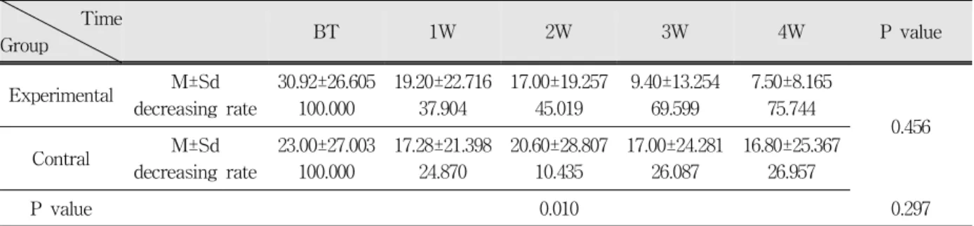
Table 1. Means and standard deviations of VAS score measured using a tactile test according to time after laser irradiation and results of repeated measures 2-way ANOVA
energy level of 2.2 mJ and a repetition rate of 1,000 Hz according to the instructions given by the manufacturer. The laser beam was handled in a non-contact manner about 5 mm away from a target area. The diameter of probe emitting laser was 5 mm and the area irradiated 5 mm apart from a probe of laser system used in this study was about 32 mm
2(4×8 mm). The treatment time was 3 minutes per tooth by irradiating both cervical and apical areas of tooth buccally (experimental group).
The irradiated energy per tooth was 0.40 J and the energy density was 1.24 J/cm
2in this study. The treatment was also performed identically to all contralateral teeth (control group) except focusing laser beam on the cheek side.
To apply mechanical stimuli for tactile test, scratching the dentine surface with a sharp dental probe was used.
9,14-20)The degree of sensitivity to a cold stimulus was determined qualitatively through an ice stick contact to cervical dentine for 3 seconds (temperature range 7-8℃). Ice stick was made by filling with water in an empty lidocaine ample and freezing it. Sensitivity for mechanical and thermal stimuli was assessed using a visual analogue scale.
21)Pulp vitality tester (Parkell Inc., Edgewood, Network, USA) was used for evaluate the sensitivity to an electrical stimuli. These 3 tests were performed in the order of above description.
The data obtained were analyzed statistically using repeated measures analysis of variance (ANOVA). The level of significance for statistical analysis was set at p<0.05.
Fig. 1. A linear gram showing a change of VAS by a tactile test with time after laser irradiation.
Ⅲ. RESULTS
Table 1 shows the results of the tactile evaluation from 25 paired teeth. The examiner found that the VAS score in tactile test decreased significantly with time, although there was not statistically difference between those of groups(Fig. 1). It is thought that this result should be due to a remarkable decrease of the VAS of LLL irradiated group against a weak change of control group and due to relatively large standard deviation.
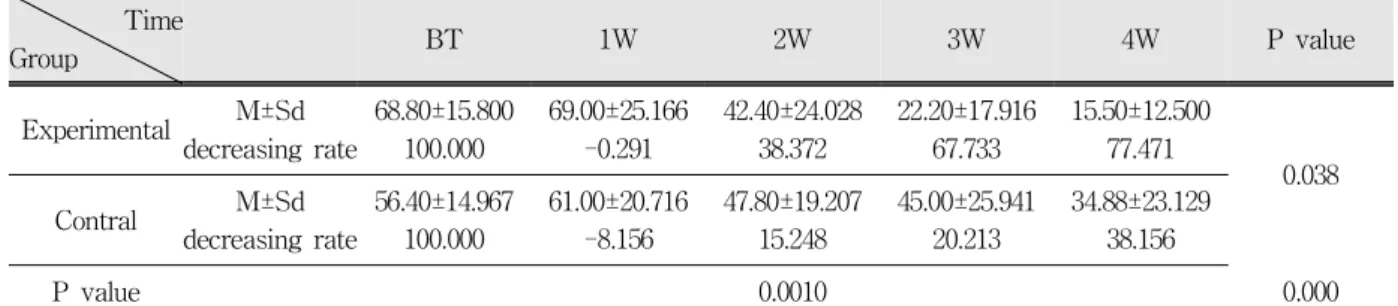
The results of thermal(cold) evaluation can be seen in Table 2. There were significant differences between groups with time. The experimental group showed that cold sensitivity significantly decreased after every LLL irradiation compared with that of control group (Fig. 2).
Table 3 and Fig. 3 showed the results of an
electrical pulp test in this study. There was a
Time
Group BT 1W 2W 3W 4W P value
Experimental M±Sd decreasing rate
68.80±15.800 100.000
69.00±25.166 -0.291
42.40±24.028 38.372
22.20±17.916 67.733
15.50±12.500 77.471
0.038
Contral M±Sd
decreasing rate
56.40±14.967 100.000
61.00±20.716 -8.156
47.80±19.207 15.248
45.00±25.941 20.213
34.88±23.129 38.156
P value 0.0010 0.000
Table 2. Means and standard deviations of VAS score measured using a cold test according to time after laser irradiation and results of repeated measures 2-way ANOVA
Fig. 2. A linear gram showing a change of VAS by a cold test with time after laser irradiation.
significant difference between the experimental and the control groups, but was no change with time.
The value of EPT in the experimental group was higher than that of control group (p =0.018). The initial EPT score(before treatment) of experimental group was slightly higher than that of control
Time
Group BT 1W 2W 3W 4W P value
Experimental M±Sd decreasing rate
3.12±1.900 100.000
2.72±2.170 12.821
2.78±1.904 10.897
3.04±2.086 2.564
2.82±1.520 9.615
0.018
Contral M±Sd
decreasing rate
2.88±1.641 100.000
2.1±1.534 27.083
2.22±1.242 22.917
2.22±1.331 22.917
2.46±1.664 14.583
P value 0.495 0.929
Table 3. Means and standard deviations of EPT score measured according to time after laser irradiation and results of repeated measures 2-way ANOVA
Fig. 3. A linear gram showing a change of EPT scores with time after laser irradiation.
group, but the EPT score of control group after 1
week decreased significantly, compared with that of
experimental group. The decrease of EPT score in
both groups after 1 week is thought to be due to
patient’s fear to a discomfort for electrical stimulus
having experienced at the first EPT test.
Ⅳ. DISCUSSION
To apply mechanical stimuli for tactile test in this study, scratching the dentine surface with a explorer probe was used.
9,14-18,20)Explorer probe use to evaluate sensitivity has been criticized. A mechanical probe introduces variability in pressure.
Ideally, one would require the same tactile pressure to be exerted on all test teeth at all time intervals during a given clinical trial.
22)The use of a sharp probe may also scratch the dentine surface.
According to a previous report
23)pressure, even from a gentle force of 5-10g, is sufficient to overcome the elastic limit of dentine, leading not only to compression and smear layer creation under the explorer tip, but also to permanent (microscopic) deformation of dentine (scratch development). This deformation of dentine may cause displacement of tubular fluid inwardly at a rapid rate, which activates mechanoreceptors, thereby triggering a pain impulse. The examiner in this study was an experienced dentist for these tests and a tactile force of the examiner was in the range of 20-30 g. This tactile force can be thought to be sufficient to deform dentine according to previous report. The authors, however, did not investigate a deformation of dentine in this study and further research is required to determine whether this large tactile force had an effect on this study.
The scratching of the dentine may also remove a therapeutic agent deposited during a clinical trial, but this does not seem to substantially influence pain threshold.
24)In this study, the teeth, which had dentine hypersensitive but had not been treated, were selected.
Several investigators have applied cold water to exposed cervical dentine.
14,25,26)Minkov et al.
15)applied cold water (7℃) from a syringe, while Uchida et al.
17)used 15 ml of cold water (7℃) utilized 20℃ cold water. Flynn et al.
3)used 15 ml of cold water (7℃) which was rinsed around the mouth for a few seconds. These investigators suggested that cold water at 7℃ was ideal for the identification of sensitive teeth as well as minimizing the
incidence of false positive responses. Sensitivity for reasons other than cervical dentine sensitivity, however, cannot be ruled out. Cold water testing, therefore, has been criticized for its lack of objectivity.
27)It is also difficult to determine how much water has been placed on the tooth and the timing of this placement.
28)Ice stick was used in this study to determine qualitatively the degree of sensitivity to cold stimulus. The surface temperature of ice stick used in this study was in the range of 7℃ and 8℃ as recommended by several researchers.
3)An ice stick, which was not melted easily for 3 seconds, was relatively stable and it is thought that it didn’t cause to stimulate gingiva or adjacent teeth during test in this study. It was believed, therefore, the use of an ice stick in a cold test should be very helpful for us to decrease false positive responses. Resultantly, the cold test used in this study showed the most significant difference after laser irradiation than other tests.
Electrical stimuli have been used by several
investigators to quantify both prepain and pain
thresholds in cervical dentine sensitivity.
29-32)Unlike
the other stimuli used to quantify cervical dentine
sensitivity, dentinal tubule fluid movement is not
necessary for transmission of the electrical stimulus,
but rather the presence of lower resistance organic
material in cementum, enamel or dentine.
33)Electrical stimuli, would, therefore, appear to be
more suitable for measuring pulpal activity than for
quantifying cervical dentine sensitivity,
22)but the
validity of such pulp testing has been called into
question.
34,35)Current leakage via the periodontal
ligament and subsequent stimulation of periodontal
nerves may also yield false positive data. A
conventional pulp tester, which was used in this
study, is battery powered, producing pulses of direct
current. The intensity of the output voltage
(stimulus intensity) may be increased by pre-setting
various numbered gradations (0-10) on a thumb
wheel. Problems, however, arise in the interpretation
of the information gathered in such a procedure,
since it is incorrect to assume a direct relationship
between stimulus intensity in volts and the number
on the thumb wheel.
36)Results from initial studies by these latter investigators clearly demonstrated that conventional pulp testers were not suitable for quantifying cervical dentine sensitivity. Further, stimulation of the pulp on the basis of applied voltage may fail to represent exact pain threshold values, in as much as the stimulating current depends on varying resistance pathways to the pulp or to other adjacent tissues.
37)The use of electrical stimuli to quantify cervical dentine sensitivity has been criticized on the basis of being non-physiology, since the response to such stimuli fails to correspond to the painful response normally experienced by cervical dentine sensitivity patients. Unlike thermal stimuli, electrical stimuli are not normally encountered in real life situations, and as such there is a question as to the relationship between the voltage values with the electrical stimulus procedure and the pain scale values obtained with normally experienced stimuli.
21)Fear of experiencing an unknown stimulus and possible discomfort, may, therefore, influence the patient’s assessment of pain and in consequence a lower pain threshold value may be recorded. The result of EPT in this study showed that the values of EPT in both groups decreased considerably after 1st EPT, especially the EPT value of control group was significantly lower than that of experimental group after 1st EPT experience. This EPT result was different from those of other tests, cold and tactile tests in this study.
The order of application when more than one kind of stimulus is used is important. Care should be taken to ensure that the 1st should not distract from the 2nd, nor the 2nd from the 3rd and so on. The least disturbing stimulus should, therefore, be applied first, with the most disturbing used last.
37)Several investigators have applied either tactile, electrical or heat stimuli prior to the application of cold air on the basis that the former do not appear to elicit a painful response which could affect the latter.
20,30,31,38,39)In this study, however, the order of test application was tactile, cold and electrical pulp tests. Because, unlike thermal stimuli, electrical
stimuli are not normally encountered in real life situations, EPT was performed last of all in this study. It is not assumed that cold test having performed in advance necessarily affect the result of EPT performed later in this study. However, because several reports suggested the order of application is important, further research for the effects of cold test on EPT results is required.
There are problems in evaluating patient subjective response to the various stimuli used in the assessment and treatment of cervical dentine sensitivity. Opinions vary as to the reliability of some of these methods of assessment
27,39)although more recently efforts have been made to develop controlled reproducible stimuli more suited to the evaluation of cervical dentine sensitivity.
20,38-40)Although Tarbet et al.
30)claimed that their methodology was objective, the examiner determined a threshold through the patient’s response on perceiving discomfort in this study and methodology employed in this study, may therefore, not be as objective as claimed by them. Currently, no single method of eliciting and assessing cervical dentine sensitivity may be considered ideal. The absence of suitably objective methodology of assessing cervical dentine sensitivity and the lack of standardized measurement of the subjective response following application of stimuli still gives cause for concern. In this study, therefore, three tests such as tactile, cold and electrical pulp tests were used to obtain as much as a reliability of test results from a subjective test and the cold test using an ice stick, among three subjective tests used in this study, would be recommended for researchers to use in the assessment of dentine hypersensitivity based on the results of this study. Further research is, however, required to evaluate suitable methodology for the quantification of realistic test stimuli under controlled clinical conditions, whereby the subjective response may be objectively measured.
Recently, a low level laser was introduced in
treating dentin hypersensitivity comparing
desensitizing efficacy of GaAlAs laser and dentin
bonding agent.
41)They observed two treatment groups using GaAlAs laser and dentin bonding agent promoted a considerable decrease in sensitivity after 15 days of the first application and continued desensitizing effects were observed 30 days later. In a study on the desensitizing effects of an Er:YAG laser for hypersensitive dentine,
11)it was concluded that Er:YAG laser is effective and the maintenance of the positive result was more than with a desensitizing aqueous solution containing polyurethane-isocyanate and methylenechloride.
It is possible that the two kinds of lasers may reduce DH by different mechanisms. If DH results from the movements of fluid in the tubules, fusing the tubules should result in a predictable elimination of DH. Thus, it could be suggested that dentin fused by a high power laser such as Er:YAG, Nd:YAG
42-44)and CO
245,46)lasers are responsible for an obturation of the dentinal tubules. The clinical effect of high power laser on DH, therefore, occurs immediately as reported by Schwarz et al. and can be remained for long time. Whereas, a diode laser irradiaton also provided reductions in dentinal pain elicited by thermal, tactile, and air blast stimulation. The clinical effect of low-level lasers on DH relies upon an immediate analgesic effect as a consequence of the interruption of the nerve impulse path in the affected nerve fiber, concluding that laser acts as a reversible suppressor directly on the neuronal activity
47)and an increase in the nerve ending threshold for pain, attributed to the maintenance of the receptor membrane potential and the suppression of the nerve ending fiber pulp potential.
48)This study, however, showed a long lasting effects of GaAs diode laser irradiation on DH during 4 weeks like previous studies such as Sicilia et al. (2 months)
49)and Vieira et al. (3 months).
50)Gerschman et al. investigated the effectiveness of four applications of the GaAlAs laser using the wavelength of 830 nm to both the apex and the cervical areas of hypersensitive teeth at 1 wk, 2 wk, and 8 wk intervals. It was observed that the decrease in pain severity became more evident over time and that at 8 wk, the percent reduction in
sensitivity to probe and air stimuli based on a VAS was 65% and 67%, respectively. These results have some similarity from the findings obtained in the present study. The present study, although the duration of examination, the method of cold test (ice stick instead of air blast) and the laser diode used were different those of that study, showed that the sensitivity of dentin decreased more and more over time and that at 4 wk, the percent reduction in sensitivity to probe and cold (ice stick) test based on a VAS was larger than their results: 75.7% and 77.5% respectively.
It can be assumed that this long clinical effects of LLLT on DH should be a delayed obliteration of dentinal tubules by tertiary dentin, caused by increased metabolic activity of odontoblasts.
51)Many authors have questioned if the laser light actually reached the dental pulp. Sommer et al. have clarified this point.
52)These authors also observed that the light reaches the pulp without losing practically any energy, thereby clearly showing that laser light is conducted through the inter-tubular dentine and the highly orderly structure of the dentine. It is important to note that light travels parallel to the tubuli, but not through the tubuli.
52.53)It is, therefore, suggested that LLLT, when used with the appropriate treatment parameters and method such as irradiating perpendicularly on a cervical dentine, is effective in treating DH as it quickly reduces pain and maintains a prolonged pain-free status. Further clinical investigations are needed in order to evaluate the long-term stability of the positive results obtained with various diode lasers.
Ⅴ. CONCLUSION
Treatment modality used in this study ( 1week
interval irradiation of 904 nm GaAs laser ) provided
statistically significant gradual reductions in
dentinal hypersensitivity for 4 weeks. Conclusively,
it can be suggested that the 904 nm GaAs laser
irradiation could be positively used as an effective,
reversible method in treating cervical dentine
hypersensitivity.
REFERENCES
1. Addy M, Mostafa P, Absi EG, Adams D. Cervical dentine hypersensitivity. Etiology and management with particular reference to dentifrices. In: Rowe NH, ed. Proceedings of symposium on Hypersensitive Dentin. Origin and Management. University of Michigan, Ann Arbor, MI 1985, pp147-167.
2. Graf H, Galasse R. Morbidity, prevalence and intraoral distribution of hypersensitive teeth. J Dent Res 1977;56(special issue):162, abstr. 479.
3. Flynn J, Galloway R, Orchadson R. The incidence of hypersensitive teeth in the west of Scotland. J Dentistry 1985;13:230-236
4. Scherman A, Jacobsen PL. Managing dentin hyper- sensitivity: what treatment to recommend to patients.
JADA 1992;123:57-61.
5. Absi EG, Addy M, Adams D. Dentine hyper- sensitivity. A study of the patency of dentinal tubules in sensitive and non-sensitive cervical dentine. J Clin Periodontol 1987;14:280-284.
6. Yoshiyama M, Masada J, Uchida A, Ishida H.
Scanning electron microscopic characteristics of sensitive vs. insensitive human radicular dentin. J Dent Res 1989;68:1498-1502.
7. Yoshiyama M, Noiri Y, Ozaki K et al. Transmission electron microscopic characterization of hyper- sensitive human radicular dentin. J Dent Res 1990;69:
1293-1297.
8. Addy M. Tooth brushing, tooth wear and dentine hypersensitivity- are they associated? Int Dent J 2005;55(suppl 1):261-267.
9. Herandez F, Mohammed C, Shannon I, Volpe A, King W. Clinical study evaluating the desensitizing effect and duration of two commercially available dentifrices. J Period 1972;43:367-372.
10. Gedalia I, Brayer L, Kalter N, Richter M, Stabholz A.
The effect of fluoride and strontium application on dentine: in vivo and in vitro studies. J Periodontol 1978;49:269-272.
11. Schwarz F, Arweiler N, Georg T, Reich E.
Desensitizing effects of an Er:YAG laser on hypersensitive dentine: A controlled, prospective clinical study. J Clin Periodontol 2002;29:211-215.
12. Canadian Advisory Board on Dentin Hypersensitivity.
Consensus-based recommendations for the diagnosis and management of dentin hypersensitivity. J Can Dent Assoc 2003;69(4):221-6.
13. Walsh LJ. The current status of low-level laser
therapy in dentistry Part 2. Hard tissue application.
Australian Dental Journal 1997;42:302-306.
14. Cohen A. Preliminary study of the effects of a strontium chloride dentifrices for the control of hypersensitive teeth. Oral Surgery, Oral Medicine, Oral Pathology 1961;14:1046-1052.
15. Minkov B, Marmari I, Gedalia I, Garfunkel A. The effectiveness of sodium fluoride treatment with or without iontophoresis on the reduction of hypersensitive dentin. J Period 1975;46:246-249.
16. Zinner DD, Duany LF, Lutz HJ. A new desensitizing dentifrice. Preliminary report. JADA 1977;95:982-985.
17. Uchida A, Wakano Y, Fukuyama O, Miki T, Iwayama Y, Okada H. Controlled clinical evaluation of a 10%
strontium chloride dentifrice in treatment of dentin hypersensitivity following periodontal surgery. J Period 1980;51:578-581.
18. Carlo GT, Giancio SG, Seyrek SK. An evaluation of iontophoretic application of fluoride for tooth desensitization. JADA 1982;105:452-454.
19. Guo-Hua L, Morimoto M. Magnesium sulphate as a desensitizing agent. Oral Rehabil 1991;18:363-372.
20. Person P, Demand EE, Koltun L, Spindel M. A microprocessor temperature-controlled air delivery system for dentinal hypersensitivity testing. Clinical Preventive Dentistry 1989;11:3-9.
21. Gillam DG, Newman HN. Assessment of pain in cervical dentinal sensitivity studies: A review. J Clin Periodontol 1993;20:383-394.
22. Clark GE, Troullos ES. Designing hypersensitivity clinical studies. Dental Clinics of North America 1990;34:531-44.
23. Pashely DH. Mechanisms of dentin sensitivity. Dental Clinics of North America 1990;34:449-73.
24. Smith BA, Ash MM. Evaluation of a desensitizing dentifrice. JADA 1964;68:639-47.
25. Levin MP, Yearwood LL, Carpenter WN. The desensitizing effect of calcium hydroxide and magnesium hydroxide on hypersensitive dentin. Oral Surgery, Oral Medicine, Oral Pathology 1973;35:
741-6.
26. Miller JT, Shannon IL, Kilgore WG, Bookman JE. Use of a water free stannous fluoride-containing gel in the control of dental hypersensitivity. J Period 1969;40:490-1.
27. Green BL, Green KL, McFall WT. Calcium hydroxide and potassium nitrate as desensitizing agents for hypersensitive root surfaces. J Period 1977;48:667-72.
28. Gangarosa LP. Letters to the Editor. JADA 1986;112:
808-10.
29. Starck MM, Kempler D, Pelzner RB, Rosenfeld J, Leung RL, Mintatos S. Rationalization of electric pulp testing methods. Oral Surgery, Oral Medicine, Oral Pathology 1977;43:588-606.
30. Tarbet WJ, Silverman G, Stolman JM, Fratarcangelo PA. An evaluation of two methods for the quanti- fication of dentinal hypersensitivity. JADA 1979;98:
914-918.
31. Tarbet WJ, Silverman G, Stolman JM, Fratarcangelo PA. Clinical evaluation of a new treatment for dentinal hypersensitivity. J Period 1980;51:535-40.
32. Tarbet WJ, Silverman G, Fratarcangelo PA, Kanapka JA. Home treatment for dentinal hypersensitivity. A comparative study. JADA 1982;105: 227-30.
33. Kleinberg I, Kaufman HW, Confessore F. Methods of measuring tooth hypersensitivity. Dental Clinics of North America 1990;34:515-29.
34. Seltzer S, Bender IB. The nerve supply of the pulp and pain perception. 2nd edition., hiladelphia, 1975, JB Lippincott Company, pp. 131-151.
35. Seltzer S, Bender IB, Ziontz M. The diagnosis of pulp inflammation, correlations between diagnosis data and actual histological findings in the pulp. Oral Surgery, oral Medicine, Oral Pathology 1963;16:
846-71.
36. Kanapka JA, Colucci SV. Clinical evaluation of dentinal hypersensitivity. A comparison of methods.
Endodontics & Dental Traumatology 1986;2:157-64.
37. Ash MM. Quantification of stimuli. Endodontology &
Dental Traumatology 1986;2:153-56.
38. Minkoff S, Axelrod S. Efficacy of strontium chloride in dental hypersensitivity. J Period 1987;58:470-74.
39. Addy M, Mostafa R, Newcombe R. Dentine hyper- sensitivity. A comparison of five toothpastes used during a 6-week treatment period. British Dental Journal 1987;163:45-51.
40. Kern DA, McQuade MJ, Scheidt MJ, Hanson B, Van Dyke TE. Effectiveness of sodium fluoride on tooth hypersensitivity with or without iontophoresis. J Period 1989;60:386-89.
41. Tengrungsun T, Sangkla W. Comparative study in desensitizing efficacy using the GaAlAs laser and dentin bonding agent. J Dentistry 2008;36:392-395.
42. Renton-Harper P, Midda M. Nd:YAG laser treatment of dentinal hypersensitivity. British Dental Journal 1992;172:13-16.
43. Gelskey SC, White JM, Pruthi VK. The effectiveness of the Nd:YAG laser in the treatment of dental
hypersensitivity. Scientific Journal 1993;59:377-386.
44. Gutknecht N, Moritz A, Dercks HW, Lampert F.
Treatment of hypersensitive teeth using Nd:YAG laser: A comparison of the use of various settings in an in vivo study. Journal of Clinical laser Medicine &
Surgery 1997;15:171-174.
45. Moritz A, Gutknecht N, Schoop U, Wernisch J, Lampert F, Sperr W. Effects of CO2 laser irradiation on treatment of hypersensitive dental necks: results of an in vitro study. Journal of Clinical laser Medicine
& Surgery 1995;13: 397-400.
46. Moritz A, Gutknecht N, Schoop U et al. The advantage of CO2 treated dental necks, in comparison with a standard method: Results of an in vivo study.
Journal of Clinical laser Medicine & Surgery 1996;14:27-32.
47. Kasai S, Kono T, Yamamoto Y et al. Effect of low power laser irradiation on impulse conduction in anesthetized rabbits. J Clin Laser Med Surg 1996;14:107-113.
48. Wakabayashi H, Hamba M, Matsumoto K, Tachibana H. Effect of irradiation by semiconductor laser on responses evoked in trigeminal caudal neurons by tooth pulp stimulation. Lasers Surg med 1993;13:
605-610.
49. Sicilia A, Cuesta-Frechoso S, Suarez A et al.
Immediate efficacy of diode laser application in the treatment of dentine hypersensitivity in periodontal maintenance patients: a randomized clinical trial. J Clin Periodontol 2009;36:650-660.
50. Viera AHM, Passos VF, Silva de Assis J, Mendonca JS, Santiago SL. Clinical evaluation of a 3%
potassium oxalate gel and a GaAlAs laser for the treatment of dentinal hypersensitivity. 2009;27:
807-812.
51. Kimura Y, Wilder-Smith P, Yonaga K, Matsumoto K.
Treatment of dentine hypersensitivity by lasers: a review. J Clin Periodontol 2000;27:715-721.
52. Sommer AP , Gente M. Light-induced control of polymerization shrinkage of dental composites by generating temporary hardness Gradients. Biomed Tech 1999;44:290-293.
53. Brugnera Junior A, Garrini AE, Pinheiro A et al.
Laser therapy in the treatment of dental
hypersensitivity-A histologic study and clinical
application. Laser Therapy 2000;12:16-21.
국문초록
과민치아에 대한 904nm GaAs 반도체레이저의 효과