CASE REPORT
DOI: 10.4174/jkss.2011.80.Suppl 1.S26
JKSS
Journal of the Korean Surgical Society pISSN 2233-7903ㆍeISSN 2093-0488
Received August 18, 2010, Accepted November 10, 2010 Correspondence to: Yong Jin Kim
Department of Surgery, Soonchunhyang University Seoul Hospital, Soonchunhyang University College of Medicine, 657 Hannam-dong, Yongsan-gu, Seoul 140-743, Korea
Tel: +82-2-709-9479, Fax: +82-2-795-1687, E-mail: [email protected]
cc Journal of the Korean Surgical Society is an Open Access Journal. All articles are distributed under the terms of the Creative Commons Attribution Non-Commercial License (http://creativecommons.org/licenses/by-nc/3.0/) which permits unrestricted non-commercial use, distribution, and reproduction in any medium, provided the original work is properly cited.
Sarcomatoid carcinoma of colon: extremely poor prognosis
Yoon Young Choi, Yoon Mi Jeen
1, Yong Jin Kim
Departments of Surgery and 1Pathology, Soonchunhyang University College of Medicine, Seoul, Korea
Colonic sarcomatoid carcinomas are known to be extremely rare tumors, with only 22 cases reported either as sarcomatoid carcinomas or carcinosarcomas in the literature. The known characteristics are rapid growth, a high recurrence rate, and an extremely poor prognosis. Herein we report a case of a patient who had a sarcomatoid carcinoma of the sigmoid colon and died only 22 days after surgery due to rapid tumor growth.
Key Words: Carcinosarcoma, Sarcomatoid carcinoma, Colon
INTRODUCTION
Sarcomatoid carcinoma is a rare histopathological en- tity with uncertain histogenesis that is composed of mixed carcinoma cells and mesenchymal cells. It is known to oc- cur in various organs, but is most common in the head and neck, lung, and female genital tract [1]. Sometimes it arises in the gastrointestinal tract, predominantly in the esoph- agus or the stomach [2,3]. Colonic sarcomatoid carcino- mas are rare tumors, with 22 cases documented either as sarcomatoid carcinomas or carcinosarcomas in the liter- ature [4-25]. This tumor is generally aggressive with a poor prognosis. Here, we present a case of sarcomatoid carcino- ma in the sigmoid colon which was extremely aggressive.
CASE REPORT
A 59-year-old man who had no significant past medical history was presented to the hospital with lower abdomi- nal pain and discomfort which had begun 2 weeks earlier.
Physical examination revealed a nontender, fetal-head- sized hard mass on palpation of the lower abdomen.
Laboratory evaluation revealed anemia (9.5 g/dL) with elevated serum levels of carcinoembryonic antigen and carbohydrate antigen 19-9 (13.59 ng/mL, 56.07 U/mL).
Abdominal sonogram showed a large mass in the pelvic cavity with ascites and a metastatic liver nodule in S8.
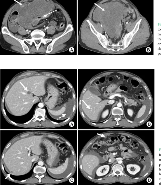
One day after admission, the patient complained of se- vere abdominal pain, and the serum hemoglobin level de- creased to 7.15 g/dL. Computed tomography (CT) showed a ruptured 14 × 15 × 12 cm sized heterogenous low density mass in the pelvic cavity with fluid collection (Fig. 1), and there were multiple liver nodules and an enhanced nodule
Fig. 1. Preoperative computed tomography findings. (A) Huge mass on pelvic cavity. Arrow, internal bleeding on tumor; Dotted arrow, gas forming abscess on tumor due to rupture. (B) Huge mass on pelvic cavity.
Fig. 2. Pre-operative computed tomography findings. (A) Arrow, nodule on liver S4; Dotted arrow, perisplenic hematoma. (B) Arrow, hepatic nodule on S6. (C) Arrow, hepatic nodule on S7. (D) Arrow, enhanced nodule with fat infiltra- tion on omentum.
with fat infiltration in the omentum (Fig. 2). Urgent lapa- rotomy was performed and multiple liver metastases and peritoneal and omental seeding of the tumor were found.
The mass originated from the sigmoid colon. Sigmoid co- lectomy and tumorectomy were performed with an end colostomy for survival. Worsening abdominal distension and vomiting occurred 10 days after operation, and a re- peat CT showed a 10 × 13 cm sized new mass in the pelvic cavity with small bowel obstruction. Repeat laparotomy was performed for palliative cytoreductive treatment, with small bowel resection and end ileostomy with debulking. The patient tolerated a slight advance in diet
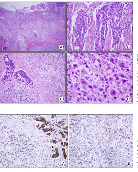
after the second surgery for three days, but ultimately ab- dominal distension recurred with a hard palpable mass, and the patient died of abdominal compartment syn- drome due to rapid tumor growth 22 days after the initial operation. Histologic examination revealed an invasive tumor with two components: the luminal surface of the tu- mor was mainly composed of moderately differentiated adenocarcinoma, and in the deeper area there were spin- dle and epithelioid cells showing high grade anaplasia, which morphologically corresponded to the sarcoma (Fig.
3). The immunohistochemical study showed pancytoker- atin expression in the adenocarcinoma component. The
Fig. 3. Histological findings. (A) The tumor was composed of two components. (B) The adenocar- cinoma area was located mainly in the luminal surface and was moderately differentiated. (C) In the deeper area, the sarcomatous area was juxtaposed to the carcinoma area. (D) The high power view revealed spindle to epithelioid cells with high grade anaplasia.
Fig. 4. Immunohistochemical stains showed (A) strong positivity for cytokeratin in the carcinoma area and focal positivity in the sar- comatous area. (B) The vimentin was expressed in the sarcomatous tumor cells.
sarcomatous cells were focally positive for vimentin and cytokeratin (Fig. 4). A few variable tumor nodules were seen in the separately sent mesentery and omentum. They consisted mostly of the sarcomatous component. Five of eight lymph nodes isolated in the pericolic fat were pos- itive for tumor metastasis.
DISCUSSION
Sarcomatoid carcinoma is widely considered a rare, highly aggressive tumor which arises most commonly in
the head and neck or female genital tract [1]. It sometimes involves the upper aerodigestive tract such as the stomach and esophagus, and most have polypoid growth patterns and can be diagnosed early in their course, so are asso- ciated with a relatively favorable prognosis. However, since the time that Weidner and Zekan [4] reported the first case of colonic sarcomatoid carcinoma, most sarco- matoid carcinomas of the colon have been found to have an aggressive clinical course, often presenting with symp- toms or signs related to distant metastasis.
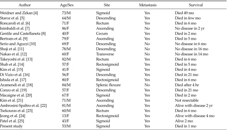
To our knowledge, there have been 22 cases of colon car- cinosarcomas and sarcomatoid carcinomas reported in the
Table 1. Previous cases of colon carcinosarcomas reported in literature
Author Age/Sex Site Metastasis Survival
Weidner and Zekan [4] 73/M Sigmoid Yes Died 49 mo
Staroz et al. [5] 64/M Descending Yes Died in few mo
Roncaroli et al. [6] 71/F Rectum Yes Died in 6 mo
Isimbaldi et al. [7] 86/F Ascending No No disease in 2 yr
Gentile and Castellaneta [8] 40/F Cecum Yes Died in 2 mo
Bertram et al. [9] 79/F Ascending Yes Died in 5 mo
Serio and Aguzzi [10] 69/F Descending No No disease in 6 mo
Shoji et al. [11] 78/M Descending No No disease in 16 mo
Nakao et al. [12] 60/F Transverse No No disease in 14 mo
Takeyoshi et al. [13] 82/M Rectum Yes Died in 6 mo
Shah et al. [14] 57/F Rectosigmoid Yes Died in 5 mo
Kim et al. [15] 41/F Sigmoid Yes Died in 4 mo
Di Vizio et al. [16] 56/F Descending Yes Died in 21 mo
Ishida et al. [17] 80/F Rectosigmoid Yes Died in 6 mo
Aramendi et al. [18] 84/M Splenic flexure No Died after 4 hr
Conzo et al. [19] 57/F Descending Yes Died in 21 mo
Macaigne et al. [20] 67/F Sigmoid Yes Died in 2 mo
Kim et al. [21] 71/M Ascending Yes Not resectable
Ambrosini-Spaltro et al. [22] 81/M Ascending Yes Alive with disease 2 yr
Tsekouras et al. [23] 60/M Rectum Yes Died in 6 mo
Jeong et al. [24] 13/F Rectosigmoid Yes Alive with disease 4 mo
Patel et al. [25] 43/F Sigmoid Yes Alive 2 mo
Present study 53/M Sigmoid Yes Died in 1 mo
literature [4-25], and are summarized in Table 1. Mean age of diagnosis was 63.70 ± 17.64 years (male, 71.78 ± 10.79; fe- male, 58.50 ± 19.51) with an age range of 13 to 86 years.
Eighteen patients (78%) have metastasis with the liver be- ing the most common location. There were 5 cases of right side colon (cecum and ascending colon), 2 cases of trans- verse colon, 5 cases of left side colon, and 11 cases (48%) of sigmoid colon and rectal sarcomatoid carcinoma. The prognosis is poor, with patients dying within 6 months of diagnosis in 13 (56%) of 23 cases. The longest published survival has been 49 months after initial diagnosis [4].
The histogenesis of sarcomatoid carcinoma is contro- versial. Morphologically, the presence of distinct carci- nomatous and sarcomatous components suggests a differ- ent origin. However, the molecular analysis perfomed in the current case supports the hypothesis of a common cell precursor since the same mutation (R282W in exon 8 of the p53 gene) and the same allelic status (with the loss of 18q21) were identified in both the carcinomatous and sar- comatous components.
It is known that lymph nodes and distant metastases
show a predominance of the carcinomatous component, and only one case has been documented that shows meta- stasis with the sarcomatous component [13].
There are no specific treatment guidelines due to the limited number of cases of colonic sarcomatoid carcino- ma, however, radical surgery with adjuvant chemo- therapy and close follow-up may be an acceptable ap- proach to this disease [15].
In this case, our patient unfortunately had no chance for adjuvant chemotherapy. Despite two operations, the pa- tient did not survive past 22 days due to rapid tumor growth. The present case may be the most aggressive case of colonic sarcomatoid carcinoma reported so far.
CONFLICTS OF INTEREST
No potential conflict of interest relevant to this article was reported.
REFERENCES
1. Wick MR, Swanson PE. Carcinosarcomas: current per- spectives and an historical review of nosological concepts.
Semin Diagn Pathol 1993;10:118-27.
2. Madan AK, Long AE, Weldon CB, Jaffe BM. Esophageal carcinosarcoma. J Gastrointest Surg 2001;5:414-7.
3. Tsuneyama K, Sasaki M, Sabit A, Yokoi K, Arano Y, Imai T, et al. A case report of gastric carcinosarcoma with rhabdo- myosarcomatous and neuroendocrinal differentiation.
Pathol Res Pract 1999;195:93-7.
4. Weidner N, Zekan P. Carcinosarcoma of the colon: report of a unique case with light and immunohistochemical studies. Cancer 1986;58:1126-30.
5. Staroz F, Botton A, Potet F. Malignant tumors of the colon with two components (carcinosarcoma): report of a case.
Ann Pathol 1995;15:457-8.
6. Roncaroli F, Montironi R, Feliciotti F, Losi L, Eusebi V.
Sarcomatoid carcinoma of the anorectal junction with neu- roendocrine and rhabdomyoblastic features. Am J Surg Pathol 1995;19:217-23.
7. Isimbaldi G, Sironi M, Assi A. Sarcomatoid carcinoma of the colon: report of the second case with immunohisto- chemical study. Pathol Res Pract 1996;192:483-7.
8. Gentile R, Castellaneta A. Carcinosarcoma of the colon, one or two tumors? Pathologica 1997;89:62-8.
9. Bertram P, Treutner KH, Tietze L, Schumpelick V. True car- cinosarcoma of the colon: case report. Langenbecks Arch Chir 1997;382:173-4.
10. Serio G, Aguzzi A. Spindle and giant cell carcinoma of the colon. Histopathology 1997;30:383-5.
11. Shoji M, Dobashi Y, Iwabuchi K, Kuwao S, Mikami T, Kameya T. Sarcomatoid carcinoma of the descending co- lon: a histological, immunohistochemical and ultrastruc- tural analysis. Acta Oncol 1998;37:765-8.
12. Nakao A, Sakagami K, Uda M, Mitsuoka S, Ito H.
Carcinosarcoma of the colon: report of a case and review of the literature. J Gastroenterol 1998;33:276-9.
13. Takeyoshi I, Yoshida M, Ohwada S, Yamada T, Yanagisawa A, Morishita Y. Skin metastasis from the spindle cell com- ponent in rectal carcinosarcoma. Hepatogastroenterology
2000;47:1611-4.
14. Shah S, Kim DH, Harster G, Hossain A. Carcinosarcoma of the colon and spleen: a fleshy purple mass on colonoscopy.
Dig Dis Sci 2001;46:106-8.
15. Kim JH, Moon WS, Kang MJ, Park MJ, Lee DG.
Sarcomatoid carcinoma of the colon: a case report. J Korean Med Sci 2001;16:657-60.
16. Di Vizio D, Insabato L, Conzo G, Zafonte BT, Ferrara G, Pettinato G. Sarcomatoid carcinoma of the colon: a case re- port with literature review. Tumori 2001;87:431-5.
17. Ishida H, Ohsawa T, Nakada H, Hashimoto D, Ohkubo T, Adachi A, et al. Carcinosarcoma of the rectosigmoid colon:
report of a case. Surg Today 2003;33:545-9.
18. Aramendi T, Fernández-Aceñero MJ, Villanueva MC.
Carcinosarcoma of the colon: report of a rare tumor. Pathol Res Pract 2003;199:345-8.
19. Conzo G, Giordano A, Candela G, Insabato L, Santini L.
Colonic carcinosarcoma. J Gastroenterol Hepatol 2003;18:
748-9.
20. Macaigne G, Aouad K, Boivin JF, Bellaïche A, Auriault ML, Picard D, et al. Sarcomatoid carcinoma of the colon: report of a case and review of the literature. Gastroenterol Clin Biol 2004;28(6-7 Pt 1):600-4.
21. Kim N, Luchs JS, Halpern D, Davis E, Donovan V, Weston SR, et al. Radiology-Pathology Conference: carcinosarco- ma of the colon. Clin Imaging 2005;29:259-62.
22. Ambrosini-Spaltro A, Vaira V, Braidotti P, Rovati MP, Ferrero S, Bosari S. Carcinosarcoma of the colon: report of a case with morphological, ultrastructural and molecular analysis. BMC Cancer 2006;6:185.
23. Tsekouras DK, Katsaragakis S, Theodorou D, Kafiri G, Archontovasilis F, Giannopoulos P, et al. Rectal carcino- sarcoma: a case report and review of literature. World J Gastroenterol 2006;12:1481-4.
24. Jeong YJ, Lee MR, Kim JC, Hwang PH, Moon WS, Chung MJ. Carcinosarcoma of the rectosigmoid colon in a 13-year-old girl. Pathol Int 2008;58:445-50.
25. Patel DH, Dang S, Bentley FR, Julka RN, Olden KW, Aduli F. Carcinosarcoma of the colon: a rare cause of colovesical fistula. Am Surg 2009;75:335-7.