https://doi.org/10.4174/astr.2016.91.6.273 Annals of Surgical Treatment and Research
Silencing the livin gene enhances the cytotoxic effects of anticancer drugs on colon cancer cells
Bo Young Oh, Kwang Ho Kim1, Soon Sup Chung1, Ryung-Ah Lee1
Department of Surgery, Samsung Medical Center, Sungkyunkwan University School of Medicine, Seoul, 1Department of Surgery, Ewha Womans University School of Medicine, Seoul, Korea
INTRODUCTION
Colorectal cancer is one of the most common malignant tu
mors worldwide and the incidence rate has steadily increased.
It is the third leading cause of death in cancer patients [1].
The complete cure rate is 70% to 80% at diagnosis; however, meta stasis develops in 50% of patients [1]. Therefore, systemic chemotherapy, in addition to surgical treatment, has been widely applied to improve the survival rate [2]. Currently, the com monly used regimens combine drugs, such as oxaliplatin (LOHP) or irinotecanbased on 5fluorouracil (FU). However, these treatments have not led to a satisfactory survival rate
in metastatic colorectal cancer patients [1]. Therefore, new therapies have been developed to improve the treatment outcome for colorectal cancer patients. Targeted therapy has attracted attention as a potential new treatment option that targets apoptotic genes, oncogenes, and cell cycle regulatory genes.
The inhibition of apoptosis is one of the most important mechanisms in carcinogenesis. Apoptosisrelated genes and proteins take a significant role in the growth and proliferation of cancer cells [3]. Inhibitor of apoptosis proteins (IAPs) is a typical antiapoptotic protein that suppresses apoptosis by binding caspases and inhibiting proteolytic processes [4]. The Purpose: Livin is associated with drug response in several cancers. The aim of this study was to investigate the effect of silencing the livin gene expression on anticancer drug response in colorectal cancer.
Methods: siRNA was transfected at different concentrations (0, 10, and 30nM) into HCT116 cells, then cells were treated with either 5-fluorouracil (FU)/leucovorin (LV) or oxaliplatin (L-OHP)/5-FU/LV. Cellular viability and apoptosis were evaluated following silencing of livin gene expression combined with treatment with anticancer drugs.
Results: Livin gene expression was effectively suppressed by 30nM siRNA compared with control and 10nM siRNA. The 3-(4, 5-dimethylthiazol-2-yl)-2, 5-diphenyltetrazolium bromide assay showed that proliferation was effectively inhibited in cells treated with a combination of both siRNA and an anticancer drug, compared to cells treated with siRNA-Livin or anticancer drug alone. In particular, the combination of 30nM siRNA and L-OHP/5-FU/LV resulted in a 93.8% and 91.4%
decrease, compared to untreated control or L-OHP/5-FU/LV alone, respectively. Cellular proliferation was most effectively suppressed by a combination of 30nM of siRNA and L-OHP/5-FU/LV compared to other combinations.
Conclusion: siRNA-mediated down-regulation of livin gene expression could significantly suppress colon cancer growth and enhance the cytotoxic effects of anticancer drugs such as 5-FU and L-OHP. The results of this study suggest that silencing livin gene expression in combination with treatment with anticancer drugs might be a novel cancer therapy for colorectal cancer.
[Ann Surg Treat Res 2016;91(6):273-277]
Key Words: Livin protein, Oxaliplatin, Apoptosis, Colorectal neoplasms
Reviewed January February March April May June July August September October November December
Received August 17, 2016, Reviewed August 19, 2016, Accepted August 22, 2016
Corresponding Author: Ryung-Ah Lee
Department of Surgery, Ewha Womans University School of Medicine, 1071, Anyangcheon-ro, Yangcheon-gu, Seoul 07985, Korea
Tel: +82-2-2650-2659, Fax: +82-2-2644-7984 E-mail: ralee@ewha.ac.kr
Copyright ⓒ 2016, the Korean Surgical Society
cc Annals of Surgical Treatment and Research is an Open Access Journal. All articles are distributed under the terms of the Creative Commons Attribution Non- Commercial License (http://creativecommons.org/licenses/by-nc/4.0/) which permits unrestricted non-commercial use, distribution, and reproduction in any medium, provided the original work is properly cited.
most wellknown IAPs are CIAP1, CIAP2, NAIP, Survivin, Xlinked IAP (XIAP), Bruce, ILP2, and Livin. Livin and Survivin are the most representative members. Of special interest is Livin, which is a member of the IAP family. Livin is abundantly expressed in cancer cells, but is rarely detected in normal cells.
It has a high affinity for cancer cells, so it is expected to be a potential target of chemotherapy [5]. In this study, we aimed to investigate whether silencing the livin gene affects the antitumor effect of anticancer drugs in colorectal cancer.
METHODS
Cell culture and transfection
The HCT116 colon cancer cell line was purchased from the Korean cell line bank. The cells were grown in RPMI1640 medium (Life Technologies Inc., Grand Island, NY, USA) supplemented with 10% fetal bovine serum (Life Technologies Inc.), 100 U/mL of penicillin, and 100 g/mL of streptomycin at 37oC in a humidified incubator under an atmosphere of 5% CO2. The HCT116 cells were seeded at a density of 1×10⁵ cells per well in 6well plates and transfected with different concentrations (10nM and 30nM) of siRNA (Dharmacon Inc., Lafayette, CO, USA) against the livin gene using Lipofectamine 2000 (Life Technologies Inc.) according to the manufacturer’s instructions [6]. Reverse transcriptionpolymerase chain reaction (RTPCR) was performed as in the previous study to verify target knock
down [6]. The primers used to amplify the livin gene were as follows: forward 5’CTGGTCAGAGCCAGTGTTCC3’, and reverse 5’TCATAGAAGGAGGCCAGACG3’, for internal control gene β–
actin, forward 5’GACCTGACTGACTACCTCATGAA3’, reverse 5’CTTCATGATGGAGTTGAAGGTAG3’.
Drug treatment
LOHP, 5FU, and leucovorin (LV) were purchased from SigmaAldrich (St. Louis, MO, USA). Stock solutions of these drugs were prepared in sterile distilled water with filtration.
We chose concentrations of these drugs that caused 80%–90%
growth reduction when used alone for HCT116 cell line: 2.5 μM of LOHP, 25 μM of 5FU, and 25 μM of LV. Cells were exposed to FL regimen (5FU/LV) or folfox regimen (LOHP/5FU/LV) at 18 hours after transfection.
MTT assay
Cell viability was examined by routine 3(4, 5dimethylthiazol
2yl)2, 5diphenyltetrazolium bromide (MTT) assay. The HCT116 cells were seeded at a density of 2×10⁴ cells per well in 96
well plates with RPMI1640 in a final volume of 100 μL. On the following day, cells were treated with 10 or 30 nM of siRNA and incubated for 6 hours and then treated with LOHP, 5FU, and LV, and then incubated for 18 hours. For MTT assay, 5mg/mL MTT solution was added into each well. Following incubation
at 37oC for 4 hours, the reaction was stopped by the addition of dimethyl sulfoxide. After the crystals dissolved, the absorbency of the samples was determined at 550 nm.
Caspase activity analysis
Additionally, we analyzed the apoptotic activity of caspase 3 and caspase 7. The HCT116 cells were seeded at a density of 1×10⁵ cells per well in 6well plates. The cells were then treated with 30nM of siRNA against livin, LOHP/5FU/LV, or siRNA with LOHP/5FU/LV as previously described. The degree of apoptosis was determined based on the expression of caspase 3 and caspase 7 measured using RTPCR. The primers were as follows: caspase 3, forward 5’GACTCTAGACGGCATCCAGC3’
and reverse 5’TGACAGCCAGTGAGACTTGG3’, caspase 7, forward 5’AGTGACAGGTATGGGCGTTC3’ and reverse 5’CGG CATTTGTATGGTCCTCT 3’.
Statistical analysis
Each experiment was repeated two or more times. Bands from RTPCR were quantified with UNSCANIT gel version 6.1 software (Silk Scientific Inc., Orem, UT, USA). mRNA levels were calculated using βactin levels as the reference. Statistical com
parisons between different treatments were analyzed by Mann
Whitney test and analysis of variance. All statistical analyses were performed using SPSS ver. 16.0 (SPSS Inc., Chicago, IL, USA). Statistical significance was considered when Pvalues were less than 0.05.
RESULTS
Transfection of HCT116 cells with siRNA-Livin
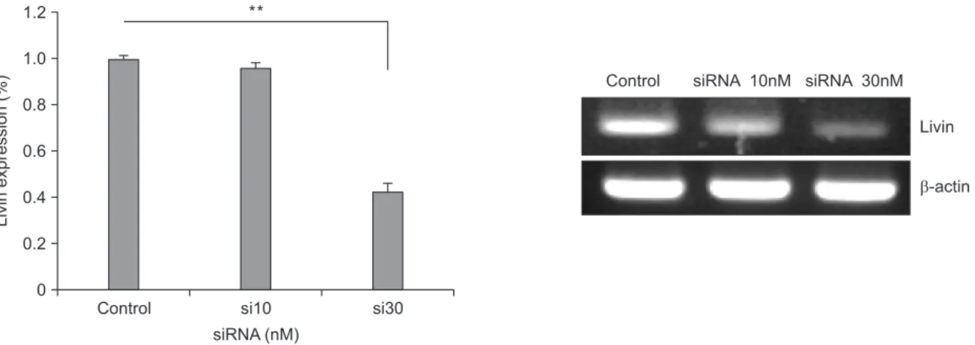
We verified that livin gene expression was suppressed by siRNA in our previous study [5]. In this study, siRNA was transfected at different concentrations (10 and 30nM) into HCT116 cells, and then livin gene expression was detected by RTPCR 18 hours after transfection. Transfection of siRNA against livin was successful. Control HCT116 cells expressed livin mRNA and this expression decreased after siRNA trans
fection. Livin expression was effectively suppressed by 30nM siRNA compared with control and 10nM of siRNA (Fig. 1).
Antiproliferative effect of silencing livin gene in combination with anticancer drugs in HCT116 cells
To identify whether silencing livin affected the antitumor effect of anticancer drugs, cells were transfected with siRNA against livin and treated with anticancer drugs. siRNA was transfected at different concentrations (0, 10, and 30nM) into HCT116 cells, and then cells were treated with either 5FU/LV or LOHP/5FU/LV after 18 hours. To quantify the cellular viability, MTT assay was performed.
The MTT assay showed that the proliferation of cells treated with a combination of siRNA and anticancer drug was effec
tively inhibited, compared with cells treated with siRNALivin or an anticancer drug alone. Although the proliferation of cells treated with 5FU/LV in combination with 10nM of siRNA was similar to cells treated with 5FU/LV alone, the proliferation of cells treated with the 30nM siRNA combination was significantly inhibited. When cells were treated with LOHP/5
FU/LV with 10nM of siRNA, cellular proliferation was inhibited by 20.9% compared to treating with LOHP/5FU/LV alone. The combination of 30nM of siRNA and LOHP/5FU/LV resulted in
a 93.8% and 91.4% decrease compared to untreated control or LOHP/5FU/LV alone, respectively. As shown in Fig. 2, cellular proliferation was most effectively suppressed by a combination of 30nM of siRNA and LOHP/5FU/LV.
Determination of caspase 3 and caspase 7 activities
We investigated whether livin silencing with chemotherapy induces apoptosis, resulting in suppression of cellular prolife
ration. The expression of caspase 3 and caspase 7, common markers for apoptosis, was evaluated by RTPCR. The expression of caspase 3 and 7 decreased more with the combination of 30nM siRNA and LOHP/5FU/LV compared with either siRNA or LOHP/5FU/LV alone (Fig. 3). This result suggests that livin silencing enhances the cytotoxic effect of anticancer drugs by inducing apoptosis.
Control si10 si30
Livinexpression(%)
siRNA (nM) 0
1.2 1.0 0.8 0.6 0.4 0.2
Control siRNA 10nM siRNA 30nM Livin
-actin
**
Fig. 1. Silencing of livin gene expression after siRNA transfection. Livin gene expression was effectively suppressed after trans
fection of 30nM siRNA in HCT116 cells. Livin mRNA detected by reverse transcriptionpolymerase chain reaction. Columns = means ± standard deviation (n = 3). *P < 0.05. **P < 0.01. ***P < 0.001.
Contro l
si10 si30
Cellviability(%)
0 1.2 1.0 0.8 0.6 0.4 0.2
*
FL si10+FL
si30+FL Folfo
x
si10+Folfo x
si30+Folfo x
*
*
***
Fig. 2. Antiproliferative effect of silencing livin gene in com
bination with anticancer drug treatment. Proliferation of cells treated with combination of siRNALivin and anticancer drug was effectively inhibited. This effect was most prominent with combination of 30nM siRNA and oxaliplatin/5
fluorouracil/leucovorin. Cell vi ability was evaluated by 3(4, 5dimethylthiazol2yl)2, 5diphenyltetrazolium bromide assay. Columns = means ± standard deviation (n = 3). *P <
0.05. **P < 0.01. ***P < 0.001.
Fig. 3. Caspase activity after siRNA transfection and treatment with anticancer drugs. Expression of caspase 3 and 7 showed greater decrease in cells treated with combination of 30nM siRNA and oxaliplatin/5fluorouracil/leucovorin (LOHP/5
FU/LV) compared with either siRNA or LOHP/5FU/LV alone. Expression of caspase 3 and caspase 7 was evaluated by reverse transcriptionpolymerase chain reaction.
Contro l
Folfo x siRNA
30nM
Caspase-3
-actin Caspase-7 siRNA
30nM +Folfo
x
DISCUSSION
In this study, we investigated the effect of a combination treatment of siRNALivin and anticancer drugs in colon cancer.
Compared with singleagent treatments, the combination of the siRNALivin and anticancer drugs displayed significantly greater inhibition of growth in HCT116 cells. Above all, the proliferation of cells was remarkably inhibited by up to 93.8%
compared to the untreated control when cells were treated with LOHP/5FU/LV with 30nM of siRNA. Our results showed that treatment with LOHP/5FU/LV and siRNALivin produced a synergistic effect in HCT116 cells.
Many studies have revealed that silencing the livin gene inhibits cell proliferation and induces apoptosis in lung cancer, gastric cancer, and liver cancer [710]. Especially, some studies have investigated the role of Livin on drug response in several types of cancer. A study observed that knockdown of livin gene expression increased apoptosis in colon cancer cells that were resistant to several chemoagents [11]. In other studies, silencing of livin reduced drug resistance to chemotherapy in glioblastoma, melanoma, and lung cancer [1214]. In addi tion, several studies have reported a synergetic anticancer effect when silencing of the livin gene is combined with chemo
therapy [1519].
Livin, a member of the IAP family, is composed of 280 amino acids and has a single baculovirus IAP repeat domain and a COOHterminal RING domain [20]. Livin inhibits apoptosis by binding to caspase 3, caspase 7, and caspase 9, and protects cells from various proapoptotic stimuli [20,21]. Livin has been the focus of cancer therapy because it is easy to handle due to its small size, and it is rarely detected in normal tissues [20]. siRNA transfection targeting specific genes is relatively easy and thus has been used widely as a therapeutic strategy for numerous diseases including cancer [22]. In our previous study, we reported the use of siRNA targeting of livin as a treatment for colon cancer [6]. We used siRNA against livin at different concentrations of 25, 50, 100, 150, and 200nM in HCT116 colon cancer cells, and then detected expression of livin mRNA at 18, 24, and 30 hours after transfection. We found that the expression of livin was effectively suppressed by 25nM of siRNA at 18 hours after transfection. In this study, we transfected siRNALivin at 10 and 30nM, and then detected
livin mRNA levels at 18 hours after transfection.
Chemotherapy has contributed to the improvement in the survival rate of cancer patients, but toxicity and resistance are major obstacles to successful treatment. Exposure of cancer cells to a single cytotoxic agent can cause resistance to unrelated agents. Therefore, the combination of chemotherapy and other modalities is often attempted to enhance the efficacy of chemotherapy [23]. It is possible that combination therapy with siRNALivin may be useful in patients who have resistance to 5FU or LOHPbased chemotherapy. In addition to the antitumor effect, the systemic toxicity of treatment should be considered. In our previous study using a mouse xenograft model [5], we observed no significant toxic effects, so it may be that siRNA targeting of livin can be used in colon cancer therapy without systemic toxic effects in vivo. Thus, a regimen with combined use of siRNALivin and anticancer drugs may emerge as a potent and safe strategy for the treatment of colorectal cancer.
There were some limitations in our study. We used one cell line and did not confirm in vitro results with in vivo experiments. We did not evaluate the toxic effect of combined treatments with siRNALivin and chemoagents, although we identified that there was no significant toxicity of siRNA
Livin in vivo in our previous study. In addition, we did not investigate the mechanism of the synergetic antiapoptotic effect of siRNALivin and chemoagents. However, we showed that the expression of caspase 3 and 7 decreased following siRNA treatment, which indicates that siRNA transfection against Livin induces caspase dependent apoptosis in colon cancer cells.
In conclusion, siRNAmediated downregulation of livin gene expression could significantly suppress colon cancer growth and enhance the cytotoxic effects of anticancer drugs such as 5FU and LOHP. The results of this study suggest that silencing the livin gene might be a novel cancer therapy when used in combination with anticancer drugs for treating colorectal cancer.
CONFLICTS OF INTEREST
No potential conflict of interest relevant to this article was reported.
1. de CastroCarpeno J, BeldaIniesta C, Casado Saenz E, Hernandez Agudo E, Feliu Batlle J, Gonzalez Baron M. EGFR
and colon cancer: a clinical view. Clin Transl Oncol 2008;10:613.
2. Oh SY, Kim DY, Suh KW. Oncologic
outcomes following metastasectomy in colorectal cancer patients developing dis
tant metastases after initial treatment.
REFERENCES
Ann Surg Treat Res 2015;88:2539.
3. Bremer E, van Dam G, Kroesen BJ, de Leij L, Helfrich W. Targeted induction of apoptosis for cancer therapy: current pro gress and prospects. Trends Mol Med 2006;12:38293.
4. Schimmer AD. Inhibitor of apoptosis pro
teins: translating basic knowledge into clini cal practice. Cancer Res 2004;64:7183
90.
5. Chang H, Schimmer AD. Livin/melanoma inhibitor of apoptosis protein as a poten
tial therapeutic target for the treatment of malignancy. Mol Cancer Ther 2007;6:24
30.
6. Oh BY, Lee RA, Kim KH. siRNA targeting Livin decreases tumor in a xenograft mo
del for colon cancer. World J Gastroenterol 2011;17:256371.
7. Zhao X, Yuan Y, Zhang Z, Feng X, Zhang J, Yuan X, et al. Effects of shRNAsilenced livin and survivin on lung cancer cell pro
liferation and apoptosis. J BUON 2014;19:
75762.
8. Wang SL, Deng WT, Wen GF, Li CW, Zeng YJ. Construction of a Hep2 cell line stably transfected with Livin shRNA. Bratisl Lek Listy 2016;117:2725.
9. Liu P, Wang TS, You SH, Ge HM. Expres
sion of livin in gastric cancer and effect of silencing of the livin gene on apoptosis in gastric cancer cells. Zhonghua Zhong Liu Za Zhi 2007;29:5704.
10. Wang TS, Ding QQ, Guo RH, Shen H, Sun J,
Lu KH, et al. Expression of livin in gastric cancer and induction of apoptosis in SGC
7901 cells by shRNAmediated silencing of livin gene. Biomed Pharmacother 2010;
64:3338.
11. Wang X, Xu J, Ju S, Ni H, Zhu J, Wang H.
Livin gene plays a role in drug resistance of colon cancer cells. Clin Biochem 2010;
43:65560.
12. Liu Y, Guo Q, Zhang H, Li GH, Feng S, Yu XZ, et al. Effect of siRNALivin on drug re
sistance to chemotherapy in glioma U251 cells and CD133+ stem cells. Exp Ther Med 2015;10:131723.
13. Lazar I, Yaacov B, Shiloach T, Eliahoo E, Kadouri L, Lotem M, et al. The oncolytic activity of Newcastle disease virus NDV
HUJ on chemoresistant primary mela
noma cells is dependent on the pro apo
ptotic activity of the inhibitor of apoptosis protein Livin. J Virol 2010;84:63946.
14. Sun JG, Liao RX, Zhang SX, Duan YZ, Zhuo WL, Wang XX, et al. Role of inhibitor of apoptosis protein Livin in radiation re
sistance in nonsmall cell lung cancer.
Cancer Biother Radiopharm 2011;26:585
92.
15. Yu L, Wang Z. Effects of Livin gene RNA interference on apoptosis of cer vi cal can
cer HeLa cells and enhanced sensi tivity to cisplatin. J Huazhong Univ Sci Technolog Med Sci 2009;29:62530.
16. Wang R, Lin F, Wang X, Gao P, Dong K, Zou AM, et al. Silencing Livin gene
expres sion to inhibit proliferation and enhance chemosensitivity in tumor cells.
Cancer Gene Ther 2008;15:40212.
17. CrnkovicMertens I, Wagener N, Semzow J, Grone EF, Haferkamp A, Hohenfellner M, et al. Targeted inhibition of Livin re
sensitizes renal cancer cells towards apo
ptosis. Cell Mol Life Sci 2007;64:113744.
18. Bavykin AS, Korotaeva AA, Poyarkov SV, Syrtsev AV, Tjulandin SA, Karpukhin AV. Double siRNAtargeting of cIAP2 and LIVIN results in synergetic sensitization of HCT116 cells to oxaliplatin treatment.
Onco Targets Ther 2013;6:133340.
19. Liu F, Chang H, Xu W, Zhai Y. The effects of Livin shRNA on the response to cis
pla tin in HepG2 cells. Oncol Lett 2015;10:
295761.
20. Kasof GM, Gomes BC. Livin, a novel inhibitor of apoptosis protein family member. J Biol Chem 2001;276:323846.
21. Wang L, Zhang Q, Liu B, Han M, Shan B.
Challenge and promise: roles for Livin in progression and therapy of cancer. Mol Cancer Ther 2008;7:36619.
22. Ryther RC, Flynt AS, Phillips JA 3rd, Patton JG. siRNA therapeutics: big potential from small RNAs. Gene Ther 2005;12:511.
23. Park DG. Antichemosensitizing effect of resveratrol in cotreatment with oxali
platin in HCT116 colon cancer cell. Ann Surg Treat Res 2014;86:6875.