Received July 24, 2014, Revised September 18, 2014, Accepted for publication September 29, 2014
Corresponding author: Jun-Mo Yang, Department of Dermatology, Samsung Medical Center, Sungkyunkwan University School of Medicine, 81 Irwon-ro, Gangnam-gu, Seoul 135-710, Korea. Tel:
82-2-3410-3541, Fax: 82-2-3410-3869, E-mail: junmo.yang@samsung.
com
This is an Open Access article distributed under the terms of the Creative Commons Attribution Non-Commercial License (http://
creativecommons.org/licenses/by-nc/4.0) which permits unrestricted non-commercial use, distribution, and reproduction in any medium, provided the original work is properly cited.
ORIGINAL ARTICLE
Are Podoplanin Gene Polymorphisms Associated with Atopic Dermatitis in Koreans?
Jung-Hyun Namkung1,2, Eugene Kim1, Yong-Doo Park1,3, Geontae Park4, Jun-Mo Yang1
1Department of Dermatology, Samsung Medical Center, Sungkyunkwan University School of Medicine, Seoul, Korea, 2Case Western Reserve University School of Medicine, Cleveland, OH, USA, 3Yangtze Delta Region Institute of Tsinghua University, Zhejiang, China,
4Laboratory of Cellular Neurobiology, Department of Oral Anatomy, School of Dentistry, Seoul National University, Seoul, Korea
Background: The histologic characteristics of atopic dermati- tis (AD) include perivascular edema and dilated tortuous ves- sels in the papillary dermis. A single nucleotide polymor- phism (SNP) of the fms-related tyrosine kinase 4 (FLT4) gene is associated with AD. Objective: To investigate the associa- tions between podoplanin (PDPN) gene SNPs and AD.
Methods: We genotyped 9 SNPs from 5 genes of 1,119 sub- jects (646 AD patients and 473 controls). We determined the promoter activity of 1 SNP (rs355022) by luciferase assay;
this SNP was further investigated using 1,133 independent samples (441 AD patients and 692 controls). Results: The rs355022 and rs425187 SNPs and the C-A haplotype in the PDPN gene were significantly associated with intrinsic AD in the initial experiment. The rs355022 SNP significantly af- fected promoter activity in the luciferase assay. However, these results were not replicated in the replication study.
Conclusion: Two SNPs and the C-A haplotype in the PDPN gene are significantly associated with intrinsic AD; although, the results were confirmed by luciferase assay, they could not be replicated with independent samples. Nevertheless, fur- ther replication experiments should be performed in future studies. (Ann Dermatol 27(3) 275∼282, 2015)
-Keywords-
Atopic dermatitis, Luciferases, Podoplanin protein, Gene- tic polymorphisms
INTRODUCTION
Atopic dermatitis (AD) is a genetically complex disease in- volving gene-gene and gene-environment interactions1. Genetic linkage analysis and association studies have identified several candidate genes associated with either epidermal barrier function or the immune system. Stress, bacterial, or viral infections, exposure to airborne or food allergens, and hygienic factors are thought to aggravate AD symptoms1.
The K14-IL-4 transgenic mouse model of AD demonstrates that progressive dermal lymphatic growth is a prominent feature of AD; this is characterized by increased vessel number, vessel diameter, and the percent vascularized area2. This transgenic mouse model exhibits significantly increased dermal expression of podoplanin (PDPN), lym- phatic vessel endothelial hyaluronan receptor 1 (LYVE-1), and fms-related tyrosine kinase 4 (FLT4, vascular endothe- lial growth factor receptor 3 [VEGFR 3]). The histological findings of human AD include dilated tortuous vessels within the papillary dermis, perivascular edema, mono- nuclear cell accumulation, and rare neutrophils and eosi- nophils3. However, the role of the dermal vasculature in AD pathogenesis remains poorly understood.
PDPN/T1a/aggrus/PA2.26 antigen, a transmembrane gly- coprotein, is a well-known lymphatic endothelial marker.
PDPN expression surrounding malignant tumors is a prog- nostic factor associated with lymphangiogenesis and dis- tant metastasis4. Immunostaining with D2-40 mouse mon- oclonal antibody shows that PDPN is highly expressed in
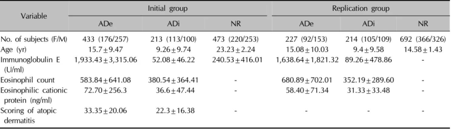
Table 1. Demographic characteristics of the initial and replication study samples
Variable Initial group Replication group
ADe ADi NR ADe ADi NR
No. of subjects (F/M) 433 (176/257) 213 (113/100) 473 (220/253) 227 (92/153) 214 (105/109) 692 (366/326)
Age (yr) 15.7±9.47 9.26±9.74 23.23±2.24 15.08±10.03 9.4±9.58 14.58±1.43
Immunoglobulin E (U/ml)
1,933.43±3,315.06 52.08±46.22 240.53±416.01 1,638.64±1,821.32 89.26±478.86 -
Eosinophil count 583.84±641.08 380.54±364.41 - 680.89±702.01 352.19±289.60 -
Eosinophilic cationic protein (ng/ml)
72.70±256.3 36.6±47.44 - 58.40±71.34 31.33±33.48 -
Scoring of atopic dermatitis
33.35±20.06 22.3±16.38 - - - -
Values are presented as number only or mean±standard deviation. ADe: extrinsic type of atopic dermatitis, ADi: intrinsic type of atopic dermatitis, NR: normal control, F: female, M: male.
lymphatic endothelial cells and the basal cell layer of se- baceous glands but not in normal human interfollicular epidermis5. Marked PDPN expression is detected in the outer root sheath of hair follicles from the mid portion to the hair bulb excluding the bulge area5. Gröger et al.6 re- port that PDPN and LYVE-1 are not only lineage markers for lymphatic endothelial cells, but also activation markers of blood endothelial cells. In normal skin, they found 2 types of vessels: vessels expressing high levels of PDPN (i.e., lymphatic vessels), and vessels negative for PDPN (i.e., blood vessels). However, within the papillary dermis in cases of eczema and psoriasis, they found a third type of vessel expressing low amounts of PDPN. Henno et al.7 report that lymphatic vessels are expressed after blood vascular development in psoriasis.
We previously reported that single nucleotide poly- morphisms (SNPs) and haplotypes of the FLT4 and VEGFA genes are associated with psoriasis8 and that the rs10085109 SNP in the FLT4 gene is associated with AD susceptibility9. These results suggest FLT4 may increase dermal vasculature in Korean patients with AD and psoriasis. Therefore, in the current study, we performed a similar experiment comparing patients with AD with nor- mal controls to determine the association between PDPN SNPs and AD in Korean.
MATERIALS AND METHODS
Subjects
This study included 1,119 samples from 646 patients with AD and 473 normal control (NR) subjects at the initial stage. The AD samples were collected from non-asthmatic patients with AD examined at Samsung Medical Center, Seoul, Korea. AD was diagnosed according to the criteria of Hanifin and Rajka, and classified as extrinsic or intrinsic
(ADe and ADi, respectively) according to serum immuno- globulin E (IgE) level and/or the presence or absence of al- lergy following the CAP test and/or skin prick test. Details regarding AD diagnosis, the criteria for classifying ADe and ADi, and the blood and prick tests for allergens are described in our previous report9. All patients with AD (357 men and 289 women, mean age: 13.58±9.62 years) met our previously reported inclusion/ exclusion cri- teria10,11. Among patients with AD, 433 (257 men and 176 women, mean age: 15.7±9.47 years) had ADe, and 213 (100 men and 113 women, mean: 9.26±9.74 years) had ADi. The 473 NR subjects included medical students and volunteers (253 men and 220 women, mean age:
23.23±2.24 years) with no history of AD skin lesions. For the replication study, 1,133 samples (227 ADe, 214 ADi, and 692 NR) independent of the initial study were in- cluded; the samples were obtained from a cohort from Jeju Island, Korea12. The demographic characteristics of the study participants are summarized in Table 1.
This study was conducted in accordance with the princi- ples of the Declaration of Helsinki, and written informed consent was obtained from all participants. The Samsung Medical Center Ethics Committee approved this study (IRB: 2008-09-044-003).
Marker selection
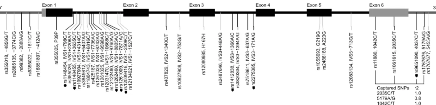
The SNP information was retrieved from the dbSNP (build 141, http://www.ncbi.nlm.nih.gov/SNP; accessed 12 Sep 2014). We selected 37 SNPs from 5 kbp upstream to 5 kbp downstream of the PDPN gene. The selected SNPs were genotyped from 48 independent samples from the general Korean population (data not shown). On the basis of these genotype results, we selected SNPs by using the linkage disequilibrium bin approach in the Tagger pro- gram (http://www.broad.mit.edu/mpg/tagger). This approach
Fig. 1. Map of the podoplanin gene on chromosome 1p36.21 (41.7 kb). Black and grey blocks indicate coding exons, and the 5′
and 3′ untranslated regions, respectively. The first nucleotide of the translation start site is denoted as nucleotide +1. Genotyped polymorphisms are marked. Black and grey lines indicate minor allele frequencies of the polymorphisms in 48 samples >10% and
<10%, respectively. Dots indicate polymorphisms genotyped in the larger population (n=1,119). Linkage disequilibrium between the tag single nucleotide polymorphisms (SNPs) and their tagged SNPs are presented as r2.
defines bins of SNPs that are in very strong linkage dis- equilibrium with a specified r2 threshold; one SNP is then selected to represent the remaining SNPs in a bin13. We used an r2 threshold of 0.8 and a minimum allele fre- quency of 0.1. Thus, a total of 9 SNPs from PDPN were selected as markers for the association study (Fig. 1).
Genotyping with fluorescence polarization detection We extracted genomic DNA from 5-ml whole blood sam- ples by using a commercially available DNA isolation kit (Gentra Genomic DNA Purification Kit; Qiagen, Minneap- olis, MIN, USA) in accordance with the manufacturer’s protocol. Genotypes were identified with the ultra-high throughput GenomeLab SNPstream system14, which uses multiplex polymerase chain reaction (PCR) in conjunction with tag array single-base extension genotyping technol- ogy (Beckman Coulter, Fullerton, CA, USA) and the SNP- stream software suite15. PCR amplifications were per- formed in a PTC-225 Peltier Thermal Cycler (MJ Research, Waltham, MA, USA) with Taq Gold DNA polymerase (Applied Biosystems, Foster City, CA, USA). The sequences of the PCR primers and extension primers are available upon request. Multiplex PCR and genotyping were per- formed in homogeneous reactions, and the assay results were read by direct two-color fluorescence on the SNP- stream Ultra-High Throughput Array Imager. Individual genotypes were generated on the basis of the relative fluo- rescent intensities for each SNP and processed for graph- ical review. All genotyping results were reviewed and confirmed manually by experienced researchers.
Luciferase assay
Double-stranded oligonucleotides were synthesized with three concatenated copies of the T or C allele for a 21-bp region centered on the polymorphism with KpnI and BglII at the 5′ and 3′ ends, respectively. The oligonucleotides
were subsequently cloned into the pGL3-promoter vector (Promega, Madison, WI, USA), which has a simian virus 40 (SV40) promoter.
HEK293 cells were transfected with 1 μg reporter constructs and 0.1 μg pRL-TK Renilla luciferase vector (Promega) with Lipofectamine 2000 (Invitrogen; Thermo Fisher Scientific, Waltham, MA, USA) according to the manu- facturer’s protocol. The transfection efficiency was nor- malized to that of Renilla luciferase activity. The medium was changed to growth medium 18 hours after trans- fection. Cells were harvested 24 hours after the medium was changed, and the luciferase activity was measured with the Dual-Luciferase Reporter Assay System (Promega).
Statistical analysis
The χ2 test was used to determine if individual variants were in Hardy-Weinberg equilibrium at each locus for normal samples. The allelic (i.e., additive) and genotypic effects of individual SNPs were tested using a logistic re- gression model adjusted for sex and age. The level of sig- nificance was set at p<0.05. Odds ratios (ORs) and 95%
confidence intervals (CIs) were also estimated from the lo- gistic regression model.
To detect the most significantly associated haplotype, the significance of overall haplotype effects was scanned for haplotypes of 2 to 4 SNPs by using a sliding-window approach. For the haplotype scan, the haplo.score func- tion in the R package (http://www.r-project.org) was used;
this function is a haplotype association test method that enables the simultaneous modeling of haplotype effects including various controlling covariates, and statistical sig- nificance is determined according to score test statistics16. Haplo.score provides global test statistics for a given hap- lotype locus in addition to results for individual haplotypes.
For loci that had a significant effect in individual SNP tests, we obtained the combined effect by using the hap-
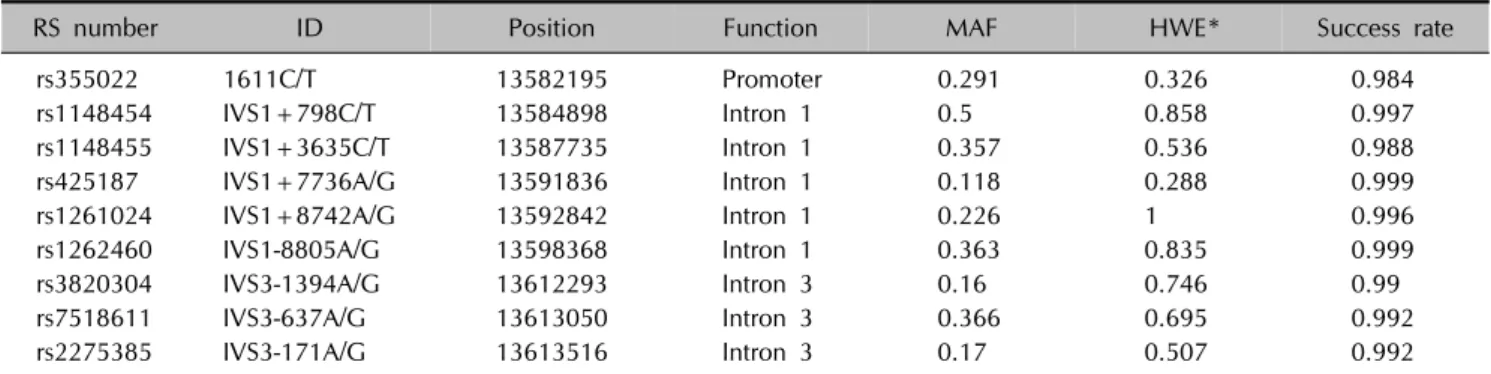
Table 2. Genotyped SNP markers (dbSNP build 141)
RS number ID Position Function MAF HWE* Success rate
rs355022 1611C/T 13582195 Promoter 0.291 0.326 0.984
rs1148454 IVS1+798C/T 13584898 Intron 1 0.5 0.858 0.997
rs1148455 IVS1+3635C/T 13587735 Intron 1 0.357 0.536 0.988
rs425187 IVS1+7736A/G 13591836 Intron 1 0.118 0.288 0.999
rs1261024 IVS1+8742A/G 13592842 Intron 1 0.226 1 0.996
rs1262460 IVS1-8805A/G 13598368 Intron 1 0.363 0.835 0.999
rs3820304 IVS3-1394A/G 13612293 Intron 3 0.16 0.746 0.99
rs7518611 IVS3-637A/G 13613050 Intron 3 0.366 0.695 0.992
rs2275385 IVS3-171A/G 13613516 Intron 3 0.17 0.507 0.992
The minor allele frequency (MAF) and Hardy-Weinberg equilibrium (HWE) were calculated from 459 normal control samples. *p-values from 2 test for HWE.
lo.glm function in the R package. ORs and the sig- nificance of effect differences with respect to the reference haplotype were calculated by haplo.glm adjusted for age and sex17. The most frequent haplotypes were used as the reference haplotype group in this analysis.
Total IgE levels were categorized as <40, 40 to 200, 200 to 500, 500 to 2,000, or >2,000 U/ml. Cumulative logis- tic regression analysis was subsequently conducted to ex- amine the associations between genotype and total IgE levels. The genetic effects on blood eosinophil counts and eosinophilic cationic protein levels (both log-transformed) in patients with AD were tested using a linear regression model. The regression model was adjusted for age, sex, and the scoring of atopic dermatitis index.
Statistical analysis was performed with SAS ver. 9.1 (SAS Institute Inc., Cary, NC, USA) and the R statistical lan- guage (http://www.r-project.org; accessed 31 Mar 2009).
RESULTS
Nine SNPs of the PDPN gene were genotyped in the 1,119 subjects at the initial stage. Information on the SNPs, including genomic function, chromosomal position, dbSNP ID, and minor allele frequency, is shown in Table 2.
All SNPs were in Hardy-Weinberg equilibrium at a sig- nificance level of 0.01. The average genotyping success rate was 99.3%. We compared the distributions of the al- lelic and genotypic frequencies for the 9 SNPs between the AD and NR groups. Statistical significance was ob- tained in logistic regression analysis adjusted for age and sex. For further analysis, haplotype association tests were conducted using a sliding window approach.
Associations of polymorphism gene single nucleotide polymorphisms with atopic dermatitis
Regarding the associations of the SNPs with AD, two SNPs
(rs355022 and rs425187) had significantly different allelic or genotypic distributions between the ADi and NR groups (Table 3); rs355022 exhibited a greater difference between groups (p=0.016, OR=0.587, 95% CI: 0.38∼
0.907). The genotypic effect test showed a significant dif- ference in the rs425187 SNP (p=0.043) with ORs of 0.616 (95% CI: 0.316∼1.204) for A-A vs. A-G and 6.444 (95% CI: 1.021∼40.678) for A-A vs. G-G (Table 3).
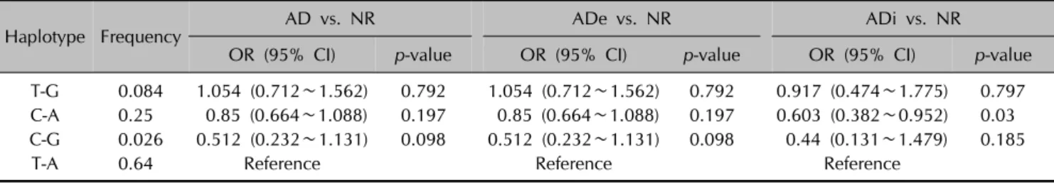
To calculate the combined effect size of the two loci, we used the haplo.glm function to estimate haplotypes simul- taneously in a generalized linear model. The haplotype C-A of the two loci was significantly associated with ADi (p=0.03), and the OR vs. T-A was 0.603 (95% CI: 0.382∼
0.952). Although no other haplotypes showed significant associations, the C-G group, which has two risk alleles, tended to have an increased effect for ADi compared to individual SNPs (Table 4).
rs355022 single nucleotide polymorphism luciferase assay
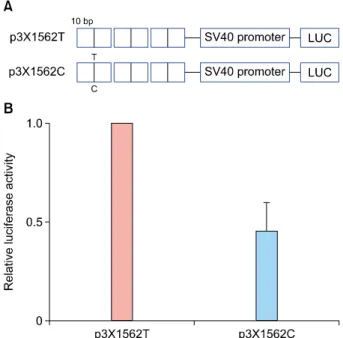
Because rs355022 is located in the promoter (−1611 C/T) region of the PDPN gene, its effect rs355022 on transcrip- tional activity was examined by luciferase reporter assay (Fig. 2). Luciferase gene constructs containing three con- catenated copies of a 21-bp region of either the T or C al- lele centered on the SNP (i.e., p3X1562T and p3X1562C) were prepared. Transfecting p3X1562C into HEK293 cells decreased luciferase activity to a greater extent than p3X1562T (0.44±0.25 fold induction) (Fig. 2). This result suggests rs355022 affects the transcriptional activity of the PDPN gene.
Replication of the single nucleotide polymorphism experiments
To confirm the effects of rs355022 and rs425187, we gen- otyped 1,133 independent samples from 441 patients with
Table 3. Allelic and genotypic frequencies of the 9 PDPN gene SNPs among the AD, AD subtype, and the normal control groups rs number
(allele A/
allele B) Group
Genotype frequency
MAF
Allele test Genotype test
AA AB BB p-value OR (95% CI) p-value* OR (95% CI)† OR (95% CI)‡
rs355022 T/C
NR 239 (0.513) 183 (0.393) 44 (0.094) 0.291
AD 334 (0.525) 266 (0.418) 36 (0.057) 0.266 0.06 0.805 (0.642∼1.009) 0.105 0.89 (0.662∼1.198) 0.544 (0.308∼0.959) Ade 220 (0.514) 182 (0.425) 26 (0.061) 0.273 0.257 0.87 (0.684∼1.107) 0.322 0.97 (0.706∼1.332) 0.631 (0.346∼1.153) ADi 114 (0.548) 84 (0.404) 10 (0.048) 0.25 0.016§ 0.587 (0.38∼0.907) 0.058 0.627 (0.365∼1.079) 0.291 (0.085∼0.993) rs1148454
C/T
NR 119 (0.253) 232 (0.494) 119 (0.253) 0.5
AD 161 (0.249) 344 (0.533) 141 (0.218) 0.485 0.643 0.954 (0.781∼1.165) 0.586 1.087 (0.772∼1.53) 0.907 (0.608∼1.353) Ade 102 (0.236) 232 (0.536) 99 (0.229) 0.497 0.902 0.987 (0.795∼1.224) 0.439 1.201 (0.828∼1.741) 0.973 (0.631∼1.502) ADi 59 (0.277) 112 (0.526) 42 (0.197) 0.46 0.351 0.846 (0.596∼1.202) 0.61 0.784 (0.437∼1.407) 0.726 (0.361∼1.459) rs1148455
C/T
NR 196 (0.42) 208 (0.445) 63 (0.135) 0.358
AD 282 (0.441) 289 (0.452) 68 (0.106) 0.333 0.267 0.887 (0.718∼1.096) 0.523 0.913 (0.675∼1.236) 0.767 (0.482∼1.222) Ade 185 (0.434) 192 (0.451) 49 (0.115) 0.34 0.551 0.933 (0.744∼1.171) 0.702 1.002 (0.724∼1.388) 0.817 (0.495∼1.351) NR 119 (0.253) 232 (0.494) 119 (0.253) 0.317 0.072 0.704 (0.481∼1.031) 0.167 0.636 (0.369∼1.095) 0.55 (0.238,1∼274) rs425187
A/G
NR 366 (0.772) 104 (0.219) 4 (0.008) 0.118
AD 519 (0.805) 117 (0.181) 9 (0.014) 0.105 0.701 0.94 (0.684∼1.291) 0.296 0.838 (0.59∼1.189) 2.224 (0.571∼8.662) Ade 347 (0.803) 79 (0.183) 6 (0.014) 0.105 0.741 0.944 (0.671∼1.329) 0.787 0.901 (0.62∼1.309) 1.351 (0.304∼5.996) ADi 172 (0.808) 38 (0.178) 3 (0.014) 0.103 0.611 0.858 (0.474∼1.551) 0.043§ 0.616 (0.316∼1.204) 6.444 (1.021∼40.678) rs1261024
A/G
NR 282 (0.597) 166 (0.352) 24 (0.051) 0.227 0.65 0.868 (0.642∼1.173) 0.918 (0.478∼1.762) AD 399 (0.621) 207 (0.322) 36 (0.056) 0.217 0.426 0.908 (0.715∼1.152) 0.743 0.887 (0.642∼1.225) 0.879 (0.44∼1.752) Ade 273 (0.636) 131 (0.305) 25 (0.058) 0.211 0.466 0.91 (0.705∼1.173) 0.714 0.798 (0.459∼1.387) 0.83 (0.256,2.691) ADi 126 (0.592) 76 (0.357) 11 (0.052) 0.23 0.459 0.847 (0.545∼1.315)
rs1261009 A/G
NR 193 (0.409) 216 (0.458) 63 (0.133) 0.362
AD 277 (0.429) 281 (0.435) 88 (0.136) 0.354 0.682 0.958 (0.78∼1.177) 0.919 0.961 (0.71∼1.3) 0.915 (0.586∼1.429) Ade 190 (0.439) 176 (0.406) 67 (0.155) 0.358 0.819 0.975 (0.783∼1.213) 0.783 0.899 (0.65∼1.244) 1.007 (0.63∼1.608) ADi 87 (0.408) 105 (0.493) 21 (0.099) 0.345 0.704 0.929 (0.634∼1.36) 0.368 1.212 (0.709∼2.074) 0.637 (0.252∼1.611) rs3820304
A/G
NR 272 (0.576) 170 (0.36) 30 (0.064) 0.16
AD 346 (0.541) 242 (0.379) 51 (0.08) 0.169 0.106 1.254 (0.953∼1.648) 0.432 1.129 (0.836∼1.524) 1.4 (0.797∼2.457) Ade 228 (0.534) 159 (0.372) 40 (0.094) 0.173 0.094 1.285 (0.959∼1.722) 0.383 1.093 (0.791∼1.511) 1.507 (0.835∼2.72) ADi 118 (0.557) 83 (0.392) 11 (0.052) 0.161 0.581 1.151 (0.699∼1.895) 0.515 1.364 (0.804∼2.313) 1.106 (0.366∼3.345) rs7518611
A/G
NR 331 (0.703) 129 (0.274) 11 (0.023) 0.366
AD 439 (0.689) 181 (0.284) 17 (0.027) 0.349 0.464 0.924 (0.748∼1.142) 0.254 1.218 (0.886∼1.673) 1.798 (0.712∼4.539) Ade 289 (0.678) 127 (0.298) 10 (0.023) 0.363 0.558 0.934 (0.744∼1.173) 0.245 1.292 (0.92∼1.814) 1.612 (0.598∼4.342) ADi 150 (0.711) 54 (0.256) 7 (0.033) 0.322 0.598 0.905 (0.623∼1.313) 0.497 0.998 (0.558∼1.784) 2.459 (0.547∼11.05) rs2275385
A/G
NR 187 (0.398) 222 (0.472) 61 (0.13) 0.171
AD 268 (0.419) 297 (0.464) 75 (0.117) 0.169 0.376 0.886 (0.679∼1.158) 0.607 0.997 (0.735∼1.353) 0.803 (0.507∼1.27) ADe 167 (0.391) 210 (0.492) 50 (0.117) 0.165 0.366 0.876 (0.658∼1.167) 0.618 1.028 (0.74∼1.428) 0.809 (0.493∼1.328) ADi 101 (0.474) 87 (0.408) 25 (0.117) 0.178 0.79 0.936 (0.575∼1.523) 0.86 0.935 (0.546∼1.603) 0.795 (0.35,1∼809) ORs and p-values were obtained from a logistic regression model adjusted for age and sex. PDPN: podoplanin, SNP: single nucleotide polymorphism, AD: atopic dermatitis, AA: homozygous genotype of A allele, AB: heterozygous genotype, BB: homozygous genotype of B allele, MAF: minor allele frequency, OR: odds ratio, CI: confidence interval, NR: normal control, ADe: extrinsic type of atopic dermatitis, ADi: intrinsic type of atopic dermatitis. *p-value of the type III effect of the genotype; †OR between individuals with AB and BB; ‡OR between individuals with AA and BB. §p<0.05.
Table 4. Analysis of haplotypes of the rs355022 and rs425187 loci to test for associations with AD and AD subtypes
Haplotype Frequency AD vs. NR ADe vs. NR ADi vs. NR
OR (95% CI) p-value OR (95% CI) p-value OR (95% CI) p-value
T-G 0.084 1.054 (0.712∼1.562) 0.792 1.054 (0.712∼1.562) 0.792 0.917 (0.474∼1.775) 0.797 C-A 0.25 0.85 (0.664∼1.088) 0.197 0.85 (0.664∼1.088) 0.197 0.603 (0.382∼0.952) 0.03 C-G 0.026 0.512 (0.232∼1.131) 0.098 0.512 (0.232∼1.131) 0.098 0.44 (0.131∼1.479) 0.185
T-A 0.64 Reference Reference Reference
ORs and p-values were calculated by the haplo.glm function using age and sex as adjusting covariates. The most frequent haplotypes were used as the reference haplotype group. Bold type indicates p<0.05. AD: atopic dermatitis, NR: normal control, ADe: extrinsic type of atopic dermatitis, ADi: intrinsic type of atopic dermatitis, OR: odds ratio, CI: confidence interval.
Table 5. Logistic regression analysis of the rs355022 SNP in the PDPN gene using combined data from the initial and replicate samples rs number
(allele A/
allele B) Group
Genotype frequency
MAF
Allele test Genotype test
AA AB BB p-value OR (95% CI) p-value* OR (95% CI)† OR (95% CI)‡
rs355022 T/C
NR 628 (0.544) 443 (0.384) 84 (0.073) 0.265
AD 551 (0.52) 439 (0.415) 69 (0.065) 0.272 0.911 1.008 (0.87,1.169) 0.832 1.047 (0.864,1.269) 0.949 (0.654,1.378) ADe 334 (0.515) 269 (0.414) 46 (0.071) 0.278 0.721 1.031 (0.873,1.217) 0.883 1.057 (0.849,1.315) 1.017 (0.673,1.539) ADi 217 (0.529) 170 (0.415) 23 (0.056) 0.263 0.49 0.92 (0.726,1.166) 0.709 0.963 (0.712,1.302) 0.766 (0.407,1.442) The model was adjusted for age, sex and sample source (i.e., initial or replicate). SNP: single nucleotide polymorphism, PDPN:
podoplanin, AA: homozygous genotype of A allele, AB: heterozygous genotype, BB: homozygous genotype of B allele, MAF: minor allele frequency, OR: odds ratio, CI: confidence interval, NR: normal control, AD: atopic dermatitis, ADe: extrinsic type of atopic dermatitis, ADi: intrinsic type of atopic dermatitis. *p-value for the type III effect of the genotype. †OR of G-C vs. C-C. ‡OR of G-G vs. C-C.
Fig. 2. Effect of the rs355022 single nucleotide polymorphism (SNP) of podoplanin on transcriptional activity. (A) Reporter gene constructs. Three concatenated oligonucleotides of the rs355022 T or C SNP allele were inserted into a pGL3-SV40 promoter vector.
(B) The relative luciferase activity of the p3X1562C construct is presented as the ratio to that of the p3X1562T construct. The experiment was repeated three times in duplicate. HEK293 cells were used for transfection.
AD and 692 controls (Table 1). However, rs425187 did not exhibit any association, while rs355022 exhibited a significant association in the replicate samples (data not shown). Thus, the direction of the effect was opposite to that of the initial study. Therefore, the association became non-significant for the merged dataset (Table 5).
DISCUSSION
PDPN is a sensitive marker for identifying lymphatic vessels. Transgenic mice exhibit significantly elevated der-
mal expression of PDPN, LYVE-1, and VEGFR-3 (FLT4)5, suggesting lymphatic vessels may be involved in AD pathogenesis in the animal model. VEGF and effector cells of skin inflammation (i.e., mast cells, basophils, eosino- phils, macrophages lymphocytes, etc.) are major sources of the vast array of angiogenesis and lymphangiogenesis in AD. However, the role of lymphangiogenesis in AD is largely unknown3.
We previously reported that a SNP in the FLT4 gene is as- sociated with AD susceptibility in Koreans9. On the basis of these reports, we hypothesized PDPN is associated with AD and therefore evaluated the association between the SNPs of PDPN and AD susceptibility. We initially identified two SNPs, rs355022 and rs425187, in the PDPN gene that were significantly associated with in- trinsic AD in Koreans. As the rs355022 SNP is located in the promoter region of PDPN (−1611 C/T), we performed a luciferase assay; the results show that rs355022 can af- fect PDPN transcription, which indicates PDPN SNP T→C is associated with lower transcriptional activity of PDPN.
To confirm these findings, we replicated the experiment with a different set of patients and controls. However, the replication study did not confirm the results of the initial experiment. The reason for the contrasting results of our two experiments is unknown. It is possible that the SNPs in PDPN gene weakly influence the whole-protein effect of this gene in vivo. This discrepancy highlights the im- portance of replication studies for SNPs.
The role of dermal vasculature in AD pathogenesis is con- troversial3. Our previous results suggest there might be dif- ferences between the K14-IL-4 transgenic mouse model and humans5. In the K14-IL-4 transgenic mouse model, in- terleukin (IL)-4-triggering macrophage recruitment has been suggested to be closely associated with lymphangiogenesis in AD. However, in comparison to K14-IL-4 transgenic mice, the active action time of IL-4 is not long enough in humans to sustain macrophage recruitment.
A substantial amount of research is performed worldwide to search for genetic factors in the etiology of AD; three main approaches are being used: candidate gene associa- tion, selecting genes for study based on a hypothesis of a known biological function, and genome-wide linkage screening. Efforts to identify candidate genes for AD through genome-wide linkage screening and DNA micro- arrays have identified at least 20 genes significantly asso- ciated with AD. However, only six of these genetic associ- ations−IL-4, IL-4R, IL-13, mast cell chymase, serine pro- tease inhibitor Kazal-type 5 (SPINK5), and filaggrin (FLG) genes−have been replicated in at least two independent studies18. At least four genome-wide association stud- ies19-22 have been performed since 2009. In each study, the authors identified 1 to 8 different candidate genes in- cluding FLG; however, most of the reported genes’ func- tions have not yet been verified in AD. Regarding SNP studies in AD, there are many reports of different SNPs in several genes in different ethnicities. We previously re- ported associations between AD and SNPs in sphingomye- linase 223, IL-1824, IL-5 & IL-5R25, defensin 126, IL-12 &
IL-12R27, SPINK528, IL-9 & IL-9R29, IL-4, IL-13 & IL-13R30, and FLT49 in the Korean population. Other groups in Korea report associations of AD with SNPs in the FLG gene31, FCεRI gene32, and the haplotype of the IL-10 gene33. Among those reports, the FLT4 gene is the only gene that was studied in replication experiments with dif- ferent samples.
In summary, we genotyped 9 SNPs from the PDPN gene in 1,119 samples and found two SNPs associated with ADi. In addition, the rs355022 SNP affects PDPN trans- cription. However, we could not replicate these results.
Despite these conflicting results, replication experiments are critical for SNP studies.
ACKNOWLEDGMENT
This study was supported by “The Environmental Health Action Program” of the Korean Ministry of the Environ- ment and by a grant from the Samsung Biomedical Research Institute (SMX1131301).
REFERENCES
1. Bieber T. Atopic dermatitis. Ann Dermatol 2010;22:125-137.
2. Shi VY, Bao L, Chan LS. Inflammation-driven dermal lym- phangiogenesis in atopic dermatitis is associated with CD11b+ macrophage recruitment and VEGF-C up-regulation in the IL-4-transgenic mouse model. Microcirculation 2012;19:567-579.
3. Genovese A, Detoraki A, Granata F, Galdiero MR, Spadaro
G, Marone G. Angiogenesis, lymphangiogenesis and atopic dermatitis. Chem Immunol Allergy 2012;96:50-60.
4. Durchdewald M, Guinea-Viniegra J, Haag D, Riehl A, Lichter P, Hahn M, et al. Podoplanin is a novel fos target gene in skin carcinogenesis. Cancer Res 2008;68:6877-6883.
5. Honma M, Minami-Hori M, Takahashi H, Iizuka H. Podo- planin expression in wound and hyperproliferative psoriatic epidermis: regulation by TGF-β and STAT-3 activating cytokines, IFN-γ, IL-6, and IL-22. J Dermatol Sci 2012;65:
134-140.
6. Gröger M, Niederleithner H, Kerjaschki D, Petzelbauer P. A previously unknown dermal blood vessel phenotype in skin inflammation. J Invest Dermatol 2007;127:2893-2900.
7. Henno A, Blacher S, Lambert CA, Deroanne C, Noël A, Lapière C, et al. Histological and transcriptional study of angiogenesis and lymphangiogenesis in uninvolved skin, acute pinpoint lesions and established psoriasis plaques: an approach of vascular development chronology in psoriasis.
J Dermatol Sci 2010;57:162-169.
8. Lee JH, Cho EY, Namkung JH, Kim E, Kim S, Shin ES, et al.
Single-nucleotide polymorphisms and haplotypes in the VEGF receptor 3 gene and the haplotype GC in the VEGFA gene are associated with psoriasis in Koreans. J Invest Dermatol 2008;128:1599-1603.
9. Namkung JH, Lee JE, Kim E, Huh IS, Park T, Shin ES, et al.
Single nucleotide polymorphism in the FLT4 gene is asso- ciated with atopic dermatitis in Koreans. Cytokine 2013;62:
110-114.
10. Jeong CW, Ahn KS, Rho NK, Park YD, Lee DY, Lee JH, et al. Differential in vivo cytokine mRNA expression in lesional skin of intrinsic vs. extrinsic atopic dermatitis patients using semiquantitative RT-PCR. Clin Exp Allergy 2003;33:1717- 1724.
11. Rho NK, Kim WS, Lee DY, Lee JH, Lee ES, Yang JM.
Immunophenotyping of inflammatory cells in lesional skin of the extrinsic and intrinsic types of atopic dermatitis. Br J Dermatol 2004;151:119-125.
12. Kim HK, Jang TW, Jung MH, Park HW, Lee JE, Shin ES, et al. Association between genetic variations of the transforming growth factor β receptor type III and asthma in a Korean population. Exp Mol Med 2010;42:420-427.
13. Carlson CS, Eberle MA, Rieder MJ, Yi Q, Kruglyak L, Nickerson DA. Selecting a maximally informative set of single-nucleotide polymorphisms for association analyses using linkage disequilibrium. Am J Hum Genet 2004;74:
106-120.
14. Bell PA, Chaturvedi S, Gelfand CA, Huang CY, Kochersperger M, Kopla R, et al. SNPstream UHT: ultra-high throughput SNP genotyping for pharmacogenomics and drug discovery.
Biotechniques 2002;Suppl:70-72, 74, 76-77.
15. Denomme GA, Van Oene M. High-throughput multiplex single-nucleotide polymorphism analysis for red cell and platelet antigen genotypes. Transfusion 2005;45:660-666.
16. Schaid DJ, Rowland CM, Tines DE, Jacobson RM, Poland GA. Score tests for association between traits and haplo- types when linkage phase is ambiguous. Am J Hum Genet 2002;70:425-434.
17. Lake SL, Lyon H, Tantisira K, Silverman EK, Weiss ST, Laird NM, et al. Estimation and tests of haplotype-environment interaction when linkage phase is ambiguous. Hum Hered 2003;55:56-65.
18. Brown SJ, McLean WH. Eczema genetics: current state of knowledge and future goals. J Invest Dermatol 2009;129:
543-552.
19. Esparza-Gordillo J, Weidinger S, Fölster-Holst R, Bauerfeind A, Ruschendorf F, Patone G, et al. A common variant on chromosome 11q13 is associated with atopic dermatitis.
Nat Genet 2009;41:596-601.
20. Sun LD, Xiao FL, Li Y, Zhou WM, Tang HY, Tang XF, et al.
Genome-wide association study identifies two new susceptibility loci for atopic dermatitis in the Chinese Han population. Nat Genet 2011;43:690-694.
21. Paternoster L, Standl M, Chen CM, Ramasamy A, Bønnelykke K, Duijts L, et al. Meta-analysis of genome-wide association studies identifies three new risk loci for atopic dermatitis.
Nat Genet 2011;44:187-192.
22. Hirota T, Takahashi A, Kubo M, Tsunoda T, Tomita K, Sakashita M, et al. Genome-wide association study identifies eight new susceptibility loci for atopic dermatitis in the Japanese population. Nat Genet 2012;44:1222-1226.
23. Kim HT, Lee JY, Han BG, Kimm K, Oh B, Shin HD, et al.
Association analysis of sphingomyelinase 2 polymorphisms for the extrinsic type of atopic dermatitis in Koreans. J Dermatol Sci 2007;46:143-146.
24. Kim E, Lee JE, Namkung JH, Park JH, Kim S, Shin ES, et al.
Association of the single-nucleotide polymorphism and haplotype of the interleukin 18 gene with atopic dermatitis in Koreans. Clin Exp Allergy 2007;37:865-871.
25. Namkung JH, Lee JE, Kim E, Cho HJ, Kim S, Shin ES, et al.
IL-5 and IL-5 receptor alpha polymorphisms are associated
with atopic dermatitis in Koreans. Allergy 2007;62:934-942.
26. Kim E, Lee JE, Namkung JH, Kim PS, Kim S, Shin ES, et al.
Single nucleotide polymorphisms and the haplotype in the DEFB1 gene are associated with atopic dermatitis in a Korean population. J Dermatol Sci 2009;54:25-30.
27. Namkung JH, Lee JE, Kim E, Kim S, Kim S, Shin ES, et al.
Association of single nucleotide polymorphisms in the IL-12 (IL-12A and B) and IL-12 receptor (IL-12Rbeta1 and beta2) genes and gene-gene interactions with atopic dermatitis in Koreans. J Dermatol Sci 2010;57:199-206.
28. Namkung JH, Lee JE, Kim E, Byun JY, Kim S, Shin ES, et al.
Hint for association of single nucleotide polymorphisms and haplotype in SPINK5 gene with atopic dermatitis in Koreans. Exp Dermatol 2010;19:1048-1053.
29. Namkung JH, Lee JE, Kim E, Park GT, Yang HS, Jang HY, et al. An association between IL-9 and IL-9 receptor gene poly- morphisms and atopic dermatitis in a Korean population. J Dermatol Sci 2011;62:16-21.
30. Namkung JH, Lee JE, Kim E, Kim HJ, Seo EY, Jang HY, et al.
Association of polymorphisms in genes encoding IL-4, IL-13 and their receptors with atopic dermatitis in a Korean population. Exp Dermatol 2011;20:915-919.
31. Park MK, Kim HK, Lee JW, Yoo KH, Park KY, Seo SJ, et al.
Identification of filaggrin gene polymorphisms in Korean atopic dermatitis patients. Korean J Dermatol 2010;62(Suppl 2):151-152.
32. Park KY, Park MK, Kim EJ, Lee MK, Seo SJ. FCεRI gene promoter polymorphisms and total IgE levels in suscepti- bility to atopic dermatitis in Korea. J Korean Med Sci 2011;
26:870-874.
33. Shin HD, Park BL, Kim LH, Kim JS, Kim JW. Interleukin-10 haplotype associated with total serum IgE in atopic dermatitis patients. Allergy 2005;60:1146-1151.