MK Choi, et al
S404 Ann Dermatol
Received February 7, 2011, Revised July 18, 2011, Accepted for publication August 5, 2011
†This work was supported for 2 years by a Pusan National University Research Grant.
*These two authors equally contributed to this work.
Corresponding author: Jeong Heo, M.D., Department of Internal Medicine, Pusan National University School of Medicine, 1-10 Ami-dong, Seo-gu, Busan 602-739, Korea. Tel: 82-51-240-7215, Fax:
82-51-244-8180, E-mail: [email protected]
This is an Open Access article distributed under the terms of the Creative Commons Attribution Non-Commercial License (http://
creativecommons.org/licenses/by-nc/3.0) which permits unrestricted non-commercial use, distribution, and reproduction in any medium, provided the original work is properly cited.
Ann Dermatol Vol. 23, Suppl. 3, 2011 http://dx.doi.org/10.5021/ad.2011.23.S3.S404
CASE REPORT
Toxic Epidermal Necrolysis Associated with Sorafenib and Tosufloxacin in a Patient with Hepatocellular
Carcinoma
Mun Ki Choi, M.D.*, Hyun Young Woo, M.D.*, Jeong Heo, M.D., Mong Cho, M.D., Gwang Ha Kim, M.D., Geun Am Song, M.D., Moon Bum Kim, M.D.1
Departments of Internal Medicine, 1Dermatology, Pusan National University School of Medicine, Busan, Korea
This is the first case report to describe a 44-year-old woman with a history of advanced hepatocellular carcinoma who developed toxic epidermal necrolysis (TEN) clinically after taking 400 mg sorafenib (NexavarⓇ, BAY 43-9006) and tosufloxacin orally once per day. Both sorafenib and tosufloxacin were eventually discontinued, and the TEN resolved with corticosteroids and supportive treatment.
Clinical physicians should be aware of this possible complication so that early interventions can be made. (Ann Dermatol 23(S3) S404∼S407, 2011)
-Keywords-
Hepatocellular carcinoma, Sorafenib, Tosufloxacin, Toxic epidermal necrolysis
INTRODUCTION
Adverse cutaneous reactions to drugs are common occur- rences in 2∼3% of hospitalized patients1. Severe drug reaction patterns include angioedema, anaphylaxis, exfo-
liative dermatitis, drug hypersensitivity syndrome, Stevens- Johnson syndrome (SJS), and toxic epidermal necrolysis (TEN). TEN, which is a rare but potentially life-threatening condition, is associated with medications, including sulfo- namides, penicillin, and other antibiotics, anticonvulsants, non steroidal anti-inflammatory drugs, allopurinol, and corticosteroids. Herein, we report a case of a patient diag- nosed with sorafenib (Nexavar, Bayer HealthCare Pharma- ceuticals-Onyx Pharmaceuticals) and tosufloxacin-induced TEN who was successfully treated with corticosteroids. To our knowledge, TEN associated with sorafenib and tosu- floxacin has never been previously discussed in the literature.
CASE REPORT
A 44-year-old woman presented 3 years ago because of chronic hepatitis B-related liver cirrhosis with advanced multifocal hepatocellular carcinoma (HCC). The stage of liver cirrhosis was Child-Pugh’s functional class A. She had received 12 sessions of transcatheter arterial chemo- embolization (TACE) repeatedly and one session of radio- frequency ablation since the HCC diagnosis. In combina- tion with TACE, she had also been treated with sorafenib, which is a tyrosine-kinase inhibitor, at the reduced dose of 200 mg orally twice per day since 2008. Two months prior to admission, she underwent percutaneous drainage combined with broad spectrum antibiotics (third genera- tion cephalosporin and cefotaxime) because of an infected intrahepatic biloma secondary to frequent TACE. At that time, the sorafenib therapy was temporarily discontinued.
The infected intrahepatic biloma was cured by the above mentioned treatment after 1 month, then administration of sorafenib was restarted at 400 mg once daily, and intrave-
Toxic Epidermal Necrolysis Associated with Sorafenib and Tosufloxacin
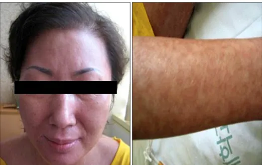
Vol. 23, Suppl. 3, 2011 S405 Fig. 1. Picture of the patient at ad- mission. (A) Confluent and erythe- matous macules on the face (A) with numerous and widespread sheets of flaccid blistering with positive Nikolsky’s sign on the arm and leg (B∼D).
nous antibiotics were switched to oral tosufloxacin (Ozex, SK Chemicals, Seoul, Korea) at 150 mg twice daily.
About 2 weeks after restarting the sorafenib, she was admitted to the emergency department for a generalized erythematous rash and fever. On physical examination, erythematous macules were confluent on >80% of the total body surface with numerous and widespread sheets of flaccid blistering (Fig. 1). Nikolsky’s sign was positive.
There were also hemorrhagic crusts on the lips and erosions involving the tongue, oral mucosa, but not the conjunctiva. She evolved rapidly with increased epider- mal detachment in the face, neck, thorax, dorsum, and limbs, affecting more than 30% of her body. The patient presented with a temperature of 38.2oC without localized signs of infection. Laboratory studies were normal except for a slightly high C-reactive protein of 3.55 mg/dl (nor- mal, 0∼0.5 mg/dl) and an elevated lactate dehydrogenase of 1,017 IU/L (normal range, 95∼280 IU/L). Her other medications on admission, included lamivudine, ursode- oxycholic acid, mecobalamin, thiamine, gabapentin, oxy- codone, and tosufloxacin, which she had taken for over 2 years without any recent change in dose except for the tosufloxacin. The diagnosis of drug-induced TEN was made based on the clinical history and sorafenib and tosufloxacin administration, which was strongly suspected
as the offending medication, and was withdrawn imme- diately. Tosufloxacin was also discontinued because of the possibility of being the culprit drug. The exposed dermis and eroded mucosal surface were protected with polyure- thane foam (Medifoam, DongSung Biopol, Seoul, Korea) and wet gauze. Intravenous hydration and total parenteral nutrition was continued until oral intake was possible without oral pain. She received corticosteroid therapy, given initially as intravenous 5 mg dexamethasone daily and later changed to oral prednisolone. New epithelializa- tion appeared within 15 days, and the lesions gradually resolved (Fig. 2), but hyperpigmentation in the involved areas remained. The patient was discharged with normal oral feeding 8 weeks after admission. She has remained in good health without recurrence of symptoms during 8 months of follow-up.
DISCUSSION
TEN, also known as Lyell’s syndrome, is a rare disease of unclear pathophysiology, and its incidence is estimated to be 1.5∼2 new cases per million population per year1. The disorder seems to be more common among patients with human immunodeficiency virus infection, systemic lupus erythematosus, and malignancy2. TEN is a cutaneous
MK Choi, et al
S406 Ann Dermatol
Fig. 2. Picture of the patient 2 weeks after beginning steroid treat- ment. The skin lesions had resol- ved but hyperpigmentation remain- ed.
reaction to various precipitating agents, characterized by sudden apoptosis of keratinocytes leading to widespread erythema, mucous membrane erosion, extensive epider- mal detachment, and severe constitutional symptoms1,3. Within this spectrum of epidermal necrolysis, detachment of <10% of the total body surface area defines SJS. When
>30%, it is defined as TEN, whereas intermediate cases are an overlap of SJS-TEN4. Many factors have been proposed as causes of TEN, including adverse reactions to drugs, infections, malignant disorders, and graft-versus-host disease5,6. However, most case reports and studies suggest that TEN is usually an idiosyncratic hypersensitivity to medication7,8. TEN is a serious diseases, considering the severity of systemic manifestations, the unpredictable evo- lution, and the absence of specific therapy.
The diagnosis should be confirmed by skin biopsy showing full thickness necrosis of the epidermis, but, in this case, a skin biopsy was missed unfortunately. Instead, the diagnosis of TEN in our case was based on clinical features which showed skin detachment of >30% of the total body surface area and involvement of mucous membranes. The cause of TEN in our case was considered to be a drug. The most suspected drugs in our patient were sorafenib and tosufloxacin, considering a close temporal correlation between drug exposure and the onset of symptoms. The typical interval between the first drug intake and onset of symptoms is between 1 and 3 weeks9. In this case, the characteristic symptoms appeared 2 weeks after restarting sorafenib and tosufloxacin. It remains doubtful why TEN did not occur during the 1.5 years between the initial sorafenib administration and the temporal discontinuation of sorafenib. This was too long
to accept as a period of sensitization. Concomitant oral tosufloxacin may have increased the blood level of sorafenib by inhibiting sorafenib metabolism in the liver.
Both drugs are metabolized by cytochrome P-450 family enzymes in the liver. Other drugs might be questioned, but the patient had received these drugs for 2 years and these medications were well tolerated and no adverse cutaneous reaction had developed. Sorafenib and tosu- floxacin were the only drugs added before the cutaneous lesions appeared.
The pathophysiological mechanisms of TEN remain un- known, but several studies support the hypothesis of an immune mediation. TEN is thought to be initiated by an immune response to an antigenic complex formed by the reaction of drug metabolites with certain host tissues10-12. Genetic susceptibility including HLA genotype might also play a role13.
There is no specific treatment for TEN. Discontinuation of the culprit drug is the first step for managing these patients. Additionally, supportive care, including wound care, hydration, and nutritional support forms the basis of treatment. The role of corticosteroids in TEN remains controversial. Although some studies have suggested that systemic corticosteroids are mandatory and life-saving14, other studies do not recommend their, because corticos- teroids may increase the risk for infection, delay healing, mask early signs of sepsis, and induce severe gastroin- testinal bleeding15. We observed the efficacy and safety of corticosteroids administered at an early stage of TEN in our patient.
The SCORTEN, a severity-of-illness score for TEN, is an accurate predictor of mortality in patients with TEN16.
Toxic Epidermal Necrolysis Associated with Sorafenib and Tosufloxacin
Vol. 23, Suppl. 3, 2011 S407 Seven predictive factors are included, such as age, malig-
nancy, body surface area detached, tachycardia, serum urea, glucose and bicarbonate. The calculated SCORTEN during the first 24 hours of admission was 3 with a predicted mortality of 35.3%.
Sorafenib, which has gained widespread use in the treatment of advanced HCC, has a good safety profile. The most common cutaneous side effects include skin rash with or without desquamation, alopecia, pruritus, dry skin, and hand-foot skin reactions17. Severe skin manifes- tations such as TEN, as shown in this case, have never been reported. We suspect that tosufloxacin may have contributed to the development of the skin manifestation by altering sorafenib metabolism. Therefore, physicians should be aware of this possible complication, so that early interventions can be made.
REFERENCES
1. Roujeau JC, Stern RS. Severe adverse cutaneous reactions to drugs. N Engl J Med 1994;331:1272-1285.
2. Roujeau JC, Chosidow O, Saiag P, Guillaume JC. Toxic epidermal necrolysis (Lyell syndrome). J Am Acad Dermatol 1990;23:1039-1058.
3. Lyell A. Toxic epidermal necrolysis (the scalded skin syn- drome): a reappraisal. Br J Dermatol 1979;100:69-86.
4. Bastuji-Garin S, Rzany B, Stern RS, Shear NH, Naldi L, Roujeau JC. Clinical classification of cases of toxic epidermal necrolysis, Stevens-Johnson syndrome, and erythema multi- forme. Arch Dermatol 1993;129:92-96.
5. Parsons JM. Toxic epidermal necrolysis. Int J Dermatol 1992;
31:749-768.
6. Becker DS. Toxic epidermal necrolysis. Lancet 1998;351:
1417-1420.
7. Roujeau JC, Kelly JP, Naldi L, Rzany B, Stern RS, Anderson T, et al. Medication use and the risk of Stevens-Johnson syn-
drome or toxic epidermal necrolysis. N Engl J Med 1995;
333:1600-1607.
8. Guillaume JC, Roujeau JC, Revuz J, Penso D, Touraine R.
The culprit drugs in 87 cases of toxic epidermal necrolysis (Lyell's syndrome). Arch Dermatol 1987;123:1166-1170.
9. French LE. Toxic epidermal necrolysis and Stevens Johnson syndrome: our current understanding. Allergol Int 2006;55:
9-16.
10. Villada G, Roujeau JC, Clérici T, Bourgault I, Revuz J.
Immunopathology of toxic epidermal necrolysis. Keratino- cytes, HLA-DR expression, Langerhans cells, and mononu- clear cells: an immunopathologic study of five cases. Arch Dermatol 1992;128:50-53.
11. Correia O, Delgado L, Ramos JP, Resende C, Torrinha JA.
Cutaneous T-cell recruitment in toxic epidermal necrolysis.
Further evidence of CD8+ lymphocyte involvement. Arch Dermatol 1993;129:466-468.
12. Nassif A, Bensussan A, Dorothée G, Mami-Chouaib F, Bachot N, Bagot M, et al. Drug specific cytotoxic T-cells in the skin lesions of a patient with toxic epidermal necrolysis.
J Invest Dermatol 2002;118:728-733.
13. Chung WH, Hung SI, Hong HS, Hsih MS, Yang LC, Ho HC, et al. Medical genetics: a marker for Stevens-Johnson syn- drome. Nature 2004;428:486.
14. van der Meer JB, Schuttelaar ML, Toth GG, Kardaun SH, Beerthuizen G, de Jong MC, et al. Successful dexametha- sone pulse therapy in a toxic epidermal necrolysis (TEN) patient featuring recurrent TEN to oxazepam. Clin Exp Der- matol 2001;26:654-656.
15. Khoo AK, Foo CL. Toxic epidermal necrolysis in a burns centre: a 6-year review. Burns 1996;22:275-278.
16. Bastuji-Garin S, Fouchard N, Bertocchi M, Roujeau JC, Revuz J, Wolkenstein P. SCORTEN: a severity-of-illness score for toxic epidermal necrolysis. J Invest Dermatol 2000;115:149-153.
17. Llovet JM, Ricci S, Mazzaferro V, Hilgard P, Gane E, Blanc JF, et al; SHARP Investigators Study Group. Sorafenib in advanced hepatocellular carcinoma. N Engl J Med 2008;
359:378-390.