INTRODUCTION
Behcet’s disease is a systemic vasculitis characterized by recurrent oral and genital ulcers and ocular inflammation, and may involve the joint, skin, central nervous system and gastrointestinal tract. The etiology of the disease remains unknown. There is no specific test for Behcet’s disease, and the diagnosis is based upon clinical criteria. Although mul- tiple diagnostic criteria have been established, there is no universally accepted definition of Behcet’s disease.
The prevalence of intestinal Behcet’s disease has been vari- able in several reports and accounts for about 1-2% of Behcet’s disease (1, 2). The clinical symptoms are wide and include anorexia, vomiting, dyspepsia, diarrhea, and abdominal pain.
The typical endoscopic findings are segmental mucosal in- flammation, punch-out, fissuring or aphthoid ulcers in the ileocecal area (3, 4). Although strictures are unusual, trans- mural inflammation and fistulae are frequently observed (5).
These findings are similar to those of Crohn’s disease. How- ever, longitudinal ulcers, cobblestone appearance, and gran- uloma formation are very rare findings in intestinal Behcet’s disease. We experienced a rare case of systemic Behcet’s dis- ease with intestinal involvement similar to Crohn’s colitis.
CASE REPORT
A 39-yr-old female presented with recurrent oro-genital ulcers, erythema nodosum, and arthralgia. She was diagnosed
as having Behcet’s disease. She had taken the maintenance therapy with colchicines. Two years later, she was admitted to our hospital because of oral and genital ulcer, lower abdom- inal pain, and frequent diarrhea for 15 days. She had suffered from intermittent hematochezia and cramping abdominal pain for the previous two years. Further history taking reveal- ed she had a post-traumatic epilepsy by traffic accident 13 yr before and had taken anti-epilepsy drugs, carbamazepine and sodium valproate.
On admission, her blood pressure was 90/60 mmHg, her pulse rate was 70/min, body temperature was 36.5℃, and respiration rate was 20/min. Her abdominal pain was locat- ed in the bilateral lower quadrants without rebound tender- ness. She felt cramping pain intermittently. She appeared chronically ill and reported that her stools were maroon, fol- lowed by mucoid. On examination of external genitalia, a linear to ovoid shaped ulcerating wound was observed (Fig. 1).
Laboratory values showed a white blood cell count of 8,600/
L, hemoglobin of 9.1 g/dL, and a platelet count of 172,000/
L. Her liver function test showed aspartate transaminase of 13 IU/L, alanine transaminase of 6 IU/L, albumin of 2.3 g/
dL, alkaline phosphatase of 357 U/L, and total bilirubin of 0.2 mg/dL. The other laboratory findings showed fast blood sugar of 119 mg/dL, blood urea nitrogen of 5.2 mg/dL, cre- atinine of 0.8 mg/dL, sodium of 136 mEq/L, and potassium of 3.3 mEq/L.
An esophagogastroduodenoscopy (EGD) was performed to rule out the presence of a massively bleeding upper gas- trointestinal lesion. The EGD was normal. Colonoscopic exam-
Eun-Sun Kim, Woo-Chul Chung, Kang-Moon Lee, Bo-In Lee, Hwang Choi, Sok-Won Han, Kyu-Yong Choi, In-Sik Chung
Department of Internal Medicine, College of Medicine, The Catholic University of Korea, Suwon, Korea
Address for correspondence Woo-Chul Chung, M.D.
Department of Internal Medicine, The Catholic University of Korea, St. Vincent Hospital, 93-6 Ji-dong, Paldal-gu, Suwon 442-723, Korea
Tel : +82.31-249-7137, Fax : +82.31-253-8898 E-mail : [email protected]
918
A Case of Intestinal Behcet’ s Disease Similar to Crohn’ s Colitis
Behcet’s disease is a multi-systemic vasculitis and characterized by systemic organ involvement. Although the gastrointestinal and systemic features of Behcet’s dis- ease and inflammatory bowel disease overlap to a considerable extent, they are generally viewed as two distinct diseases. A 39-yr-old female was diagnosed as having Behcet’s disease. She was admitted to our hospital because of oral and genital ulcer, lower abdominal pain, and frequent diarrhea. Colonosopy showed dif- fuse involvement of multiple longitudinal ulcers with inflammatory pseudopolyps with a cobblestone appearance and ano-rectal fistula was suspected. These find- ings are extremely rare in Behcet’s disease. However, there were no granulomas, the hallmark of Crohn’s colitis. Microscopically, perivasculitis and multiple lymph follicles compatible with Behcet’s disease were seen. Although being rarely encoun- tered, multiple longitudinal ulcers, cobblestone appearance, and ano-rectal fistula can develop in Behcet’s disease, as in Crohn’s colitis. Therefore, Behcet’s disease and Crohn’s disease may be closely related and part of a spectrum of disease.
Key Words : Behcet Syndrome; Crohn Disease; Colonoscopy
Received : 20 April 2006 Accepted : 9 August 2006
ination revealed grossly normal-appearing mucosa in the rec- tum and the sigmoid colon. However, descending, transverse, and ascending colon, including the cecum, showed multiple longitudinal ulcers and inflammatory pseudopolyps with a cobblestone appearance. The ileocecal valve and terminal ileum were preserved from the inflammation. A suspicious fistular opening just above the anus was observed (Fig. 2).
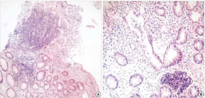
Microscopically, specimens from ulcers of the colon showed shallow ulcerations with inflammatory infiltration consisting of lymphocytes and plasma cells. There were no granulmas (Fig. 3).
Further studies during her hospitalization showed that the patient was negative for HIV. She was negative for antinulear antibody and HLA-B51. Under sterile condition, intrader- mal injection of the skin with a 20-guage needle was done.
Within 48 hr, an erythematous papule developed. The pa- thergy test was positive. Histological examination of the perineal lesion revealed chronic ulcer with acute and chron- ic inflammatory cell infiltration and increased small blood vessels (Fig. 4). On ophthalmologic examination, there was no ocular manifestation of Behcet’s disease.
Total parenteral nutrition with nothing per oral and intra- venous administration of methylprednisolone, 62.5 mg were started. In addition, metronidazole was injected for anal fistu- la. After 1 week, the patient responded well clinically and was tolerable with diet. Treatment with prednisone, 40 mg and 5-aminosalicylate (mesalazine), 4.0 g by mouth was started.
The abdominal symptoms and vulva ulcer improved gradu- ally, and she was discharged 2 months after admission. The dose of prednisone was tapered and discontinued. One year later, she was doing well without recurrence of symptoms.
DISCUSSION
Behcet’s disease was originally described in 1937 and char- acterized by oral and genital ulceration and ocular inflamma- tion (6). It is now acknowledged that this disorder has a wide spectrum of clinical manifestation. Although many diagnostic criteria have been established, there is no universally accept- ed definition. The diagnosis is clinical and is now based on criteria suggested by an international study group for Behcet’s disease (7). In the present case, oro-genital ulceration and skin lesions led to the diagnosis of Behcet’s disease. The eti- ology remains unknown, but the most widely held hypoth- esis of pathogenesis is that a profound inflammatory response is triggered by an infectious agent in a genetically suscepti- ble host (6). Other possible mechanism is that Behcet’s dis- ease may be autoimmune in origin (8, 9). An inflammatory response to several autoantigens is found, and generalized aberrant T cell responses results in enhanced nonspecific inflammation. An increased production of interferon gamma (IFN- ) by T cells has been demonstrated in active Behcet’s disease, and circulating T cells have the T-helper phenotype (Th1) predominantly (10). In Crohn’s disease, murine and human studies have demonstrated an increased expression of Th1 cytokines by lamina propria lymphocytes (11).
Oshima et al. (12) reported that over 40% of Behcet’s dis- ease patients had gastrointestinal complaints. Symptoms included abdominal pain, diarrhea, nausea, anorexia, and abdominal distension. Although gastrointestinal symptoms are common, the demonstration of gastrointestinal ulcer is rare. This so-called intestinal Behcet’s disease accounts for only 1-2% of cases (1, 2). In intestinal Behcet’s disease, ulcer- ation of the gastrointestinal tract can be found throughout the intestine, but the most frequent area of involvement is the ileocecal region. Only 15% of intestinal Behcet’s disease diffusely involve the colon (13). Behcet’s colitis can appear as ulcerative colitis or Crohn’s disease when there are skip lesions with rectal sparing. The ulcers are usually large, dis- crete, punch-out appearing lesions and can extend to the serosa. Formation of fistula, hemorrhage, or perforation occurs in up to 50% of cases involving the intestine. The ulcers are found within normal or minimally inflamed mucosa. Micro- scopic examination reveals vasculitis involving small and medium sized vessels. The lymphocytes are infiltrated, and dense perivascular infiltrate is frequently seen (3, 4).
There are many extra-intestinal findings of Crohn’s disease, such as oral and genital ulcers, erythema nodosum, uveitis and arthritis, resembling the manifestations of Behcet’s dis- ease. It is also very difficult to distinguish the intestinal Beh- cet’s disease from that of Crohn’s disease in some patients. It is possible that a patient with Crohn’s disease meets the cri- teria for Behcet’s disease. There are several reports on the coexistence of Behcet’s disease and Crohn’s disease (14, 15).
In the present case, we made the diagnosis of Behcet’s dis- ease as described above. However, the gastrointestinal mani-
Fig. 1.The external genitalia showed a linear to ovoid shaped ulcerating wound at the perineum and vulva area.
festation of this case is quite atypical for Behcet’s colitis and more similar to Crohn’s colitis - longitudinal ulcers, cobble- stone appearance, and ano-rectal fistula are usual findings in Crohn’s colitis. However, we cannot conclude that the patient had Crohn’s disease because granulomas, the hallmark of Crohn’s colitis, were absent. The lack of this pathologic find- ing is against the diagnosis of Crohn’s disease.
The findings in the family reported by Yim and White (16), suggest that inflammatory bowel disease and Behcet’s disease may be closely related and part of a spectrum of dis- ease rather than distinct disease entities. In our patient, clin-
ical and pathological findings are characteristic of intestinal Behcet’s disease. Patient’s bowel symptoms, endoscopic ap- pearance, and the response to medical treatment were com- patible with Crohn’s colitis. These findings suggest that Behcet’s disease may be a part of the spectrum of chronic inflammatory bowel disease.
REFERENCES
1. Kasahara Y, Tanaka S, Nishino M, Umemura H, Shiraha S, Kuyama Fig. 2.Colonoscopic findings revealed grossly normal-appearing mucosa in the terminal ileum (A). Multiple longitudinal ulcers and inflam- matory pseudopolyps with a cobblestone appearance were observed in the ascending, transverse, and descending colon. However, the Ileocecal valve was preserved from the inflammation (B, C). A suspicious fistular opening just above the anus was observed (D).
A B
C D
T. Intestinal involvement in Behcet’s disease: review of 136 surgical cases in the Japanese literature. Dis Colon Rectum 1981; 24: 103-6.
2. Masugi J, Matsui T, Fujimori T, Maeda S. A case of Behcet’s disease with multiple longitudinal ulcers all over the colon. Am J Gastroen- terol 1994; 89: 778-80.
3. Kim JH, Choi BI, Han JK, Choo SW, Han MC. Colitis in Behcet’s disease: characteristics on double-contrast barium enema examina- tion in 20 patients. Abdom Imaging 1994; 19: 132-6.
4. Lee RG. The colitis of Behcet’s syndrome. Am J Surg Pathol 1986;
10: 888-93.
5. Kyle SM, Yeong ML, Isbister WH, Clark SP. Behcet’s colitis: a dif- ferential diagnosis in inflammations of large intestine. Aust N Z J Surg 1991; 61: 547-50.
6. Behcet H. Uber rezidivierende, aphthose, durch ein virus verursachte Geschwure am Mund, am Auge und an den Genitalien. Dermatol Wochenschr 1937; 105: 1152-7.
Fig. 4.Microscopic examination of the perineal lesion revealed chronic ulcer with acute and chronic inflammatory cell infiltration and in- creased small blood vessels (A: H&E stain, ×100; B: H&E stain, ×400).
A B
Fig. 3.Microscopic examination from ulcers of the colon showed shallow ulcerations with inflammatory infiltration consisting of lymphocytes and plasma cells. There were no granulmas (A: H&E stain, ×100; B: H&E stain, ×200).
A B
7. International Study Group for Behcet’s Disease. Criteria for diagno- sis of Behcet’s disease. Lancet 1990; 335: 1078-80.
8. Benoist C, Mathis D. Autoimmunity provoked by infection: how good is the case for T cell epitope mimicry? Nat Immunol 2001; 2: 797- 801.
9. Yazici H. The place of Behcet’s syndrome among the autoimmune diseases. Int Rev Immunol 1997; 14: 1-10.
10. Freysdottir J, Lau S, Fortune F. Gammadelta T cells in Behcet’s dis- ease (BD) and recurrent aphthous stomatitis (RAS). Clin Exp Im- munol 1999; 118: 451-7.
11. Cobrin GM, Abreu MT. Defects in mucosal immunity leading to Crohn’s disease. Immunol Rev 2005; 206: 277-95.
12. Oshima Y, Shimizu T, Yokohari R, Matsumoto T, Kano K, Kagmani
T, Nagaya T. Clinical studies on Behcet’s syndrome. Ann Rheum Dis 1963; 22: 36-45.
13. Smith JA, Siddiqui D. Intestinal Behcet’s disease presenting as a massive acute lower gastrointestinal bleeding. Dig Dis Sci 2002; 47:
517-21.
14. Tolia V, Abdullah A, Thirumoorthi MC, Chang CH. A case of Behcet’s disease with intestinal involvement due to Crohn’s disease. Am J Gastroenterol 1989; 84: 322-5.
15. Naganuma M, Iwao Y, Kashiwagi K, Funakoshi S, Ishii H, Hibi T.
A case of Behcet’s disease accompanied by colitis with longitudinal ulcers and granuloma. J Gastroenterol Hepatol 2002; 17: 105-8.
16. Yim CW, White RH. Behcet’s syndrome in a family with inflamma- tory bowel disease. Arch Intern Med 1985; 145: 1047-50.