INTRODUCTION
Although cerebrovascular diseases are rare in childhood, moyamoya disease is a relatively important and common dis- ease, especially among East Asian children. The pathophysi- ology of the moyamoya disease is that of progressive narrow- ing of the major medium- and/or large-sized cerebral arter- ies; however, the cause of the arterial narrowing is not known.
Moyamoya disease was named for the abnormal vascular network at the base of the brain that is typically found on cerebral angiography (1). The disease was first reported by Takeuchi and Shimizu in 1957 and the term ‘‘moyamoya’’
means ‘‘something hazy just like a puff of cigarette smoke in the air’’ (1, 2).
In children, the disease generally presents as either tran- sient ischemic attacks manifesting as a motor disturbance (70 to 80%), or with epileptic seizures (20 to 30%) (3). Once the diagnosis of moyamoya disease is suspected on clinical evaluation, confirmation by neuroradiological studies follows.
Use of either invasive angiography or magnetic resonance imaging (MRI) and magnetic resonance angiography (MRA) are commonly employed for diagnosis (3, 4). The most con- sistent finding on the neuroradiological examination is the stenosis or occlusion of the terminal portion of the bilateral
internal carotid arteries that is associated with an abnormal vascular network at the base of the brain. Therefore, several test modalities, such as the single photon emission comput- ed tomography (SPECT) or MRA, have been used for diag- nosis (5-7).
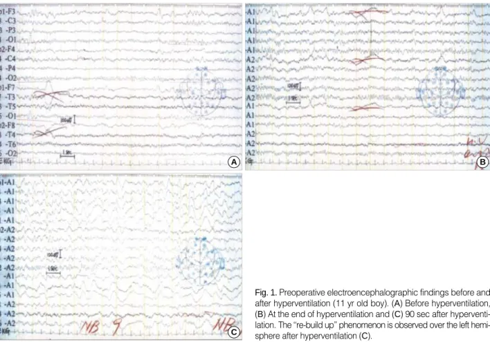
The electroencephalogram (EEG) is not commonly used as a diagnostic tool for this disorder. The EEG findings asso- ciated with moyamoya disease are high-amplitude slow waves, called hemispheric posterior slowing or centrotemporal slow- ing; these findings often appear in association with cerebral ischemia (8). The characteristic EEG findings associated with moyamoya have been reported to be a ‘‘build up’’ phenomenon, that is, the appearance of monorhythmic generalized and high voltage slow waves during hyperventilation followed by the ‘‘re-build up’’ phenomenon, the reappearance of poly- morphous high amplitude slow waves several minutes after the end of hyperventilation (9, 10).
The aim of this study was to evaluate the usefulness of the EEG as a follow-up test for the postoperative status of patients with moyamoya disease. We studied the clinical characteris- tics of patients who underwent surgical revascularization for moyamoya disease and analyzed the correlationship among clinical, EEG and SPECT findings before and after the sur- gical procedure.
Deok-Soo Kim, Tae-Sung Ko*, Young-Shin Ra�, Choong-Gon Choi�
Department of Pediatrics, Kangbuk Samsung Hospital, Sungkyunkwan University School of Medicine, Seoul;
Departments of Pediatrics*, Neurosurgery�and Radiology�, Asan Medical Center, University of Ulsan College of Medicine, Seoul, Korea
Address for correspondence Tae-Sung Ko, M.D.
Department of Pediatrics, Asan Medical Center, University of Ulsan College of Medicine, 388-1 Pungnap-dong, Songpa-gu, Seoul 138-736, Korea Tel : +82.2-3010-3381, Fax : +82.2-473-3725 E-mail : [email protected]
495
Postoperative Electroencephalogram for Follow up of Pediatric Moyamoya Disease
It is well known that the electroencephalographic finding in patients with moyamoya disease demonstrates the characteristic ‘‘re-build up’’ phenomenon a few minutes after hyperventilation. To evaluate the usefulness of an electroencephalogram (EEG) in the postoperative management of children with moyamoya disease, we studied the presence or absence of improvement in the clinical, single photon emission computed tomography (SPECT) and EEG findings, before and after surgery. Twen- ty-two patients, who underwent indirect revascularization surgery for moyamoya disease, were included in our study. Clinical improvement was assessed as the disappearance or decrease of a transient ischemic attack or headache. The findings on the EEG and SPECT were considered improved when the re-build up pheno- menon was absent and when there was improvement in the cerebrovascular reserve as a result of the acetazolamide challenge test. The statistical correlation analysis for both clinical and EEG improvement were consistent (kappa value=0.409, p<0.05).
However, the result from the clinical and SPECT improvement as well as that from EEG and SPECT improvement were not statistically significant. Our results suggest that EEG can be used as a noninvasive and simple follow-up test for moyamoya disease after indirect revascularization surgery if the hyperventilation procedure is effectively performed during EEG recording.
Key Words : Moyamoya Disease; Electroencephalography; Follow-up Studies
Received : 16 June 2005 Accepted : 1 December 2005
cerebral infarction on MRI or with continuous focal slowing on the EEG were included. We recruited twenty-two patients for this study.
We performed the EEG or SPECT evaluations 6-12 months after the surgery; clinical evaluation for the recurrence of sym- ptoms was done every three months after the surgery. The EEG was done on an international 10-20 system and was recorded 2 to 5 min after hyperventilation for 3 min (Fig. 1).
We obtained SPECT images from the Triad XLT 20 (Trion- ix, OH, U.S.A.) using the Tc-99m ethyl cysteinate dimer with acetazolamide stress.
Clinical improvement was defined as the decrease in fre- quency or intensity of the TIAs or headache. Improvement of EEG findings was based on the disappearance of the re-
Clinical characteristics
Patients presented initially with the following symptom and sign: TIA (n=19), seizure (n=1), hemiplegia (n=1) and headache (n=1). The ratio of male to female patients was 1:1.
The median age of the patients was six years old. The dura- tion from the onset of symptoms to the operation ranged from one month to nine years (median duration, 16 months).
Before the surgery, cerebral infarction was detected in 8 pati- ents and associated with hydrocephalus in one case. One pati- ent had a cerebral infarction after the first operation. The follow-up duration ranged from 7 months to 60 months with a median duration of 26 months (Table 1). Clinical improve-
Fig. 1. Preoperative electroencephalographic findings before and after hyperventilation (11 yr old boy). (A) Before hyperventilation, (B) At the end of hyperventilation and (C) 90 sec after hyperventi- lation. The ‘‘re-build up’’ phenomenon is observed over the left hemi- sphere after hyperventilation (C).
A
C
B
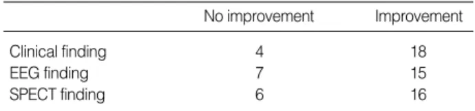
ment was found in about 82% of patients. Improvement in EEG and SPECT was about 68%, 73% respectively (Table 2).
Agreement among clinical, EEG and SPECT improvement
Based on the statistical analysis for clinical and EEG imp- rovement, the kappa value was 0.409, which signifies good agreement (p<0.05). In the case of clinical and SPECT imp- rovement, there were poor agreement and no significant result.
In addition, agreement between the postoperative EEG and SPECT improvement did not show statistical significance.
Out of four cases with no clinical improvement, three cases showed no improvement in EEG findings, whereas there was no SPECT improvement in one case. In seven cases of no EEG improvement, SPECT improvement did not observed in two cases.
DISCUSSION
Moyamoya disease was first reported by Takeuchi and Shi- mizu (2). The disease is characterized by hypoplasia of the bilateral internal carotid arteries. The abnormal vascular net- work is found on angiography (1, 2). The prevalence of the disease is high in East Asian countries, especially in Japan, China and Korea. Although the etiology of moyamoya dis- ease is still not known, it is considered to be multifactorial condition that is affected by elements such as vascular growth factors, cytokines, basic-fibroblast growth factor or genetic abnormality (11-15).
In children, the disease generally presents with either fre- quent TIAs manifesting as hemiparesis, monoparesis and/or sensory disturbance (70 to 80%), or with epileptic seizures (20 to 30%) (3). In addition, headache or involuntary move- ments can also be a presenting symptom. In our study, hemi-
paresis was observed in about 86.4%; the presence of seizure or headache was noted in a small number of patients.
The diagnosis of moyamoya disease can be confirmed by conventional angiography or MRA; the findings from these studies can be useful for planning surgical treatments (3, 4).
Cerebral blood flow is evaluated to determine the clinical severity of moyamoya disease, the effectiveness of indirect revascularization surgery and it can be measured by 133xenon computed tomography, SPECT, or positron emission tomog- raphy (5-7, 16, 17).
In children, EEG may provide definite information on moyamoya disease even when clinical symptoms are miss- ing (10). The EEG findings associated with moyamoya dis- ease shows high-amplitude slow waves in association with cerebral ischemia and the characteristic ‘‘re-build up’’ phe- nomenon. A ‘‘re-build up’’ phenomenon is the reappearance of polymorphous slow waves several minutes after cessation of the hyperventilation (8-10). About 95% of all reported cases where a hyperventilation test was performed during EEG recording showed this ‘‘re-build up’’ phenomenon (10).
In this study we obtained the re-build up phenomenon in about 80% of the patients who were capable of performing an effective hyperventilation. When a patient presented with seizure and characteristic EEG changes, moyamoya disease was considered as a possible etiology.
The mechanism that is responsible for the EEG re-build up phenomenon is still unclear, but several studies have sug- gested a possible mechanism. Electrophysiological build-up during hyperventilation is based on a reduction of the arte- rial partial pressure of carbon dioxide as a result of hyperven- tilation and the consequent reduction of cerebral perfusion (18). The concentration of oxygenated hemoglobin progres- sively increases during hyperventilation and subsequently decreases after hyperventilation for 5 to 7 min. The re-build up phenomenon occurs when oxygenated hemoglobin decreas- es and the deoxygenated hemoglobin increases (19). In a region where cerebrovascular reactivity is disturbed, a reduc- tion of regional cerebral blood flow after hyperventilation results. Therefore, the regional cerebral hypoxia and the dis- turbance in oxygen metabolism play important roles in the occurrence of the re-build up phenomenon after hyperventi- lation (20, 21).
Surgical revascularization procedures are used to augment cerebral blood flow and indirect revascularization surgery is
Sex
Male 11 (50.0%)
Female 11 (50.0%)
Age at operation 4-15 yr
Chief complaint at first visit
Transient ischemic attacks 19
Headache 1
Seizure 1
Hemiplegia 1
Preoperative
Duration of symptom 1 month-9 yr (median, 16 months)
Cerebral infarction 8 (36.4%)
Abnormal EEG findings* 17 (77.2%)
Postoperative
Follow-up period 7-60 months (median, 26 months) Table 1.The clinical characteristics of patients with moyamoya disease
EEG, electroencephalogram. *, Abnormal EEG findings included slow wave discharges or re-build up phenomenon.
EEG, electroencephalogram; SPECT, single photon emission comput- ed tomography.
No improvement Improvement
Clinical finding 4 18
EEG finding 7 15
SPECT finding 6 16
Table 2.The improvement in clinical, EEG and SPECT findings after revascularization surgery
early detection of areas with impaired hemodynamics is crit- ical in the management of moyamoya disease. It is also impor- tant to evaluate whether there is the improvement of cere- bral perfusion after the operation. Improved blood supply to the ischemic brain from the external carotid artery branches has been demonstrated. Determination of the correlation bet- ween clinical outcome and the degree of revascularization from surgery, as evaluated by neurological investigation, is difficult (24). Therefore, effective methods to monitor the change of regional cerebral perfusion reserve after surgery is required. SPECT is valuable in assessing the cerebral perfu- sion reserve and predicting a long-term prognosis, but the procedure is somewhat inconvenient to perform in children.
Unlike SPECT, the EEG cannot directly reflect the blood per- fusion or vascular reserve in cortical areas. However, the EEG is a noninvasive diagnostic tool that is commonly used, and well tolerated, in children. We performed EEG and SPECT 6-12 months after surgery. The postoperative EEG findings showed the disappearance or the reduction in the duration and distribution of the ‘‘re-build up’’ phenomenon; these find- ings are consistent with other reports (9). We found that there was good agreement between clinical and EEG improvement using the kappa statistic and that EEG may be useful tool for predicting the disappearance or recurrence of clinical sym- ptoms in children with moyamoya disease. In conclusion, we suggest that an EEG, a simple and noninvasive study, may be useful as the primary follow-up test after surgery for moyamoya disease in children.
REFERENCES
1. Suzuki J, Takaku A. Cerebrovascular ‘‘moyamoya’’ disease: disease showing abnormal net-like vessels in base of brain. Arch Neurol 1969;
20: 288-99.
2. Takeuchi K, Shimizu K. Hypoplasia of the bilateral internal carotid arteries [in Japanese]. No To Shinkei 1957; 9: 37-43.
3. Handa H, Yonekawa Y, Goto Y. Analysis of the filing data bank of 1500 cases of spontaneous occlusion of the circle of Willis and fol- low-up study of 200 cases for more than 5 years. In: Handa H (eds):
Annual Report (1984) of Research Committee on Spontaneous Occlu- sion of the Circle of Willis (Moyamoya Disease). Tokyo, Ministry of
sion-weighted MR imaging: initial experience. Am J Neuroradiol 2003; 24: 741-7.
8. Kodama N, Aoki Y, Hiraga H, Wada T, Suzuki J. Electroencephalo- graphic findings in children with moyamoya disease. Arch Neurol 1979; 36: 16-9.
9. Kuroda S, Kamiyama H, Isobe M, Houkin K, Abe H, Mitsumori K.
Cerebral hemodynamics and ‘‘re-build-up’’ phenomenon on elec- troencephalogram in children with moyamoya disease. Child’s Nerv Syst 1995; 11: 214-9.
10. Kurlemann G, Fahrendorf G, Krings W, Sciuk J, Palm D. Charac- teristic EEG findings in childhood moyamoya syndrome. Neurosurg Rev 1992; 15: 57-60.
11. Takahashi A, Sawamura Y, Houkin K, Kamiyama H, Abe H. The cerebrospinal fluid in patients with moyamoya disease (spontaneous occlusion of the circle of Willis) contains high level of basic fibro- blast growth factor. Neurosci Lett 1993; 160: 214-6.
12. Suzui H, Hoshimaru M, Takahashi JA, Kikuchi H, Fukumoto M, Ohta M, Itoh N, Hatanaka M. Immunohistochemical reactions for fibroblast growth factor receptor in arteries of patients with moyamoya disease. Neurosurgery 1994; 35: 20-5.
13. Ikeda H, Sasaki T, Yoshimoto T, Fukui M, Arinami T. Mapping of a familial moyamoya disease gene to chromosome 3p24.2-p26. Am J Hum Genet 1999; 64: 533-7.
14. Inoue TK, Ikezaki K, Sasazuki K, Matsushima T, Fukui M. Linkage analysis of moyamoya disease on chromosome 6. J Child Neurol 2000; 15: 179-82.
15. Yamauchi T, Tada M, Houkin K, Tanaka T, Nakamura Y, Kuroda S, Abe H, Inoue T, Ikezaki K, Matsushima T, Fukui M. Linkage of familial moyamoya disease (spontaneous occlusion of the circle of Willis) to chromosome 17q25. Stroke 2000; 31: 930-5.
16. Nakashima H, Meguro T, Kawada S, Hirotsune N, Ohmoto T. Long- term results of surgically treated Moyamoya disease. Clin Neurol Neurosurg 1997; 99 (Suppl 2): 156-61.
17. Fukui M. Guidelines for the diagnosis and treatment of spontaneous occlusion of the circle of Willis (‘Moyamoya’ disease). Clin Neurol Neurosurg 1997; 99 (Suppl 2): 238-40.
18. Konishi T. The standardization of hyperventilation on EEG record- ing in childhood. The quantitative analysis of build-up. Brain Dev 1987; 9: 21-5.
19. Lin Y, Yoshiko K, Negoro T, Watanabe K, Negoro M. Cerebral oxy- genation state in childhood moyamoya disease: a near-infrared spec- troscopy study. Pediatr Neurol 2000; 22: 365-9.
20. Kazumata K, Kuroda S, Houkin K, Abe H, Mitumori K. Regional cerebral hemodynamics during re-build-up phenomenon in child- hood moyamoya disease: An analysis using 99mTc-HMPAO SPECT.
Child’s Nerv Syst 1996; 12: 161-5.
21. Yonekawa Y, Taub E. Moyamoya disease: status 1998. The Neu- rologist 1999; 5: 13-23.
22. Kinugasa K, Mandai S, Kamata I, Sugiu K, Ohmoto T. Surgical treatment of Moyamoya disease: Operative technique for encepha-
lo-duro-arterio-myo-synangiosis, its follow-up, clinical results, and angiograms. Neurosurgery 1993; 32: 527-31.
23. Kim DS, Yoo DS, Huh PW, Kim JK, Cho KS, Kang JK. Recent sur- gical treatment of moyamoya disease. J Korean Neurosurg Soc 2001;
30: 800-4.
24. Caldarelli M, Di Rocco C, Gaglini P. Surgical treatment of moyamoya disease in pediatric age. J Neurosurg Sci 2001; 45: 83-91.