경락경혈학회지 Vol.28, No.3, pp.141 150, 2011
∼ Korean Journal of Acupuncture
Hye-Yeon Yun , Sun-Oh Kwon , Seung-Tae Kim , Hi-Joon Park Dae-Hyun Hahm , Hye-Jung Lee
Acupunture & Meridian Science Research Center, Kyung-Hee University Dept. of Meridian and Acupoint, College of Korean Medicine, Kyung-Hee University
Dept. of Oriental medicine, Graduate School of Kyung-Hee University
Division of Meridian and Structural Medicine, School of Korean Medicine, Pusan National University
Abstract
Objectives : The aims of this study were to evaluate a blister caused by cupping.
Methods : We searched relevant case reports, survey, and review articles using databases of online bibliography.
Results :
1. The fluid in the blister caused by cupping therapy is normal substance by laboratory analysis.
The fluid has no signs of infection in the culture, Gram stain, or tissue biopsy
2. In histological finding, the blister caused by cupping therapy is made by dermo-epidermal seperation at subcellular level. Suction blistering was neither inflammatory nor autolysis activation of lysosomal hydrolases.
3. Blistering times directly, related to suction pressure. Suction blister formation time is accelerated in older subjects compared with younger individuals and higher temperature was more susceptable to the blister compared with lower temperature. The flexor aspect of forearm is a easy site for suction blister formation compared with leg and abdominal site.
4. Blister caused by cupping therapy is treated by regular and judicious changes of sterile dressing over several weeks. The vesicles healed well and left no visible scar.
Conclusions : Blister caused by cupping therapy is artificially controlled by doctor’s therapeutic purpose. Blister is not histologically injurious to health and the blister is a natural concomitant after cupping therapy.
Key words : Cupping therapy, Suction Blister, Bullae, Wet cupping, Dry cupping
- 142 -
Table 1. ( , Immunobullous disease)
18)Table 2. ( )
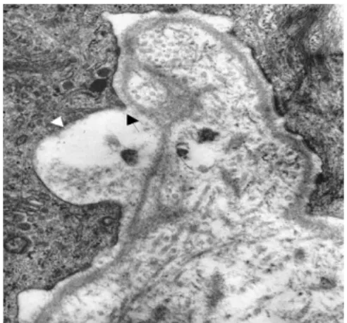
18)- 144 - Fig. 1. Two suction microblister (0.2mm in diameter) on top of dermal papillae (arrowhead).
Fig. 2. Initial events of dermo (white arrowhead)- epidermal (arrow) separation at subcellular level.
Fig. 3. Base of suction blister with two flat epidermal
cells (arrow).
Fig. 4. Schematic representation of partial separation
of epidermis (arrow) and dermis (arrowhead) by
vacuum.
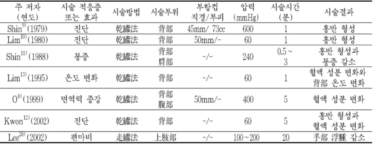
Table 3. 24 34 p=410mmHg
23)