| Abstract |
1)PURPOSE: The aim of this study was to determine how biofeedback training with Shaker exercise effects the activation of the cervical myocardial muscle in stroke with swallowing disorder.
METHODS: From June 2018 to September 2018, 30 patients who were hospitalized at C hospital and K hospital in Daegu, and K hospital in Gumi were surveyed to meet the criteria for selection. The participants were classified into three groups, and Shaker exercise was applied three times a day, five times a week, for four weeks; visuoauditory
본 논문은 신윤아(2019)의 박사 학위 논문의 요약본임.
†Corresponding Author : Hyak-Cheol Kwon
[email protected], https://orcid.org/0000-0003-4628-9236 This is an Open Access article distributed under the terms of the Creative Commons Attribution Non-Commercial License (http://creativecommons.org/licenses/by-nc/3.0) which permits unrestricted non-commercial use, distribution, and reproduction in any medium, provided the original work is properly cited.
biofeedback group (VABG), visual biofeedback group (VBG), and self-exercises group (SG). In addition, the suprahyoid muscle activity was performed three times (before intervention, after intervention, and after two weeks).
RESULTS: The pre and post-test comparisons, revealed a significant difference between the three groups (p<.01);
VABG had the highest suprahyoid muscle activity. The post-test and follow-up test produced similar results in, the three groups (p>.05). The mean comparison showed the smallest difference in VABG, indicating that muscle persistence was the best.
CONCLUSION: Shaker exercise has an effect on suprahyoid muscle activation. Biofeedback training, which provides an input of multi-sensory information in swallowing disorder treatment is recommended because it has the greatest effect when combined with visuoauditory biofeedback.
Key Words: Biofeedback training, Shaker exercise, Suprahyoid muscle, Swallowing disorder
Research Article Open Access
Shaker운동을 병행한 바이오피드백 훈련이 삼킴장애가 있는 뇌졸중환자의 설골상근 근활성도에 미치는 영향
신윤아⋅권혁철 1† ⋅김환 1
구미대학교 작업치료과, 1 대구대학교 작업치료학과
Effects of Biofeedback Training with Shaker Exercise on the Suprahyoid muscle Activity of Stroke with Swallowing Disorder
Yoon-A Shin, OT⋅Hyak-Cheol Kwon 1† ⋅Hwan Kim 1 Department of Occupational Therapy, Gumi University
1 Department of Occupational Therapy, Daegu University Received: November 21, 2018 / Revised: November 27, 2018 / Accepted: December 29, 2018
ⓒ 2019 J Korean Soc Phys Med
37~78%로 뇌졸중 후 발생하는 주요한 신경학적 증상이 라 할 수 있다[3].
삼킴장애란 삼킴의 복합적인 과정 중에 발생되는 문제로 신체적, 신경학적 기능에 의해 삼킴에 어려움을 갖는 것이다[4]. 정상 삼킴의 단계는 구강준비단계, 구 강단계, 인두단계, 식도단계로 나뉜다[5]. 이 중 인두단 계에서는 후두개 ․ 설골 ․ 후두 이동, 후두 통과와 흡인 과 같은 다양한 증상들을 나타내는데[6], 특히 흡인은 음식물의 일부 또는 전부가 기도로 들어가서 발생하게 되는 삼킴장애의 주요한 증상 중 하나이다[3]. 흡인을 막아주는 생리학적 기전으로는 후두에서 일어나는 기 도 폐쇄와 설골의 움직임이 있으며, 설골의 움직임은 전 ․ 상방으로 발생하여 후두 상승을 만들고 , 후두 상승 이 상부식도괄약근을 열어주어 음식물을 식도로 보내 므로, 인두부 구조물의 움직임 중 설골의 움직임은 갑 상연골의 상승과 상부식도괄약근의 개구를 유도하는 매우 중요한 역할을 한다고 할 수 있다[7]. 상부식도괄 약근이 열리지 않으면 위로 음식물이 쌓여 음식물이 후두를 통과하여 진성대 아래로 내려가는 흡인이 발생 하므로 정상적인 삼킴과정에서 설골의 움직임은 흡인 또는 침습을 방지함에 있어 매우 중요하고, 이러한 움 직임은 설골상 ․ 하근에 의하여 조절된다[8]. Shaker가 제시한 머리들기 운동은 설골상근을 강화시키고 상부 식도괄약근을 잘 열리게 하여 흡인의 위험을 감소시킨 다 [9]. Shaker운동은 등척성의 방법과 등속성의 방법을 사용하여 바로 누운 자세에서 머리를 들어 발끝을 바라 보게 하는 운동을 계획적으로 반복함으로 설골상근과 상부식도괄약근을 강화할 수 있다[3].
최근 연구되고 있는 뇌졸중 환자의 치료에 긍정적인 효과를 줄 수 있는 방법으로 바이오피드백 훈련을 제시 하는데[10], 이는 기계장치를 사용하여 모니터를 통해 인체의 신경생리학적 신호를 시 ․ 청각적으로 확인하여
동을 시각적 또는 청각적으로 정보를 제공하여 환자 스스로 근 긴장 수준을 조절할 수 있도록 학습을 가능하 게 해준다[13]. 시각적 정보는 환경과 움직임에 대한 정보를 제공하여 적절한 신체의 운동 조절을 가능하게 하며[14], 청각적 정보는 신체의 직접적이며 지속적인 감각 정보를 제공한다[15]. 시각적 바이오피드백으로 진행된 연구들이 재활분야에서 진행되고 있으며[16], 최근 근전도 바이오피드백 훈련이 국내와 국외 재활의 영역에서 뇌손상 환자와 다양한 질환들을 대상으로 효 과적으로 사용되고 있다[17-19].
현재까지 운동을 통한 삼킴기능에 미치는 효과에 관한 연구가 다양하게 이루어졌고[9,4,20], 근전도를 이 용한 연구들도 진행되었다[21,22]. 그러나 삼킴장애가 있는 뇌졸중 환자가 자발적으로 운동을 하면서 시 ․ 청 각적으로 자극을 받는 바이오피드백 훈련이 삼킴과 관 련된 근활성도에 미치는 영향에 대한 연구는 미비한 실정이다 . 이에 본 연구에서는 삼킴장애가 있는 뇌졸중 환자를 대상으로 인두단계 삼킴에 주요한 근육인 설골 상근을 강화하기 위하여 운동 -기반 접근인 Shaker 운동 을 이중감각 또는 단일감각 바이오피드백 훈련과 병행 하였다. 이러한 훈련이 인두단계의 근활성도에 어떠한 영향을 미치는지 확인하여 삼킴장애 재활에서 임상적 으로 더욱 효과적인 중재방법을 제안하고자 한다.
Ⅱ. 연구방법
1. 연구 대상 및 기간
본 연구는 Shaker운동을 병행한 바이오피드백 훈련
이 삼킴장애가 있는 뇌졸중 환자의 설골상근 근활성도
에 미치는 영향을 알아보기 위하여 2018년 6월부터
2018년 9월까지 대구광역시에 위치한 C병원, K병원,
그리고 구미시에 있는 K병원에 입원한 환자를 대상으
로 하였다. 참가자는 뇌졸중 후 인두단계 삼킴장애가 영상학적 평가를 통하여 증명된 환자 30명을 대상으로 실시하였다. 인두단계를 강화하는 운동-기반의 접근으 로 환자들이 스스로 할 수 있는 운동인 Shaker 운동(등 속성 방법)을 선정하여 시 ․ 청각적 바이오피드백 훈련 을 병행한 그룹(VABG)과 시각적 바이오피드백을 병행 한 그룹(VBG) 그리고 바이오피드백 없이 자가운동만 실시한 그룹(SG)으로 나누고 각각 10명씩 숫자뽑기를 사용하여 무작위로 배분하였다.
2. 연구 절차
설골상근의 근활성도는 표면근전도 측정 장비인 2EM( 2EM 4D-MT, Relive, Gimhae, Korea)를 사용하였 고 설골상근에 중 이복근과 이설골근에 부착하여 근활 성도를 측정하였다(Fig. 1). 연구대상자가 수의적으로 하는 자가운동임을 감안하여 최대 수의적 등척성 근수 축(maximal voluntary isometric contraction: MVIC)에 대 한 비율로 정규화하여 비교하는 MVIC 값을 사용하였 다 . 중재 전 바로 누운 대상자에게 평가자는 “저의 구령 에 맞추어 고개를 들어 발끝을 보세요. 3회 실시하겠습 니다.”라고 지시하였고, 3회 실시하여 설골상근의
%MVIC값을 측정하였다. 중재기간 동안 Shaker 운동의 등속성 방법을 바이오피드백과 병행 또는 하지 않고 1일 3회, 주 5회, 4주간, 총 20회기 동안 실시한 후 설골 상근 근활성도를 중재 전과 같은 방법으로 측정하였다.
중재 종결 2주 후 각 그룹의 변화된 설골상근의 근활성 도의 지속을 살펴보기 위하여 동일한 절차로 추이검사 를 실시하였다. 모든 결과 측정은 측정자간에 발생할
수 있는 오염변인을 최소화하기 위하여 동일한 연구자 에 의하여 시행되었다.
3. 측정 방법
설골상근의 근활성도를 측정하기 위하여 근전도 측 정 장비인 2EM (2EM 4D-MT, Relive, Gimhae, Korea)를 사용하여 자료를 수집 , 처리하였다. 근전도 신호의 표 본 추출률(sampling rate)은 1000 Hz로 설정하였으며, 주파수 대역폭(bandpass)은 0-500 Hz를 사용하였고, 전 기 신호에 의한 잡음을 제거하기 위하여 60 Hz의 여과 필터(notch filter)를 적용하였다. 5초간 최대 수의적 등 척성 수축을 통해 측정된 자료의 처음과 마지막 1초를 제외한 3초 동안의 평균 근전도 신호량의 3회 반복 측정 한 평균값을 기준으로 %MVIC 값을 구하였다. 근전도 신호의 수집을 위해 전도성 젤이 도포된 염화은 (Ag-AgCl) 합금의 24 mm 크기의 둥근 모양의 표면 전극 (Covidien kendell diaposable surface EMG electrode)을 사용하였고, 연구에 사용된 장비의 사진은 다음과 같다 (Fig. 1). 전극 부착은 Woo 등[23]의 연구와 동일하게 2개의 이극표면전극(bipolar surface electrode)은 설골상 근 중 이복근과 이설골근이 겹쳐지는 부위의 좌 ․ 우에 부착하였고 , 다른 한 개의 접지전극(ground electrode)은 턱뼈각(angle of mandible)에 부착하였다. 전극을 부착 하기 위한 설골상근의 위치는 다음과 같다(Fig. 2).
4. 실행 도구
바이오피드백 훈련은 시 ․ 청각적 바이오피드백 그 룹 (visuoauditory biofeedback group: VABG)과 시각적 바
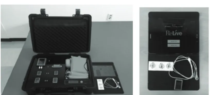
Fig. 1. sElectromyography (sEMG) system 2EM (2EM 4D-MT, Relice, Gimhae, Korea)
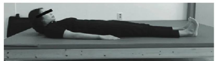
이오피드백 그룹(visual biofeedback group: VBG)으로 나누어 진행하였다. 두 그룹 모두2EM (2EM 4D-MT, Relive, Gimhae, Korea)을 태블릿 모니터에 연결하여 바 이오피드백 훈련 소프트웨어인 4D-MT Private을 사용 하였으나 VABG는 모니터의 그래프를 보면서 환경 설 정을 소리 켬으로 하여 진행하였다. VBG는 장비의 소 리 끔으로 환경 설정을 하여 동일하게 훈련하였고 , 연 구 장비, 각 운동 그룹의 세팅 및 훈련 모습은 다음과 같다(Fig. 3). 자가운동 그룹(self-exercise group: SG)은 바이오피드백 없이 운동을 절차에 맞게 수행하였다.
5. 운동 방법
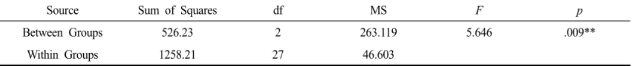
Shaker 운동은 등척성 운동방법과 등속성 운동방법 이 있는데, 등척성 운동방법은 근력 유지가 더 필요하 여 피로도를 높일 수 있기에 본 연구에서는 등속성 운동 방법으로 1일 3회, 주 5회, 4주간, 총 20회기 동안 실시하
였다 . 1회의 운동에서 대상자는 편평한 바닥이나 침대에 누워 바닥에서 고개를 들었다 내리면서 자신의 발가락 바라보기를 30회 시행하되, 일정한 속도를 유지하고 고 개를 들 때 어깨를 들어올리지 않도록 하였다(Fig. 4).
6. 자료 분석
본 연구 결과 분석은 SPSS(statistical package for the social science) for window 24.0으로 모든 통계학적 유의 수준 a는 .05로 하였다. 연구대상자의 일반적 특성은 기술통계를 이용하였고, 중재 전, 후의 설골상근 근활 성도 비교는 각 그룹들의 중재 전과 후의 차이값을 구하 여 일원배치 분산분석(one-way ANOVA)을 실시한 후 사후검정은 Scheffe를 사용하였다. 중재 후와 추이검사 의 설골상근 근활성도 비교도 차이값을 구하여 중재 전, 후와 동일한 절차대로 분석하였다.
A. Digastric muscle B. Geniohyoid muscle Fig. 2. Suprahyiod muscle for the electrode position
A. Training device B. Training of biofeedback Fig. 3. Device and training of the experiment for biofeedback training
Ⅲ. 연구결과
1. 연구 대상자의 일반적 특성
본 연구에 참여한 대상자들의 30명의 성별, 좌 ․ 우 마비유형, 나이, 발병시기, 병변부위, 침습-흡인 척도에 대한 일반적인 특성은 다음과 같다(Table 1).
2. 설골상근 근활성도 비교
1) 중재 전, 후 비교
바이오피드백 훈련을 병행한 Shaker 운동의 효과를 알아보기 위하여 중재 전 평가와 4주간의 중재 적용 후 설골상근 근활성도를 비교하였다. 중재 전과 후의
설골상근 근활성도를 분석한 결과 그룹 간에 통계학적 으로 유의한 차이를 보였고(p<.01) (Table 2), 이는 3개의 그룹 사이에서 설골상근 근활성도가 다르다는 것을 의 미한다. 평균비교에서 확인한 결과 세 그룹 모두 향상되 었으나 , VABG에서 %MVIC값이 가장 높았다(Table 3).
Scheffe 사후 검정을 실시한 결과, VABG와 VBG간에 유의한 차이를 보였다 (p<.05) (Table 4). 설골상근 %MVIC 값이 높을수록 근활성도가 크다는 것을 의미하므로 중 재 후 VABG에서 근활성도가 가장 크게 나타났다.
2) 중재 후, 추이 검사 비교
3개의 그룹의 중재 후와 중재 종료 2주 후 실시한 추이 검사를 비교하여 중재로 향상된 설골상근의 유지
Characteristics Category Number Percentage (%)
Gender Male 22 73.333
Female 8 26.666
Paralysis Rt 13 43.333
Lt 17 56.666
Age
60≤ 8 26.666
61-75 18 60.000
76≥ 4 13.333
Onset period
6 ms≤ 7 23.333
7 ms-24 ms 19 63.333
24 ms≥ 4 13.333
Lesion Brain-stem 13 43.333
Extra Brain-stem 17 56.663
PAS Mean 3.533
SEG: Shaker exercise group PAS: Penetration-Aspiration Scale
Table 1. General characteristics of the subjects Fig. 4. Shaker exercise
에 대한 지속 상태를 알아보고자 하였다. 중재 후와 추이 검사 상 차이값을 분석한 결과 유의한 차이를 나타내지 않았다(p>.05) (Table 5). 평균 비교에서 살펴 보면 VABG가 가장 적은 차이를 보이고, 이는 VABG에 서 설골상근 근활성도가 가장 오래 지속된다는 것을 의미한다(Table 6). Scheffe 사후 검정을 실시한 결과, 세 그룹간에 유의한 차이가 나타나지 않았다(p<.05) (Table 7).
Ⅳ. 고 찰
뇌졸중 후 삼킴장애는 비디오투시검사와 같은 객관 적인 연구에서 64-78%의 높은 발병률을 보이며[24], 특히, 흡인(aspiration)은 흡인성 폐렴을 일으킬 수 있어 죽음과 관련이 깊으며[25], 삼킴의 단계 중 인두단계에 발생하는 문제이다. 이에 본 연구에서는 연구 대상을 인두단계에 삼킴장애를 보이는 뇌졸중 환자로 선정하 였다. 설골의 움직임과 관련이 있는 후두 상승 여부가
*p<.05, **p<.01
Group Mean SD SE
VABG 20.342 9.340 2.953
VBG 10.610 4.247 1.343
SG 12.665 5.875 1.858
SD: Standard Deviation, SE: Standard error
VABG: Visual auditory biofeedback group, VBG: Visual biofeedback group, SG: Self-exercise group Table 3. Mean of the SHM activities improvement between the pre-test and post-test
(Unit : %MVIC)
Group MD SE
p
Scheffe
VABG VBG 9.732* 3.052 .013*
SG 7.677 3.052 .058
VBG VABG -9.732* 3.052 .013*
SG -2.055 3.052 .799
SG VABG -7.677 3.052 .058
VBG 2.055 3.052 .799
MD: Mean difference, SE: Standard error
VABG: Visual auditory biofeedback group, VBG: Visual biofeedback group, SG: Self-exercise group
*p<.05, **p<.01
Table 4. Post-hoc analysis of the comparison of the SHM activities improvement between the pre-test and post-test (Unit : %MVIC)
인두단계에 흡인과 관련된 문제를 발생시키므로 운동- 기반 접근으로 설골상근을 강화시키는Shaker운동을 선정하였다. 바이오피드백은 운동학습에 기초하므로 Shaker운동과 병행하였고 이러한 훈련이 설골상근의 근활성도에 미치는 영향을 살펴보았다.
Shaker운동의 효과를 연구한 논문으로 Shaker 등[4]
이 Shaker 운동이 장관식이 환자의 상부식도괄약근 강 화에 효과가 있음을 보고하였다. 또한, Moon [26]의 연 구에서 설골상근 근력이 Shaker 운동군이 신경근전기 자극군 보다 더 큰 증가를 보였다는 연구가 있다. 본
연구의 결과에서 Shaker 운동만 시행한 대조군에서도 중재 전 , 후 설골상근 근활성도가 증가하였으므로 선행 연구를 지지한다. 바이오피드백과 삼킴장애를 연구한 것으로 Crary 등[27]이 표면근전도 바이오피드백을 삼 킴장애 환자에게 적용하였을 때 삼킴 기능의 향상을 증명한 연구가 있다 . 또한, Kang 등[18]은 근전도 바이 오피드백을 급성기 뇌졸중 환자에게 적용하였을 때 삼 킴기능의 향상을 연구하였다 . 또한, Moon 등[28]의 연 구에 따르면 표면근전도를 사용한 생체되먹임 삼킴훈 련이 급성기 뇌졸중 환자의 삼킴기능과 식이수준에서
Source Sum of Squares df MS
F p
Between Groups 3.896 2 1.948 1.790 .186
Within Groups 29.379 27 1.088
MS: Mean Squares
*p<.05, **p<.01
Table 5. Comparison of the SHM activities improvement between the post-test and follow-up test
Group Mean SD SE
VABG 1.033 .756 .239
VBG 1.521 .742 .234
SG 1.914 1.463 .462
SD: Standard Deviation, SE: Standard error
VABG: Visual auditory biofeedback group, VBG: Visual biofeedback group, SG: Self-exercise group Table 6. Comparison of the SHM activities improvement between the post-test and follow-up test
(Unit : %MVIC)
Group MD SE
p
Scheffe
VABG VBG -.488 .466 .585
SG -.881 .466 .187
VBG VABG .488 .466 .585
SG -.393 .466 .704
SG VABG .881 .466 .187
VBG .393 .466 .704
MD: Mean difference, SE: Standard error
VABG: Visual auditory biofeedback group, VBG: Visual biofeedback group, SG: Self-exercise group
*p<.05, **p<.01
Table 7. Post-hoc analysis of the comparison of the SHM activities improvement between the post-test and follow-up test (Unit : %MVIC)