J Clinical Otolaryngol 2010;21:74-78 □ 증 례□
추체골염을 동반하지 않는 급성 유양돌기염에 의한 외전신경마비 1예
메리놀병원 이비인후과
정태영·김영호·권재환
A Case of Abducens Nerve Palsy Complicated by Acute Mastoiditis without Petrositis
Tae Young Jung, MD, Young Ho Kim, MD and Jae Hwan Kwon, MD
Department of Otolaryngology-Head and Neck Surgery, Markynoll General Hospital, Busan, Korea
- ABSTRACT -
A 29-month-old boy presented with acute onset of esodeviation and limitation of ocular movement in his right eye. He had a history of right otitis media for 6 weeks, was diagnosed as abducens nerve palsy. Ventilation tube insertion was done after the eye symptom developed. At first day after surgery, esotropia of 30 prism diopters (PD) and abduction limitation (-4) was still obseved in the right eye due to abducens nerve palsy of the right side. A computed tomography of the temporal bone and magnetic resonance imaging of the brain showed severe mastoiditis without involvement of the abducens nerve pathway including petrous bone. At 6 days after onset of esotropia, simple mastoidectomy was done. An esotropia was improved gradually and at 4 months after surgery, orthotropia and normal ocular movement were acquired. We report a rare case of abducens nerve palsy presumed to be complicated by septic thrombosis of inferior petrosal sinus from mastoiditis without petrous apicitis. (J Clinical Otolaryngol 2010;21:74-78)
KEY WORDS:Abducens nerve palsy·Mastoiditis.
서 론
중이염에 의한 급성 유양돌기염과 두개내 합병증은 항 생제가 널리 보급되면서 유병율이 크게 감소되었다.1,2) 그 러나 여전히 영유아에 있어 급성 유양돌기염에 의한 두
개내 합병증의 빈도는 성인보다 높으며 그 빈도는 10~
15%에 달한다고 보고된다.3) Ludman에 의하면 중이염 의 가장 흔한 합병증은 수막염, 경막외농양, 대뇌농양이 고 비록 그 빈도가 줄어들고 있음에도 치사율은 20%나 되며, 외전신경마비 또한 중이염에 의한 드문 합병증에 속하고 추체골염, 해면정맥동, 외측정맥동 혈전증과 같이 외전신경의 주행경로와 관련한 직접적인 손상이 원인인 경우가 많다고 한다.3-6) 또한 추체골의 염증으로 인한 외전신경의 마비는 외전신경 주행경로의 손상을 동반하 는 경우가 대부분이며 많은 문헌들에 보고되어 왔다.1,7) 본 증례에서는 유양돌기염에 의한 패혈증에서 추체골의 염증을 동반하지 않은 하추체정맥동의 혈전증에 의한 외 논문접수일:2010년 2월 01일
논문수정일:2010년 2월 22일 심사완료일:2010년 3월 26일
교신저자:권재환, 600-703 부산광역시 중구 대청동 4가 12 메리놀병원 이비인후과
전화:(051) 461-2205·전송:(051) 461-0297 E-mail:[email protected]
전신경의 마비로 추정되는 환자를 경험하였고 수술적 방 법으로 치료 되었기에 문헌 고찰과 함께 보고하는 바이다.
증 례
생후 29개월 남아가 39℃ 이상의 고열과 기침, 우측 이 통을 주소로 내원하였다. 환자는 약 6주전 중이염을 진 단 받고 경구 약물 치료를 시작하였으나 약물 치료 한 달 후에도 증세 호전을 보이지 않는 상태여서 소아과에 입원 하여 Cefotaxime 정맥 주사 및 Amoxicillin 경구투여를 시작하였다. 입원 당시 기침을 동반한 상기도 감염 증세 와 함께 우측 이루를 보였으며 우측 고막과 양측 구개편 도의 충혈 소견이 관찰되었다. 말초 혈액검사에서 백혈구 30,127/uL(중성구 80.2%, 림프구 7.6%, 단핵구 10.2%), C-반응성 단백질 51,60 mg/L, 인플루엔자 A&B 항원 검사에서 음성을 보였고, 혈액배양검사상 Streptococcus pneumoniae가 배양되어 패혈증 소견을 보였다. 입원 2 일째, 반복되는 고열과 함께 우측 유양돌기의 종창 소견 보여 급성 중이염의 합병증으로 인한 유양돌기염을 진단 받았고, 입원 4일째, 열은 조절 되었지만 유양돌기 종창 부위의 통증 호소하기 시작하여 우측 고막절개술 및 환기 관 삽입술 위해 이비인후과로 전과 되었다. Cefotaxime 과 Netilmycin정맥주사를 투여하기 시작하였으며 수술 전 혈액검사에서는 백혈구 15,000/uL(중성구 84.6%, 림 프구 9.1%, 단핵구 5.9%), C-반응성 단백질 19.68 mg/L, 적혈구침강 속도 115 mm/hr, D-dimer 739 ng/mL로 패혈증 소견을 보였다. 입원 5일째 고막 절개 및 환기관 삽입술을 시행하였으며 고막 절개시 장액성 이루에 대해 균배양 검사를 시행하였으나 동정되지 않았다. 수술 다
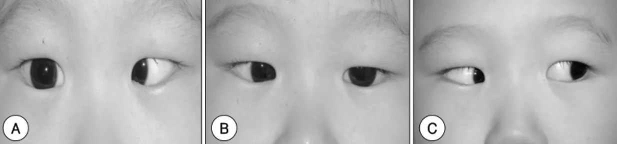
음날 우측 외전 신경마비 소견 보였으며 환자는30 PD (Prism Dipoter)의 우안 내사시 및 우안 외전장애와 안 구운동장애를 보상하기 위한 우측으로의 얼굴 돌림이 관 찰되었다(Fig. 1).
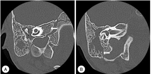
안저검사상 양안 모두 정상 안저 소견을 보였으며 조절 마비 굴절검사를 통한 이상 또한 발견되지 않았다. 중추 신경계, 추체염 및 다른 시신경 질환을 감별하기 위해 시 행한 뇌자기공명영상에서 추체골염소견을 동반하지 않는 우측 유양돌기염이 발견되었으며 횡정맥동 및 내경정맥 의 혈류 지연이 관찰 되었으나 명확한 혈전은 관찰되지 않았다(Fig. 2). 측두골 컴퓨터 단층촬영에서 유양돌기염 에 의한 S자형정맥동과 인접한 유양돌기 골막의 미란성 변화소견이 관찰되었으며, 추체골은 함기세포가 발달되어 있지 않았지만 골막 및 골파괴 소견은 보이지 않았고 반 대측과 차이가 없는등 추체골 첨부의 염증을 시사하는 소견은 발견되지 않았다(Fig. 3). 안구증상 발생 6일째, 유양돌기염에 대해 단순유양동삭개술을 시행하였고 수술 소견상 유양동이 육아조직으로 차 있었으며 S자형 정맥 동 주위의 골단 이 파괴되어 있어 측두골막으로 막았다.
수술중에 항생제 포함된 생리식염수로 수차례 수술부위 를 씻은 상태였고 수술중 육아조직밖에 보이지 않았기 때 문에 균배양 검사는 시행하지 않았다. 수술후 3주째 25 PD의 내사시와 교대 주시를 보이며 우안 외전장애는 호 전되는 양상을 보였고 술후 4달째, 정위로 호전되었으며 안구운동장애나 이상두위는 관찰되지 않았다(Fig. 4).
고 찰
외전신경은 외안근을 지배하는 신경 가운데 후천성 마
A B C
Fig. 1. Photographs show abduction defect (-4), esodeviation of 30 PD (Prism diopter) in the right eye. A:Gaze to the right. B:Gaze to the front. C:Gaze to the left.
비가 가장 흔히 발생하는 신경으로써 마비의 원인에 대 해서는 연구들 마다 차이가 있으나 미세혈관질환에 의한 허혈성 안근마비나 두부외상, 종양, 염증 등에 의해서 발 생할 수 있으며 원인 불명에 해당하는 경우도 많다.8) 소 아의 경우 성인에 비해 혈관질환의 빈도가 적고 외상이 나 종양의 빈도가 높으며 바이러스성 감염은 흔하지 않 은 것이 특징이다.9) 원인에 따른 차이는 있으나 6개월 이 내 자연적으로 호전되는 경과를 보이는 경우가 많다고 보 고된다.9)
중이염의 합병증으로 인한 외전신경의 마비에서 추체 골염이 동반되지 않은 경우는 드문데, 경뇌막염, 경막외 농양, 외측정맥동(lateral sinus)이나 하추체정맥동의 혈 전염 등에 의한 중이로부터 추체골 첨부의 경막으로의 염 증의 파급이 원인일 것으로 추정하고 있다.9,10) 해부학적
으로 하추체정맥동은 해면정맥동에서 추체골과 뇌바닥후 두골 사이를 주행하는 정맥계 구조물로서 외전신경과 매 우 인접하여 있는 반면 상추체정맥동은 외전신경과 떨어 져 있으며 해면정맥동을 통해 삼차신경의 위쪽으로 주행 한다.8,11)
본 증례는 급성중이염의 합병증으로 유양돌기염에 의 한 패혈증 상태와 동반된 편측외전신경마비로서 방사선 검사결과 추체골염이나 해면정맥동 또는 외측정맥동의 명 확한 혈전에 의한 직접적인 신경 손상의 원인을 찾을 수 없었다. 하지만 외측정맥동의 혈류 지연이 관찰되고, 측 정맥동주위 섬유소 퇴적(perisinus fibrin deposition)과 정맥염(phlebitis)에 의한 하추체 정맥동의 혈전증(throm- bosis) 증례가 보고된바 있고, 인접한 삼차신경의 침범이 동반되지 않은 점으로 보아,3,12) 상기 환자의 외전신경마 Fig. 2. A:Preoperative Axial enh- anced T2-weighted MR image shows right otomastoiditis without petrositis (White arrow head). B:Preopera- tive coronal enhanced T2-weighted MR image shows right otomastoi- ditis without petrositis (White arrow head). C:Axial enhanced T1-wei- ghted MR image shows slow flow velocity of the right transverse sinus (White arrow) D:Coronal enhanc- ed T1-weighted MR image shows slow flow velocity of the right inter- nal jugular vein (White arrow).
A B
C D
비의 원인을 하추체정맥동의 혈전증으로 추정할 수 있었 다.3) 일반적으로 혈전성정맥염의 특징은 정맥동내 염증, 감염이나 혈전 생성의 정도에 따라 다양한 양상으로 나 타날 수 있으나, 정맥동의 크기가 증가하고 조영제에 의 해 뚜렷한 조영 증강이 일어남과 동시에 정맥동내 경동 맥의 현저한 내경 감소 혹은 소실로 알려져 있다. 하지만 하추체정맥동은 매우 작은 정맥계 구조물로서 방사선학 적으로 시각화하기 힘들어 정확한 진단이 어려운 점이 그 한계점이라 할 수 있겠다.13) 감별하여야 할 질환 중 하나 인 Gradenigo 증후군은 추체골의 염증에 의해 외전신경 및 삼차신경분지인 안신경을 압박함으로써 이루, 심부안 구동통, 복시를 호소하는 것으로써 전형적인 증상이 모두 나타나는 경우는 흔하지 않다고 보고되고 있다. 본 증례 에서 방사선학적 검사상 추체염의 소견은 보이지 않아 배 제할수 있었다.
많은 항생제가 사용되는 현 시대에 중이염에 의한 두개
내 합병증의 빈도는 많이 줄었으나 발생시 치사율 또는 후유증은 여전히 높은 상태로 특히 영유아의 경우 그 발 생이 치명적일 수 있다. 치료는 균배양 감수성 검사와 함 께 충분한 항생제를 사용해야 하며 이같은 보존적 치료 에 반응하지 않고 진행하거나 주위 골파괴 소견이 보이 면 유양동삭개술 및 주위 농양 배농과 같은 적극적인 수 술적 치료가 필요하다.14)
저자들은 추체골염이 동반되지 않은 상태에서 패혈증 에 의한 하추체정맥동의 혈전에 의한 외전신경마비와 수 술적 치료 후 그 관해를 경험하였기에 이를 보고하는 바 이다.
중심 단어:외전신경마비·유양돌기염.
REFERENCES
1) Sherman SC, Sherman SC. Gradenigo syndrome: A case re- port and review of a rare complication of otitis media. The
A B
Fig. 3. A:Axial CT image shows no inflammation of the petrous bone. B:Coronal CT image shows erosive mastoid septa (White arrow).
Fig. 4. Photographs show orthotropia and no limitation of abduction in the right eye at 4 months after mastoidectomy.
J Emerg Med 2004;27(3):253-6.
2) Laurens MB, Becker RM, Johnson JK, Wolf JS, Kotloff KL.
MRSA with progression from otitis media and sphenoid si- nusitis to clival osteomyelitis, pachymeningitis and abducens nerve palsy in an immunocompetent 10-year-old patient. Int J Pediatr Otolarngol 2008;72(7):945-51.
3) Homer JJ, Johnson IJ, Jones NS. Middle ear infection and sixth nerve palsy. J Larnyngol Otol 1996;110(9):872-4.
4) Ludman H. Mawson’s disease of the ear. 5th edn. Edward Arnold. London;1998. p.479-536.
5) Christensen N, Wayman J, Spencer J. Lateral sinus throm- bosis. A review of seven cases and proposal of a manage- ment algorithm. Int J Pediatr otolarngol 2009;73(4):581-4.
6) Dorn M, Liener K, Rozsasi A, Keck T. Prolonged diplopia following sinus vein thrombosis mimicking Gradenigo’s syn- drome. Int J Pediatr otorhinolaryngol 2006;70(4):741-3.
7) Villa G, Lattere M, Rossi A, Di Pietro P. Acute onset of abdu- cens nerve palsy in a child with prior history of otitis media:
a misleading sign of Gradenigo syndrome. Brain Dev 2005;
27(2):155-9.
8) Patel SV, Mutyala S, Leske DA, Hodge DO, Holmes JM. In- cidence, associations, and evaluation of sixth nerve palsy us- ing a population-based method. Ophthalmology 2004;111 (2):369-75.
9) Knox DL, Clark DB, Schuster FF. Benign VI nerve palsies in children. J Pediatrics 1967;40(4):560-4.
10) Goodwin D. Differential diagnosis and management of ac- quired sixth nerve palsy. Optometry 2006;77(11):534-9.
11) Chole RA, Donald PJ. Petrous apicitis. Clinical considera- tions. Ann Otol Rhinol Laryngol 1983;92(6):544-51.
12) Kim BS, Do HM, Marks MP. Diagnosis and Management of Cerebral Venous and Sinus Thrombosis, Semin Cerebro- vasc Dis Stroke 2004;4(4):205-16.
13) Biousse V, Newman NJ. Venous Disease of the Central Ner- vous System. Semin Cerebrovasc Dis Stroke 2004;4(1):2-17.
14) Minotti AM, Kountakis SE. Management of abducens palsy in patients with petrositis. Ann Otol Rhinol Laryngol 1999;
108(4):897-902.