367
황금(Scutellaria baicalensis) 추출물에 의한 Streptococcus mutans의 항균 및 부착억제 효과
백종윤·김용현·권현정
1·김은님·김완종·한만덕
†순천향대학교 생명과학과, 1신흥대학 치위생과
Effects of Antibacteria and Adhesive Inhibition of Scutellaria baicalensis Extract on Streptococcus mutans
Jong-Yoon Paek, Young-Hyun Kim, Hyun-Jeoung Kwon
1, Eun-Nim Kim, Wan-Jong Kim and Man-Deuk Han
†Department of Biology, Soonchunhyang University, Asan, Chungnam, 336-745, Korea
1
Department of Dental hygiene, Shinheoung College, Uijeongbu, Gyeonggi, 480-701, Korea
Abstract
The natural products are used to be development of new antibacterial substances against human pathogenic bacteria. Adherence to the tooth surface by S. mutans is an important step in initiation of dental caries. This study was to examine antibacterial activity and anti-adhesive effect of Scutellaria baicalensis extract against S. mutans . Extracts of S.
baicalensis were tested for antimicrobial activities by paper disc methods and radial diffusion assay methods, and bacterial adherence assay using 3 type of hydroxyapatite. The antibacterial level of ethyl acetate extract, IPK-3 on the growth of S. mutans was 125 mg/ml of minimum inhibitory concentration (MIC). The maximum growth of S. mutans in medium added with IPK-3 extract (50 mg/ml) was delayed to 30 hr, while the highest at 24 hr in control medium. The pH values of the control medium was 5.63 at 18 hr, but the media supplemented with IPK-3 extract was pH 6.50 at 12 hr.
In adhesive inhibition assay, S . mutans was labelled with the fluorescent indicator DAPI and measured with fluorescence microscope. Adhesion of S. mutans on hydroxyapatite beads was inhibited by IPK-3 extracts. These results suggest that S.
baicalensis extract can be used as an effective material for antibacterial activity and adhesive inhibition against S. mutans .
Key words
Streptococcus mutans , Antibacterial activity, Bacterial adhesion, Scutellaria baicalensis
서 론
최근다양한식생활습관의변화와항생제내성균의 출 현으로치아우식증은증가되고있다1)
.
구강내대표적질병인치아우식증의주요원인균은
Streptococcus mutans
2)와 그 외에도
Lactobacillus casei , Actinomyces viscosus , S. sanguis , S. milleri , S. salivarius , S. rattus , S. cricetus
등이있다3,4)
. S. mutans
는Gram
양성균으로통성혐기성이며 구강 내에 상주하며 부착능력이 있어 치아 표면에
부착하게 된다
.
치아 표면의 세균막에 부착한S. mutans
는구강으로섭취하는음식물들의여러영양소들중포도 당 및 과당을 이용해 유기산인 젖산을 생산하는 것으로 알려져있으며
,
또한glucosyltransferase (GTF)
와같은당전이효소를 생산하여 당을 중합시키고 불용성
glucan
을합성한다
.
생성된 불용성glucan
은 구강 내 상주하는 다른미생물집단들이치아에쉽게부착하여증식하도록유 도하고치아우식증외의다른구강내질병을일으키도록 한다
.
또한젖산으로인해법랑질이 탈회되어최종적으로치아우식증을유발하게된다3)
.
치아우식증예방을위해서 는항생제사용이효과적이나항생제내성과인체에미치 는 부작용 때문에항생제를 대체할 수있는 천연 항균물 질의개발이활발히연구되고 있다5).
천연항생제는 주로식물에 존재하며 주로
alkaloid
류, flavonoid
류, phenolic compound
류, quinone
류 등의2
차 대사산물들인 것으로 알려졌다6).
그러나 합성 항생물질 보다 항균효과가 낮아실용화에 어려움이 있다
.
치아우식증을 억제하는 천연물에 대한연구로는녹차추출물은
tannin
성분이치아우식증에 효과가 있음이 보고되었고7) 오룡차8) 및
Chinese black tea, Andrographis paniculata , Cassia alata
또한치 아우식증을억제하는데효과가있다고보고되었다9).
황금
(
黃芩, Scutellaria baicalensis G.)
은 한국,
중국,
몽†
Corresponding author
Tel: +81-41-530-4702
Fax: +82-41-530-1256
E-mail: [email protected]
골 및 시베리아 동부지역에 분포하며쌍떡잎식물 통화식 물목꿀풀과에 속하는여러해살이풀로 높이는
20~60 cm
에달하고
,
뿌리는약용으로이용되어왔다10).
중국에서는고열
,
마른기침,
토혈,
변비,
태아를안정시킬때에사용되 었으며11)뿌리에는baicalein, baicalin, wohonin, wogono- side, neobaicalein
이함유되어있으며, sitosterol
등도포함되어있다12)
.
황금의약리작용으로 항균작용13),
항암작용,
항염증 효과
,
항히스타민 효과 등이 알려져 있다14).
국내황금에 대한 연구로는
Cho
등15)의 황금추출물의 항균특성 연구로황금추출물이 공시균주에 항균력이있음을 보
고하였고
, Moon
등16)은 치아우식증의 원인균 중 하나인S. mutans OMZ176
에 대해 황금추출물이 항균력이있다고보고하였다
.
그리고Lee
등17)은황금추출물이Staphy- lococcus aureus
와Bacillus cereus
에 대해, Choi
등18)은Pseudomonas aeruginosa
에 대해 항균력이 있다고 보고하였다
.
본 연구에서는 안전성이 우수한 한약재인황금을 이용하여구강내질병의 원인균에대한항균활성능력및 최소억제농도(minimal inhibitory concentration, MIC)
를 측 정하고,
황금추출물이S. mutans
의 생장에 미치는 영향과hydroxyapatite beads
에 대한 부착억제 효과를통해 치아 표면에치아우식원인균의부착억제가능성을확인하였다.
재료 및 방법 1. 황금 추출물 준비
한약재인 황금은 서울 경동시장에서 건조 상태의것을 구입하여 사용하였으며
,
여러 유기용매를 활용하여 추출 하였다.
구입한황금을분쇄한후, 3
배량의methanol
을첨가하여
72
시간냉침시켰으며methanol
추출물을aspirator (EYELA, A-3S, Tokyo Rikakikai Co.)
로 여과하였다.
여 과된methanol
추출물은 회전 감압농축기(Rotavapor, Büchi RE-111, Switzerland)
를 사용해80
oC
항온수조(Oilbath, Büchi B-485, Switzerland)
에서 감압 농축시켰다
. Methanol
추출물(IPK-6)
을 약40 ml
까지 농축한 후 분액깔때기를 이용해hexane (IPK-1), chloroform (IPK- 2), ethyl acetate (IPK-3), n-butanol (IPK-4),
물(IPK-5)
순으로용매첨가후 분리하여시료를얻었다
.
농축된추출물들은냉장보관하면서시료로사용하였다
.
2. 균주 및 배양
본 연구에 사용한 충치균은
Streptococcus mutans
ATCC 25175
균주로 한국생명공학연구원 생물자원센터(KCTC)
에서분양 받아 사용하였다.
세균의증식은brain heart infusion (BHI, Difco, USA)
에서1~2
차 계대배양후같은 배지에식균하여
37
oC CO
2incubator (5% CO
2) (Thermo electron corporation, USA)
에서 배양하였다.
항균활성용 배지는
Müeller hinton agar (MHA, Difco, USA)
를사용하였다.
3. 항균활성 측정
황금 유기용매 분획 추출물의
1
차 항균활성 검색은paper disc
방법을 사용하였다.
간략하면 보관된S.
mutans
균주를BHI
배지에 접종하여15~18
시간 활성화 시켜 항균성실험용MHA agar
배지에균주50
µl
씩도 말하였다.
도말된 배지에멸균된filter paper disc (8 mm : 1.5 mm, Toyo Roshi Kaisha, Ltd., Japan)
를 밀착시킨 후,
황금추출물을 농도별적용하였다.
황금추출물을초기 농도의
1/2
이 되도록 연속적으로4
회 희석하여 최종1000 mg/ml, 500 mg/ml, 250 mg/ml, 125 mg/ml
농도가되도록 조제하였으며
, disc
당50
µl
씩 흡수시켰다. Disc
에 추출물을 흡수시킨배지는 실온에서10
분간방치시켜 건조시킨 후
, S. mutans
를37
oC CO
2incubator(5% CO
2)
에 서18~24
시간동안배양시켰다.
배양후에는disc
주변에 생성된inhibition zone diameter (mm)
의 크기를vernier caliper (Mitutoyo Co., Japan)
로측정하여각추출물의항 균활성정도를측정하였다.
4. 최소억제농도 측정
Paper disc method
로1
차항균활성을검색하고 선별한 황금ethyl acetate
추출물(IPK-3)
은radial diffusion assay
를 통해 최소억제농도
(MIC)
를 측정하였다.
모든 균주를tryptic soy borth (TSB, Bacto, USA)
에전배양하여활성 화 시킨 후, 37
oC CO
2incubator
에서18~24
시간 배양하 였으며 본 실험에서는tryptic soy agar
를 사용하여 실험 하였다.
실험법을 간략하면,
균의O.D.
값을0.45 (4 x 10
6cells/ml)
로 맞추어underlay
배지10 ml
에 균45
µl
씩 분주하여혼합한후
, square petri dish (Falcon, USA)
에분주하여
15
분간실온에서방치하였다.
건조된배지에0.4 mm
직경의구멍을뚫고준비된황금IPK-3
추출물을 분주하였다.
최초 농도를1000 mg/ml
로 하여 다음 농도가 초기 농도의
1/2
이되도록 연속적으로17
번째까지 희 석하였다.
분주한후, 37
oC CO
2incubator (5% CO
2)
에서18~24
시간 동안 배양하였으며MIC
측정은 육안으로 관찰된생육저지대의농도로결정하였다
.
5. S. mutans 의 생장과 배지 내 환경변화
BHI
액체배지에 황금IPK-3
추출물50 mg/ml
을 첨가 한후 균을1×10
7cells/ml
이되도록접종하였다.
접종후37
oC CO
2incubator (5% CO
2)
에서18~24
시간동안배양하였으며 대조군은
BHI
액체배지를blank
로하고실험군은
IPK-3
를 첨가한BHI
액체배지를blank
로 하여Multi functional microplate reader (BMG Labtech, Germany)
를 이용해540 nm
에서 흡광도를측정하였다.
또한pH
변화는
IPK-3
추출물을50 mg/ml
로첨가하고pH
변화를6
시 간 마다 측정하였다.
배지내 총당량은phenol-sulfuric
method,
총 단백질량은BCA protein assay kit(Pierce
Co.)
을 이용하여측정하였으며,
또한glucan
생성 효과도알아보았다
.
이때 대조군은IPK-3
추출물이 첨가되지않은배지를이용하여측정하였다
.
6. Hydroxyapatite beads(HA) 부착 억제 실험 S. mutans
균의hydroxyapatite adhesion assay
는Gaines
19)의assay
를응용하여실험하였다.
우선타액은건강한성인에게서 채취하여 냉각된
conical tube
에채취한 후, 4
oC
에서15,000 rpm
으로20
분간원심분리(Mega 17R, Hanil, Korea)
하였다.
원심 분리된 타액의 상징액을취하 여60
oC
에서30
분간가온하여효소를불활성화시킨후- 20
oC
에 보관하여 실험에 사용하였다.
세균 부착 분자는hydroxyapatite beads
로써다음과같은제품을사용하였다: (a) Macro-prep ceramic hydroxyapatite type II 80
µm (Bio-Rad Laboratories, Hercules, CA); (b) Fluka crude hydroxylapatite (Fluka cat. No. 21223); (c) Hydroxyapatite (H-0252, Sigma, USA).
각 시료를30 mg
을 정량하고0.2 M NaOH 10 ml
에10
분간 전처리하여 표면을활성화시키고부유물은흡입하여 제거하였다
.
이과정을2
회반 복하고증류수로bead
를3
번세척하였다.
세척된hydrox- yapatite beads
는pH 7.0
으로 중화시키고 증류수로 한번 세척하였다.
세척된hydroxyapatite beads
에 냉장 보관된타액
1 ml
를 넣고37
oC
에서60
분간 타액을 코팅 시켰다(S-HA).
코팅된S-HA
를0.01 M potassium phosphate buffered saline (PBS, pH7.2)
로2
회 세척한후,
최종적으 로BHI
액체배지1 ml
로 세척하였다.
세척된S-HA
는96 well plate
에10
µl
씩 분주하고,
황금IPK-3
추출물125 mg/ml
을 첨가한 다음 전 배양된S. mutans
균주1×10
7cells/ml
를분주하였다.
분주된96 well plate
는CO
2incubator
에서 배양하며90
분, 12
시간, 24
시간 마다S-HA
시료를 취하였다
.
정치시킨e-tube
에서 배지를 제거하고PBS
로1
회 세척한 후,
상징액을 버리고 세척한S-HA
에DAPI (4',6-diamidino-2-phenylindole, Sigma, USA)
를첨가 하여37
oC
에서1
시간동안염색시켰다. 1
시간동안염색시킨S-HA
는PBS
로1
회세척하고20
µl
를채취하여fluorescence microscope (Olympus DP71, Japan)
상에서관찰하였다.
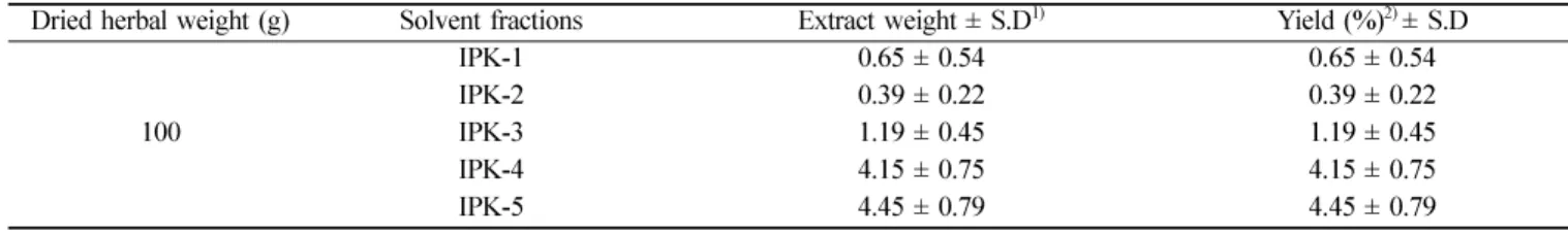
결과 및 고찰 1. 황금에 대한 유기용매 추출물의 수율
황금을
hexane, chloroform, ethyl acetate, n-butanol
의유기용매로분획추출한결과
, Table 1
과같이butanol
추 출물(IPK-4)
은4.15%, ethyl acetate
추출물(IPK-3) 1.19%, hexane
추출물(IPK-1) 0.65%, chloroform
추출 물(IPK-2) 0.39%
순으로추출물수율을얻었다.
추출최 종단계인 물 추출물(IPK-5)
은 가장 높은 수율인4.45%
로나타났다
.
2. 항균활성 측정
황금유기용매분획추출물이균주에가지는항균활성능
력을 보기 위하여
MHA
배지에 균을 도말하고paper
disc
에 추출물을1,000 mg/ml, 100 mg/ml, 10 mg/ml
농도별로 첨가해
37
oC CO
2incubator (5% CO
2)
에서 배양하여 생육 억제대를 관찰하였다
.
실험 결과는Table 2
와같이
S. mutans
균은1,000 mg/ml
의농도에서 항균활성을 나타냈으나butanol (IPK-4)
과 물 추출물(IPK-5)
에서는항균 활성을 나타내지 않았다
.
특히, IPK-3
추출물은 다른 유기용매 추출물과 비교할 때 항균활성이 가장 크게 나타났으며
,
그다음으로IPK-2
추출물이모든균주에대해 약간의 항균활성을 보였다
. 100 mg/ml
의 농도에서IPK-3
추출물에서만생육억제저지대가관찰되었다.
Table 1.
Average yield of organic solvent fractions extracted from S. baicalensis
Dried herbal weight (g) Solvent fractions Extract weight ± S.D
1)Yield (%)
2)± S.D
100
IPK-1 0.65 ± 0.54 0.65 ± 0.54
IPK-2 0.39 ± 0.22 0.39 ± 0.22
IPK-3 1.19 ± 0.45 1.19 ± 0.45
IPK-4 4.15 ± 0.75 4.15 ± 0.75
IPK-5 4.45 ± 0.79 4.45 ± 0.79
1)
S.D = Standard Deviation
Table 2.
Antibacterial effects of organic solvent fractions from S. baicalensis on pathogenic bacteria.
Bacterial Strain Extract of S. baicalensis
1)1000 mg/ml 100 mg/ml 10 mg/ml
H
2)C E B W H C E B W H C E B W
Streptococcus mutans ATCC 25175 + + ++ - - - - + - - - - - - -
1)
Concentration of organic solvent extract from
S. baicalensis.2)
Fractionated procedure of organic solvent from
S. baicalensis: H (hexane extract), C (chloroform extract), E (ethyl acetate extract), B (n-butanol extract), W (water extract)
2)
Yield(%) = Dried weight of extract fraction (g) × 100 Dried weight of
S. baicalensis(g)
3. 최소억제농도 측정
Paper disc
방법에서 높은 항균활성을 나타낸 황금IPK-3
추출물을 병원성 미생물에 대한 최소억제농도를측정하기 위하여
radial diffusion assay
로 실험하였다. S.
mutans
에 대하여 최소억제농도를 측정한 결과, IPK-3
의최소억제농도는
125 mg/ml
이었다(Fig. 1).
4. 황금의 추출물이 S. mutans 의 생장에 미치는 영향
1)
생장에미치는영향황금
IPK-3
추출물이S. mutans
가 성장하는데 미치는 효과를측정하기위해IPK-3
을50 mg/ml
의 농도로배지 에 첨가한 후 균의 생육을Multi functional microplate reader (BMG Labtech, Germany)
로측정한결과는Fig. 2
와같다
.
대조군이배양24
시간후에흡광도1.17
로최대의성장을보였으며황금
IPK-3
추출물50 mg/ml
를 투여한 배지에서는 생장이 지연되어
30
시간에흡광도가0.49
로최대성장을나타냈다
.
이결과를통해황금IPK-3
추출물은 치아우식증 원인균인
S. mutans
의 생장을 억제하 고최대생장시점을지연시키는효과가있었다.
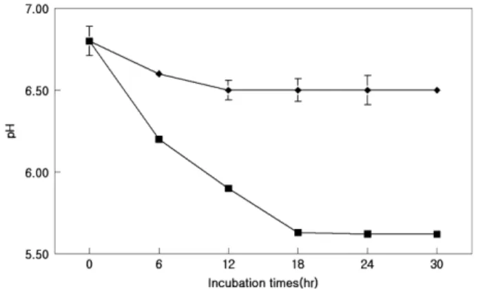
2)
배지내pH
변화에미치는영향황금
IPK-3
추출물50mg/ml
을S. mutans
의 생장배지 에 첨가하였을 때pH
변화에미치는효과는Fig. 3
과같다
.
초기pH
는6.8
로 적정하였으며,
대조군은 배양18
시간 만에
pH 5.63
로 급격한 변화를 보였으며 그 후의 시간에서는 일정한
pH
를유지하였다.
반면에IPK-3
추출물 이투여된배지에서는12
시간까지미소하게pH 6.5
로감소하였으나 그후 시간에는 더 이상 저하되지 않았다
.
따 라서 황금IPK-3
추출물은S. mutans
균의 생장을억제하 여pH
변화는거의이루어지지않았다.
3)
배지내탄수화물의변화에미치는영향황금
IPK-3
추출물을S. mutans
에첨가한후배지내의탄수화물량의변화를알아보기위해
6
시간별로측정하였 다.
초기 배지의 총 탄수화물량은ml
당0.56 mg
의 탄수화물이정량되었으며
18
시간이지난후대조군은0.24 mg/
ml
로 떨어졌으나50 mg
의 농도가첨가된 황금IPK-3
추출물이 투여된배지는
0.5 mg/ml
을 유지하였으며24
시간이 지난 후 탄수화물의 양이점차 감소하는 것을 확인할 수있었다
(Fig. 4).
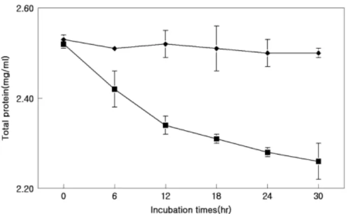
4)
배지내단백질의변화에미치는영향우수한항생제라도 생체에적용될 때
,
혈액,
타액,
장액등에 존재하는 분자
(
특히 단백질)
에 의해 활성이 감소될수 있다
.
따라서 황금의 추출물을BHI
액체배지에 넣고균체를 성장시켰을 때
,
배지 내 단백질 변화를 확인함으 로써세균의활성을확인하였다.
황금IPK-3
추출물이첨가된배지에
S. mutans
를배양하고시간별변화하는 단백질량을 측정한 결과
,
배양0
시간에서는 모든 배지에서2.52 mg/ml
이었다.
그러나균체의최대생장량을보인24
시간에서는 대조군이
2.31 mg/ml
이었고,
황금IPK-3
추Fig. 1.
Minimal inhibitory concentrations(MIC) of the ethyl acetate extract (IPK-3) from S. baicalensis on S. mutans.
Concentrations of IPK-3 extract were A) 1000 mg/ml, B) 500 mg/ml, C) 250 mg/ml, D) 125 mg/ml, E) 62.5 mg/ml, F) 31.25 mg/ml, G) 15.63 mg/ml, H) 7.81 mg/ml, I)3.90 mg/ml, J) 1.95 mg/ml, K) 0.97 mg/ml, L) 0.48 mg/ml, M) 0.24 mg/ml, N) 0.12 mg/ml, O) 0.06 mg/ml, P) 0.03 mg/ml, and Q) 0.015 mg/ml.
Fig. 2.
The growth curve of S. mutans in culture medium by addition of ethyl acetate fraction (IPK-3) extracted from S.
baicalensis.
■; control,
◆; 50 mg/ml of IPK-3.
Fig. 3.
The changes of pH in S. mutans culture medium by addition of ethyl acetate fraction (IPK-3) extracted from S.
baicalensis.
■; control,
◆; 50 mg/ml of IPK-3.
출물이 투여된 배지에서는
2.51 mg/ml
의 단백질이 정량 되어대조군보다단백질의활용이적었다(Fig. 5).
5. S. mutans 의 다당류 생성 억제 효과
S. mutans
의 다당류는치아우식증에관여하는 치면세균막을 형성하는데 가장 중요한 역할을 하는 다당류이다
.
따라서 본 연구에서는 황금
IPK-3
추출물50 mg/ml
의농도로
S. mutans
의 배양액 투여하였을 때 다당류 생성을어느 정도억제하는지확인하기 위하여액체배지에서
시간별 다당류의 양적 변화를 알아 본 결과
Fig. 6
과 같다
.
대조군에서는 배양18
시간에500 mg/100 ml
의 다당류를 얻었으나
,
황금 추출물이 투여된 배지에서는280 mg/100 ml
의 다당류가 수확되었다.
한19) 등이 연구한 바에 의하면
S. mutans
의 균체외 다당류는glucan
성 다당류인것으로 보고된바 있다.
이상의 결과로 황금 추 출물은 치면세균막 형성에 중요하게 작용하는S.
mutans
의 다당류 생성을 억제할 것으로 여겨지며 이들추출물의 활용은 새로운 항 우식물질의 요구에 적절히 대응 할 수 있는 천연물 소재로써 의미가 있는 것으로 사료된다
.
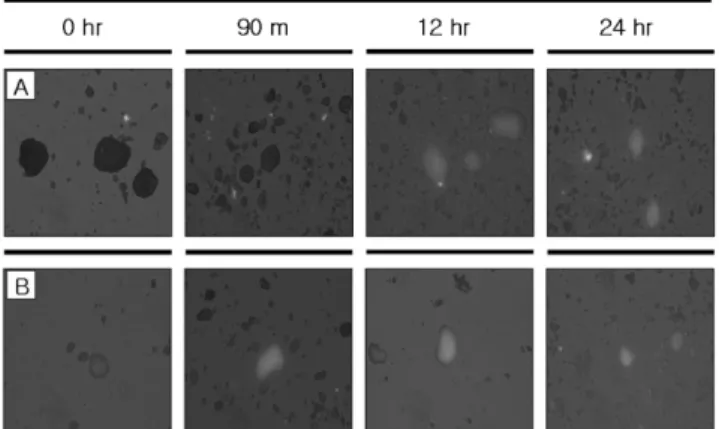
6. Hydroxyapatite(HA)에 S. mutans 부착 억제 효과
S-HA
부착 억제에대한 실험에는Macro-prep ceramic hydroxyapatite type II 80 (Bio-Rad Laboratories, Hercules, CA), Fluka crude hydroxylapatite (Fluka cat.
No. 21223), Hydroxyapatite (Sigma, USA)
등3
개 제조사의
hydroxyapatite beads
를 사용하였다.
황금ethyl acetate
추출물이S-HA
에대한S. mutans
균의부착억제 효과가있는지알아보기위하여최소억제농도인125 mg/
ml
농도에서실험한결과는Fig. 7~Fig. 9
와같다. Fig. 7
은
Macro-prep ceramic hydroxyapatite type II 80
µm (Bio-Rad Laboratories, Hercules, CA)
제품을 사용하여 실험한 결과이며Fig. 8
은Fluka crude hydroxylapatite (Fluka cat. No. 21223), Fig. 9
는Hydroxyapatite (Sigma,
USA)
제품을 사용하여 관찰한 결과이다.
균이S-HA
에부착되는 순간부터 부착된이후까지의 결과를 보기 위해
0
시간, 90
분, 12
시간, 24
시간에S-HA
를 채취하고DAPI
로염색하여관찰하였다
.
황금IPK-3
추출물이첨가된S-
Fig. 5.
The changes of total protein in S. mutans culture medium by addition of ethyl acetate fraction (IPK-3) extracted from S. baicalensis.
■; control,
◆; 50 mg/ml of IPK-3.
Fig. 6.
The polysaccharide contents in S. mutans culture medium by addition of ethyl acetate fraction (IPK-3) extracted from S. baicalensis.
■; control,
◆; 50 mg/ml of IPK-3.
Fig. 7.
Fluorescent microscope image (×400 magnification) illustrating S. mutans labelled with DAPI, hydroxyapatite under transmission light and an image overlay illustrating S.
mutans adhering to hydroxylapatite (Bio-Rad Co.). (A) control.
(B) HA beads treated with 125mg/ml of IPK-3 extracted from S. baicalensis.
Fig. 4.
The changes of total carbohydrate in S. mutans culture
medium by addition of ethyl acetate fraction (IPK-3) extracted
from S. baicalensis.
■; control,
◆; 50 mg/ml of IPK-3.
HA
에대한S. mutans
의 부착은대조군과달리시간이경 과하여도 균이부착된모습은관찰할수없었다.
이같은결과는
Gaines
20)가 실험한결과와 비슷하다는 것을 확인하였다
.
또한S. mutans
를HA
에 부착시킬 때DMSO
의처리는세균의 부착에영향을 주었다
.
즉 황금IPK-3
추출물을
20% DMSO
에 녹여처리한실험군에서는부착이확인하였으나
, DMSO
처리를 하지 않았을 경우DMSO
를처리하였을때보다부착율이떨어졌다
.
요 약
천연물 황금으로부터 여러 유기용매 추출물을 얻어 치 아우식증 원인균인
S. mutans
에대한항균활성,
배지내환경변화
,
그리고부착억제에대해알아보았다.
1.
구강 질병인치아우식증의대표적 원인균S. mutans
에대한황금
ethyl acetate
추출물(IPK-3)
의 최소억제농도는
125 mg/ml
이었다.
2.
황금IPK-3
추출물50 mg/ml
의 농도로S. mutans
의 배지내 투여하였을 때균의 생장량과최대 생장시 간은대조군보다지연되었다.
3.
황금IPK-3
추출물50 mg/ml
의 농도로S. mutans
의배지내투여하였을때
, pH
변화는 대조군은18
시간에서
pH 5.63
으로 급격한 변화를 보였으며 황금IPK-3
추출물이50 mg/ml
의 배지에서는6.50
이상을 유지하여pH
가변화가없었다.
4.
황금IPK-3
추출물을첨가한배지내탄수화물,
단백 질 및균체 외다당류의 변화는대조군에비해 매우 낮게생산되었다.
5.
균체를 형광염료(DAPI)
로 염색하여S-HA
부착억제 을 확인한 결과,
대조군은 시간이 경과함에 따라S.
mutans
의hydroxyapatite(HA)
에 부착 정도가증가되었으나
,
황금IPK-3
추출물이 첨가된 시험군에서는S-HA
에균의부착이매우적었다.
이같이황금의
IPK-3 (ethyl acetate
추출물)
이 치아우식증의 원인균인
S. mutans
에 대한 항균활성효과 뿐만아니라
hydroxyapatite
에부착억제효과도있었다.
감사의 글
이 논문은 지식경제부 지역혁신센터 사업
(MOCIE-
RIC060605)
의 지원을 받아 일부 연구되었으며,
이에 감사드립니다
.
참고문헌
1. Allaker RP, Ian Douglas CW: Novel anti-microbial therapies for dental plaque-related diseases. Int J Antimicrob Agents 33: 8-13, 2009.
2. Koga T, Askawa H, Okahashi N, Hamada S: Sucrose- dependent cell adherence and cariogenicity of serotype c Streptococcus mutans . J Gen Microbiol 10: 2873-2883, 1986.
3. Hamada S, Slade HD: Biology, immunology, and cariogenicity of Streptococcus mutans . Microbiol Rev 35:
331-384, 1980.
4. Slots J , Taubman MA: Contemporary oral microbiology and immunology. Missouri. Mosby-Year Book pp. 377-424, 1992.
5. Cohen SH, Morita MM, Bradford M: A-seven year experience with methicillin-resistant Staphylococcus aureus . Am J Med 91: 233S-237S, 1991.
6. Choi I, Cho JY, Lim SC: Antimicrobial activity of medicinal herbs against Staphylococcus aureus . J Kor Plant Res 19(4):
491-496, 2006
7. Otake S, Makimura M, Kuroki T, Nishibara Y, Hirasawa M:
Anticaries effects of polyphenolic compounds from Japanese green tea. Caries Res 25: 438-443, 1991.
8. Nakahara K, Kawabata S, Ono H, Ogura K, Tanaka T, Ooshima T, Hamada S: Inhibitory effect of oolong tea polyphenols on glycosyltransferases of mutans Streptococci .
Fig. 8.
Fluorescent microscope image (×400 magnification) illustrating S. mutans labelled with DAPI, hydroxyapatite under transmission light and an image overlay illustrating S.
mutans adhering to hydroxylapatite (Fluka Co. product). (A) control. (B) HA beads treated with 125 mg/ml of IPK-3 extracted from S. baicalensis.
Fig. 9.