108 I
Innttrroodduuccttiioonn
Behçet’s disease (BD) is a systemic inflammatory disorder of unknown etiology, characterized by recurrent oral aphthous ulcers, uveitis, genital ulcers, and skin lesions.1)2)Cardiovas- cular involvement in BD is not uncommon and can be fatal.1-3) We describe herein an extremely rare case of cardiovascular BD, who presented with a large subepicardial hematoma developed by micro-rupture of the right coronary artery dur- ing long-term warfarinization due to deep vein thrombosis (DVT).
C Caassee
A 28-year-old man was admitted to our emergency room due to chest pain for 5 hours. He denied any history of chest trauma. He was diagnosed as BD with lower extremity DVT at other tertiary hospital 4 years ago and was taking warfarin and immunosuppressive agents. His blood pressure was 120/80 mmHg, pulse rate 96 bpm, body temperature
36.4˚C, and he had a respiration rate of 20 breaths/min.
Physical examination revealed acutely ill appearance. Other physical findings were nonspecific. Electrocardiography showed sinus tachycardia (94 bpm) with incomplete right bundle branch block and T wave inversion on V1-3. Labo- ratory study revealed prolonged prothrombin time (PT) [international normalized ratio (INR), 3.69 (45.5 sec, 15.7%)], leukocytosis (15,400/µL), and only slightly elevated tropo- nin I (0.50 ng/mL). Erythrocyte sedimentation rate was 20 mm/hr and the level of highly-sensitive C-reactive protein was 74.56 mg/L.
Multidetector computed tomography (CT) revealed a 60
×60 mm-sized, round cavity-like structure compressing the right atrium (RA) with small amount of hemopericardium (Fig. 1). There was no evidence of pulmonary embolism.
Transthoracic echocardiography also showed a 59×55 mm- sized mass adjacent to the RA with small amount of pericar- dial effusion with increased echogenicity (Fig. 2A). The mass
pISSN 1975-4612/ eISSN 2005-9655 Copyright © 2010 Korean Society of Echocardiography www.kse-jcu.org DOI: 10.4250/jcu.2010.18.3.108
C
CAASSEE RREEPPOORRTT J Cardiovasc Ultrasound 2010;18(3):108-111
• Received: June 9, 2010 • Revised: August 2, 2010 �Accepted: August 17, 2010
• Address for Correspondence: Jei Keon Chae, Division of Cardiology, Department of Internal Medicine, Chonbuk National University Medical School, 634-18 Geumam-dong, Deokjin-gu, Jeonju 561-712, Korea Tel: +82-63-250-1389, Fax: +82-63-250-1680, E-mail: [email protected]
C
Ca ar rd diio ov va as sc cu ul la ar r B Be eh hç çe et t’’s s D Diis se ea as se e P
Pr re es se en nt tiin ng g a as s a a S Su ub be ep piic ca ar rd diia al l H He em ma at to om ma a: : A
An n U Un ne ev ve en nt tf fu ul l 2 2- -Y Ye ea ar r C Cl liin niic ca al l C Co ou ur rs se e
S
Suunn HHwwaa LLeeee,, MMDD,, PPhhDD11,, JJeeii KKeeoonn CChhaaee,, MMDD,, PPhhDD11,, JJoonngg BBuumm CChhooii,, MMDD,, PPhhDD22,, S
Saanngg RRookk LLeeee,, MMDD,, PPhhDD11,, KKyyoouunngg SSuukk RRhheeee,, MMDD,, PPhhDD11,, WWoonn HHoo KKiimm,, MMDD,, PPhhDD11 a
anndd JJaaee KKii KKoo,, MMDD,, PPhhDD11
1Division of Cardiology, Department of Internal Medicine, 2Department of Thoracic and Cardiovascular Surgery, Chonbuk National University Medical School, Jeonju, Korea
Cardiovascular involvement in Behçet’s disease is not uncommon and could be life-threatening. We describe here a 28-year- old man, who developed sudden onset chest pain during warfarinization due to deep vein thrombosis. Echocardiography and computed tomography showed a 60×60 mm-sized hematoma in the pericardial space compressing the right heart. Coronary angiography showed totally occluded proximal right coronary artery. The hematoma was located at the subepicardial plane of the right atrium on surgical view and successfully evacuated. Follow-up echocardiography revealed complete resolution of the hematoma. He is doing well for 24 months after surgery.
KEY WORDS:Behçet’s disease∙Hematoma∙Coronary artery.
online ©ML Comm
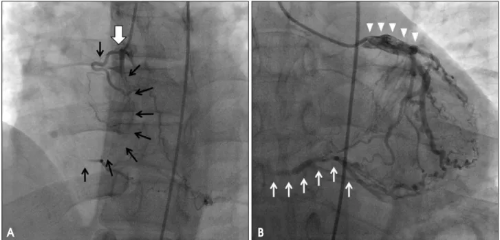
was extrinsically compressing the tricuspid annulus. Color Doppler study showed accelerated color flow from the RA to the right ventricle (RV) with peak pressure gradient of 16 mmHg (Fig. 2B and C). Inferior vena cava was dilated to 27 mm and not collapsed during inspiration (Fig. 2D). Coro- nary angiography (CAG) showed totally occluded proximal right coronary artery (RCA) with some dye staining around the mass (Fig. 3A) and collateral flows from the left coronary artery (LCA) (Fig. 3B). The LCA was normal otherwise with diffuse dilation of the proximal segments.
During stay at the coronary care unit, his vital signs and symptoms remained stable. Surgery was performed at the 10th hospital day after normalization the PT INR to 1.23.
On surgical view, a scanty amount of blood and the hema- toma were located below the epicardium at the atrioven- tricular groove. A definite bleeding focus was not found. We successfully removed the hematoma. Coronary bypass surgery was not performed. Postsurgical echocardiography showed a 24×12 mm-sized residual hematoma (Fig. 4A and B), but transtricuspid pressure gradient declined to 8 mmHg.
Subepicardial Hematoma in Behçet’s Disease|| Sun Hwa Lee, et al.
109 Fig. 1. Four-chamber view of multidetector computed tomography on
admission. It revealed a 60×60 mm-sized, round mass (H, hematoma) compressing the right atrium and the tricuspid annulus and small amount of hemopericardium (arrows).
Fig. 2. Transthoracic echocardiography on admission. A: Two-dimensional image showed a 59×55 mm-sized mass (H) adjacent to the right heart extrinsically compressing the tricuspid annulus and small amount of pericardial effusion. B and C: Color Doppler study showed accelerated color flow from the RA to the RV with peak pressure gradient of 16 mmHg. D: IVC was dilated to 27 mm and not collapsed during inspiration. H: hematoma, LV:
left ventricle, RV: right ventricle, RA: right atrium, IVC: inferior vena cava.
A B
C D
Journal of Cardiovascular Ultrasound 18 || September 2010
110
Fig. 4. Serial two-dimensional echocardiography images. Comparing with preoperative status (A), postoperative echocardiography (B) showed that a substantial volume of hematoma (H, hematoma) still remained. However, the 6th(C) and 12th(D) -month follow-up echocardiography showed complete resolution and no recurrence of the hematoma.
A B
C D
Fig. 3. Coronary angiography. A: Proximal right coronary artery (thick arrow) was totally occluded just below the ostium with some dye staining around the mass (thin black arrows). B: Grade 3 collateral flow from the left coronary artery (thin white arrows) and diffuse enlargement of the proximal segments of the left coronary artery (arrowheads) were found.
A B
The patient was discharged from the hospital 15 days after surgery without any surgical complications. The 6th-and 12th-month follow-up echocardiography revealed complete resolution of the hematoma (Fig. 4C and D). He is doing well with immunosuppressive treatment and warfarinization for 24 months.
D
Diissccuussssiioonn
This unique case of BD showed a huge subepicardial hematoma that was estimated to be formed by micro-rupture of the RCA. He was taking warfarin and immunosuppres- sives due to lower extremity DVT. The hematoma was inci- dentally found on CT that was taken to rule out pulmonary embolism, and successfully removed by surgery. We previ- ously reported this case immediately after surgery.4) The patient is undergoing an uneventful postoperative course for 2 years during anticoagulation.
The incidence of vascular BD is 7% to 38%.3-6)Vasculitis associated with BD is the major cause of morbidity and mortality, and can affect arteries and veins of all size.6-8)Venous thrombosis is the most frequent vascular complication of BD seen in 6% to 33%, and may be recurrent and not be resolv- ed despite of anticoagulation.7-10)In this case, in spite of warfa- rinization and immunosuppressive treatment for 4 years due to DVT, CT showed chronic venous thrombosis and obstruc- tion of lower extremity deep veins. Arterial involvement in BD is less common, occurring 1% to 7% of patients, but carries poorer prognosis than venous complications.3)5-8) Aorta, pulmonary, and femoral arteries are most frequently involved.5-7) It is characterized by saccular aneurysm forma- tion and obstruction.2)3)Aneurysm and subsequent rupture of the large arteries are directly related to the major cause of mortality, and stenosis or obstruction may cause ischemic symptoms or be asymptomatic.1-3)5)8)
The incidence and clinical course of cardiac BD are not clear, but it can be fatal.1-3)All 3 layers of the heart can be affected and present as endocarditis, endomyocardial fibrosis, recurrent arrhythmias, myopericarditis, valvular dysfunction, intracardiac thrombosis, and coronary artery disease (CAD) resulting in acute myocardial infarction, angina, and silent myocardial ischemia.2)3)Coronary artery involvement in BD has extremely rarely been reported.3)9)11)This patient did not experience angina before admission and presented with atypical chest pain. We initially thought that the possibility of CAD was low as the cause of chest pain and performed CT to exclude pulmonary embolism. The hematoma com- pressing the right heart was found by chance. CAG showed total occlusion of the RCA with abundant collateral circula- tion from the diffusely enlarged LCA. This finding suggests
that the occlusion of the RCA was a chronic process. We thought that the hematoma was developed by spontaneous micro-rupture of the totally occluded RCA fired by bleeding tendency during warfarinization. If patients with BD present chest pain, it should be considered the diagnosis of coronary artery involvement, even if they are young or do not have any traditional cardiovascular risk factors.
Arterial occlusive or stenotic lesions in BD have been treat- ed surgically or non-surgically by using immunosuppressive agents.8)Generally, conservative treatment is preferred to sur- gery because arterial manipulation may be complicated by aneurysm or pseudoaneurysm formation, clots, occlusion, or recurrent disease.3)8)Surgical management is usually reserved for arterial aneurysms to avoid the risk of rupture.8)Because of the same reason, bypass graft surgery or percutaneous coronary intervention have not generally been adopted in cases of severe CAD in BD, and the long-term results of those treatment strategies are not available.8)We also elected only to remove the huge hematoma for the purpose of relieving the obstruction without bypass the occlusive RCA because of the abundant collaterals and the concern of development of surgical complications.
In conclusion, it is necessary to keep in mind the possibi- lity of coronary artery involvement in young patients with BD presenting chest pain. The optimal treatment strategies should be selected according to the clinical situation.
R
Reeffeerreenncceess
1. Sismanoglu M, Omeroglu SN, Mansuroglu D, Ardal H, Erentug V, Kaya E, Guler M, Ipek G, Yakut C. Coronary artery disease and coronary artery bypass grafting in Behçet’s disease. J Card Surg 2005;20:
160-3.
2. Sakane T, Takeno M, Suzuki N, Inaba G. Behçet’s disease. N Engl J Med 1999;341:1284-91.
3. Atzeni F, Sarzi-Puttini P, Doria A, Boiardi L, Pipitone N, Salvarani C.
Behçet’s disease and cardiovascular involvement. Lupus 2005;14:723-6.
4. Kim KH, Choi JB, Kim KS. Subepicardial hematoma compressing the right atrium: spontaneous rupture of the right coronary artery. Ann Thorac Surg 2008;86:e9.
5. Ipek G, Omeroǧlu SN, Mansuroǧlu D, Kirali K, Uzun K, Sismanoǧlu M.Coronary artery bypass grafting in a 26-year-old man with total occlu- sion of the left main coronary artery related to Behçet disease. J Thorac Car- diovasc Surg 2001;122:1247-9.
6. Yakut ZI, Odev K. Pulmonary and cardiac involvement in Behçet disease:
3 case reports. Clin Appl Thromb Hemost 2007;13:318-22.
7. Al-Mutawa SA, Hegab SM. Behcet’s disease. Clin Exp Med 2004;4:103-31.
8. Alpagut U, Ugurlucan M, Dayioglu E. Major arterial involvement and review of Behcet’s disease. Ann Vasc Surg 2007;21:232-9.
9. Ahn JK, Lee YS, Jeon CH, Koh EM, Cha HS. Treatment of venous thrombosis associated with Behcet’s disease: immunosuppressive therapy alone versus immunosuppressive therapy plus anticoagulation. Clin Rheumatol 2008;27:201-5.
10. Houman MH, Ben Ghorbel I, Khiari Ben Salah I, Lamloum M, Ben Ahmed M, Miled M. Deep vein thrombosis in Behçet’s disease. Clin Exp Rheumatol 2001;19:S48-50.
11. Calamia KT, Schirmer M, Melikoglu M. Major vessel involvement in Behçet disease. Curr Opin Rheumatol 2005;17:1-8.
Subepicardial Hematoma in Behçet’s Disease|| Sun Hwa Lee, et al.
111