인간치수세포에 Mineral Trioxide Aggregate와 수산화칼슘 제재 적용 시 유전자 발현 양상 비교
김용범∙손원준∙이우철∙금기연∙백승호∙배광식*
서울대학교 치의학대학원 치과보존학교실
Comparison of gene expression profiles of human dental pulp cells treated with mineral trioxide aggregate and calcium hydroxide
Yong-Beom Kim1, Won-Jun Shon2, Woocheol Lee2, Kee-Yeon Kum2, Seung-Ho Baek2, Kwang-Shik Bae2*
1
Program in Conservative Dentistry, Seoul National University Graduate School,
2
Department of Conservative Dentistry, Seoul National University School of Dentistry and Dental Research Institute, Seoul, Korea
Objectives:
This study investigated changes in gene expressions concerning of differentiation, proliferation, mineralization and inflammation using Human-8 expression bead arrays when white Mineral Trioxide Aggregate and calcium hydroxide-containing cement were applied in vitro to human dental pulp cells (HDPCs).Materials and Methods:
wMTA (white ProRoot MTA, Dentsply) and Dycal (Dentsply Caulk) in a Teflon tube (inner diameter 10 mm, height 1 mm) were applied to HDPCs. Empty tube-applied HDPCs were used as negative control. Total RNA was extracted at 3, 6, 9 and 24 hr after wMTA and Dycal application.The results of microarray were confirmed by reverse transcriptase polymerase chain reaction.
Results:
Out of the 24,546 genes, 43 genes (e.g., BMP2, FOSB, THBS1, EDN1, IL11, COL10A1, TUFT1, HMOX1) were up-regulated greater than two-fold and 25 genes (e.g., SMAD6, TIMP2, DCN, SOCS2, CEBPD, KIAA1199) were down-regulated below 50% by wMTA. Two hundred thirty nine genes (e.g., BMP2, BMP6, SMAD6, IL11, FOS, VEGFA, PlGF, HMOX1, SOCS2, CEBPD, KIAA1199) were up-regu- lated greater than two-fold and 358 genes (e.g., EDN1, FGF) were down-regulated below 50% by Dycal.Conclusions:
Both wMTA and Dycal induced changes in gene expressions related with differentiation and proliferation of pulp cells. wMTA induced changes in gene expressions related with mineralization, and Dycal induced those related with angiogenesis. The genes related with inflammation were more expressed by Dycal than by wMTA. It was confirmed that both wMTA and Dycal were able to induce gene expression changes concerned with the pulp repair in different ways. [J Kor Acad Cons Dent 2011;36(5):397-408.]Key words:
Calcium hydroxide; Microarray; Mineral trioxide aggregate; Pulp capping; Pulp cell;Reverse transcriptase polymerase chain reaction
-Received 20 July 2011; revised 17 August 2011; accepted 21 August 2011- ABSTRACT
1Kim YB, DDS, MSD, PhD, Program in Conservative Dentistry, Seoul National University Graduate School,
2Shon WJ, DDS, MSD, PhD, Associate professor; Lee WC, DDS, MSD, PhD, Associate professor; Kum KY, DDS, MSD, PhD, Associate professor;
Baek SH, DDS, MSD, PhD, Assistant professor; Bae KS, DDS, MSD, PhD, Professor, Department of Conservative Dentistry, Seoul National University School of Dentistry and Dental Research Institute, Seoul, Korea
*Correspondence to: Kwang-Shik Bae, DDS, MSD, PhD.
Professor, Department of Conservative Dentistry, Seoul National University School of Dentistry, Dental Research Institute, 28-2 Yongon-dong, Jongno-gu, Seoul, Korea 110-749
Tel, +82-2-2072-2651; Fax, +82-2-2072-3859; E-mail, [email protected]
서 론
직접치수복조는 우식증이나 기계적인 요인에 의하여 생활 치수가 노출된 경우 치수 조직에 대한 외부자극을 차단하고 수복상아질의 형성을 촉진하여 치수의 생활력을 유지하기 위해 시행된다.1 수산화칼슘이 수 십 년 동안 직접치수복조 술에 가장 널리 사용되었다. 그러나 수산화칼슘에 의해 형 성된 수복상아질은 균일하지 않고 내부에 터널 구조를 가지 는 경우가 있으며 남은 치수에 과도한 석회화가 일어나기도 하고 수산화칼슘 자체가 치수액에 대한 높은 용해도를 가지 며 산 부식 후 분해(degradation)되는 단점을 가지고 있 다.2-4
1990년대부터 사용되기 시작한 mineral trioxide aggregate (MTA)를 치수복조술에 사용하려는 다양한 시 도가 진행되어 원숭이나 개의 치아에 MTA와 수산화칼슘을 이용한 치수복조술을 시행한 실험에서 MTA가 경조직 형성 에 더 좋은 결과를 보여 주었다.5-8또 수산화칼슘을 사용한 경우보다 MTA가 치수의 integrity를 유지하는 면에 있어 서 우수한 것으로 보고되었으며9노출된 치수에 MTA를 적 용한 경우 더 많은 경조직이 형성되며 염증반응이 더 적게 발생하는 것으로 알려져 있다.10,11그러나 또 다른 연구들에 서는 수산화칼슘에 비하여 MTA를 적용한 경우 치수조직의 초기 치유가 빠르게 일어나는 현상을 제외하고 이후에 발생 하는 조직의 치유양상은 비슷하며 임상적인 결과도 큰 차이 가 없음을 보고 하였다.12,13
이전 연구에서는 조직학적 소견에 의해 치수복조제재의 경조직 형성 능력을 평가했던 반면에 최근에는 석회화 표지 자(mineralization marker) 들의 유전자 발현을 평가하는 연구들이 보고되고 있다.14-17 이들 실험에서는 bone mor- phogenetic protein, dentin sialoprotein, nestin, osteo- pontin, bone sialoprotein, osteonectin등의 발현을 평가 하여 치수복조제재가 치수세포에 미치는 영향을 분석하였 다. 치수복조제재에 대한 치수조직의 반응에 대한 많은 연 구에도 불구하고 치수복조제재의 상아질 형성 능력이 치수 복조제재에 의해 유도되는 고유한 능력인지 외상을 입은 치 수자체의 치유과정인지에 대하여 아직 명확하게 규명되지 못하고 있다. 더욱이 치수조직에 적용된 MTA와 수산화칼 슘에 대해서 치수세포가 어떤 초기 반응과 치유양상을 보이 는 지에 대해서는 자세히 알려진 바가 없다.
최근에 인간 치수의 줄기세포(human dental pulp stem cell)와 골수 기질의 줄기세포(bone marrow stromal stem cell)의 유전자 차이를 분석한 실험, 우식증이 있는 치 아와 없는 치아의 치수조직에서 유전자 차이를 분석한 실 험, 레진 계열 봉함제가 조골세포에 미치는 영향을 분석한 실험, 염증이 있는 상태와 치유된 상태에서 치근단 조직의 유전자 차이를 비교한 실험들에서 microarray를 사용한 분
석이 시도 되었다.18-21그러나 지금까지 MTA와 수산화칼슘 으로 치수복조술을 시행하였을 때 치수세포에서 일어나는 전반적 유전자 발현 변화에 대해서 조사한 연구는 없었다.
본 연구에서는 대표적인 치수복조제재인 MTA와 수산화칼 슘제재를 인간치수세포에 적용한 후 microarray와 역전사 중합효소 연쇄반응(reverse transcriptase polymerase chain reaction [RT-PCR])을 이용하여 치수세포의 분화 와 증식, 석회화, 신생혈관형성(angiogenesis) 그리고 염증 에 관여하는 유전자들의 변화를 관찰하고자 하였다.
실험 재료 및 방법 1. 인간치수세포의 일차세포배양
서울대학교치과병원 구강악안면외과에서 2009년 12월 교정을 목적으로 발치한 성인의 건전한 제3대구치를 환자의 동의 하에 얻었으며 모든 실험과정은 서울대학교치과병원 연구윤리심의위원회의 승인 하에 진행하였다(CRI09026).
인간치수세포의 분리 및 배양은 다음과 같은 방법을 사용 하였다.22 발치 즉시 치아를 항생제가 들어있는 인산완충식 염수(phosphate-buffered saline)에 넣은 상태로 실험실 로 이동한 후 #330 bur를 사용하여 백악법랑질 경계 하방 1 mm에서 수평으로 절단하였다. 이후 치수조직을 조심스 럽게 꺼내어 blade로 작게 썰어서 4 mg/mL dispase II (Roche, San Francisco, CA, USA)와 3 mg/mL collagenase (Sigma, St Louis, MO, USA)가 1 : 1로 혼합된 용액에 넣고 37℃에서 10분간 반응시킨 후, 떨어져 나온 조직 덩어 리와 세포들을 모아서 원심 분리한 뒤 침전물(pellet)을 얻 었다. 이들을 10% 소태아혈청(fetal bovine serum, Sigma)이 포함된 Dulbecco’s Modified Eagle’s Medium (DMEM, Invitrogen, Carlsbad, CA, USA) 배지에 재 부유시켜 배양시킨 지 4일 후에 6 well culture plate (SPL life science, Pocheon, Korea)에 자라나온 세포들 이 확인되었다. 세포수와 생존도는 trypan blue 염색을 통 해 확인하였고, 5번째 계대에서 7번째 계대사이의 세포들 이 본 실험에 사용되었다. 혈구계측(Hemocytometry)의 방법을 이용하여 치수세포를 10% 소태아혈청, 100 U/mL penicillin과 100 ㎍/mL streptomycin의 항생제가 포함된 DMEM에 1 × 10⁵/well의 밀도로 분주하고 37℃, 5% CO2, 95% air상태에서 24시간 배양한 후 실험에 사용하였다.
2. 실험 재료
내경 10 mm의 테플론 튜브를 1 mm의 두께로 절단하여 ethylene oxide gas 소독을 시행하였다. 이 후 white ProRoot MTA (wMTA, Dentsply, Tulsa, OK, USA)와
수산화칼슘을 포함한 이장재인 Dycal (Dentsply Caulk, Milford, DE, USA)을 제조사의 지시대로 혼합하여 준비된 테플론 튜브에 삽입하였다. 혼합 4시간 후 시편을 0.4 ㎛의 공극크기(pore size)를 가지는 permeable transwell insertion (SPL life science)위에 올려서 치수세포에 직접 접촉이 되지 않게 6 well culture plate에 넣었다. 각 well 에 3 mL의 배양액을 담가 주었으며 빈 튜브를 넣은 well을 대조군으로 사용하였다. 시편의 적용 후 3시간, 6시간, 9시 간, 24시간에 같은 군(n=3)의 세포를 합쳐서 total RNA 의 추출에 사용하였다.
3. Microarray
Total RNA는 Trizol (Invitrogen, Carlsbad, CA, USA)을 이용하여 추출하였으며 RNeasy column (Qiagen, Chatsworth, CA, USA)을 이용하여 정제하였 다. 추출된 total RNA를 정량화하여 냉동고에서 -80℃로 보관하였다. RNA의 순도와 온전성은 각각 Nano-Drop UV Spectromter (Thermo Fisher Scientific, Wilmington, DE, USA)와 Agilent 2100 Bioanalyzer (Agilent Technologies, Palo Alto, CA, USA)을 이용하 여 OD260/280와28S/18S 비율을 확인하였다.
Microarray 실험은 Whole-genome gene expression with IntelliHyb Seal System (Illumina Inc., San Diego, CA, USA)을 이용하여 제조사가 제시한 방법에 따 라 실시하였다.23
샘플간의 비교로서, Fold-change가 사용되었으며, 유의 한 차이를 지니는 (|Fold|>2) 유전자를 대상으로 Panther
database (http://www.pantherdb.org)를 이용하여 해 당 유전자들의 특성(gene ontology)을 분류하였다.
4. RT-PCR
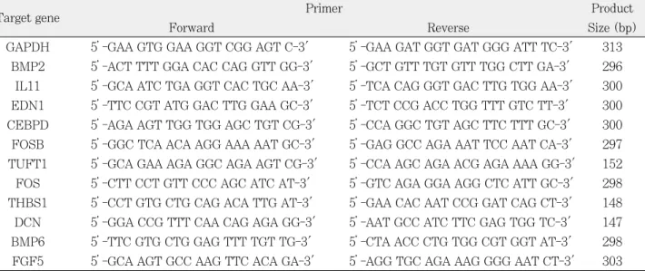
RT-PCR분석에 사용된 total RNA는 microarray 실험 에 사용하고 남은 것을 사용하였다. Maxime RT-PreMix (Intron, Suwon, Korea)를 이용하여 RNA에서 cDNA를 합성하고, 유의한 차이를 보이는 유전자 중 특징적인 몇 개 의 유전자 primer와 Maxime PCR Premix (Intron, Suwon, Korea)를 이용하여 RT-PCR을 시행하였다 (Table 1). PCR 반응은 변성(94℃, 3분), 30cycle의 증폭 (94℃, 30초; 57℃, 30초; 72℃, 30초), 비활성화(72℃, 7분)로 구성하였다. 각 증폭산물은 1.5% agarose 젤에서 전기영동되었고, 10분간 ethidium bromide용액으로 염색 시켜 Gel-Doc (Bio-rad, Hercules, CA, USA)으로 관찰 한 뒤, 이들에 대한 band intensity를 측정하였다. 유전자 의 상대적 발현 정도를 비교하기 위해 먼저 각 유전자의 band intensity를 GAPDH의 band intensity로 보정한 다음, wMTA 혹은 Dycal를 적용한 그룹의 band intensi- ty를 대조군의 band intensity로 나누어 표시하였다. PCR 은 각각 3번 시행한 후 그 평균값을 표현하였다.
결 과 1. Microarray
3시간, 6시간, 9시간, 24시간 중 어느 시간에서라도
Table 1.Primer sequence list used in RT-PCR
Target gene Primer Product
Forward Reverse Size (bp)
GAPDH 5’-GAA GTG GAA GGT CGG AGT C-3' 5’-GAA GAT GGT GAT GGG ATT TC-3' 313 BMP2 5’-ACT TTT GGA CAC CAG GTT GG-3' 5’-GCT GTT TGT GTT TGG CTT GA-3' 296 IL11 5’-GCA ATC TGA GGT CAC TGC AA-3' 5’-TCA CAG GGT GAC TTG TGG AA-3' 300 EDN1 5’-TTC CGT ATG GAC TTG GAA GC-3' 5’-TCT CCG ACC TGG TTT GTC TT-3' 300 CEBPD 5’-AGA AGT TGG TGG AGC TGT CG-3' 5’-CCA GGC TGT AGC TTC TTT GC-3' 300 FOSB 5’-GGC TCA ACA AGG AAA AAT GC-3' 5’-GAG GCC AGA AAT TCC AAT CA-3' 297 TUFT1 5’-GCA GAA AGA GGC AGA AGT CG-3' 5’-CCA AGC AGA ACG AGA AAA GG-3' 152 FOS 5’-CTT CCT GTT CCC AGC ATC AT-3' 5’-GTC AGA GGA AGG CTC ATT GC-3' 298 THBS1 5’-CCT GTG CTG CAG ACA TTG AT-3' 5’-GAA CAC AAT CCG GAT CAG CT-3' 148 DCN 5’-GGA CCG TTT CAA CAG AGA GG-3' 5’-AAT GCC ATC TTC GAG TGG TC-3' 147 BMP6 5’-TTC GTG CTG GAG TTT TGT TG-3' 5’-CTA ACC CTG TGG CGT GGT AT-3' 298 FGF5 5’-GCA AGT GCC AAG TTC ACA GA-3' 5’-AGG TGC AGA AAG GGG AAT CT-3' 303
|Fold|>2의 조건을 만족하는 유전자를 확인하였다. 여기서 fold값이란 실험군과 대조군의 값에 대해서 실제로 몇 배의 차이가 있는지를 [실험군의 값] / [대조군의 값]으로 살펴 본 수치이다. 예를 들어, “2.8”이라면 대조군에 비해 실험 군에서 2.8배 더 많이 발현된 것이고“-5.5”라면, 대조군에 비해 실험군에서 5.5배가 덜 발현됨을 의미한다.
Illumina HumanRef-8 v3 Beadchip (Illumina Inc., San Diego, CA, USA)에 표현된 24,546개의 유전자 중 wMTA를 적용한 치수세포에서는 총 68개의 유전자에서 대 조군과 2배 이상의 fold값 변화를 보였다. Dycal을 적용한 치수세포에서는 총 597개의 유전자에서 2배 이상의 fold값 변화를 보였다. wMTA에 의해 변화한 68개의 유전자 중 43개의 유전자가 2배 이상 발현이 증가하였으며 25개의 유 전자가 50% 이하로 감소하였다. Dycal에 의해 발현이 변 화한 597개의 유전자 중 239개의 유전자가 2배 이상 증가 하였으며 358개의 유전자에서 50% 이하로 감소하였다.
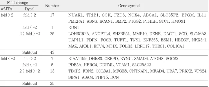
wMTA와 Dycal에 의해 발현 정도가 변화한 유전자의 증 감을 비교하였다(Table 2). 이중 wMTA와 Dycal 모두에 서 2배 이상 증가한 유전자는 17개였으며 모두 50% 이하 로 감소한 유전자는 5개였다. 이에 반해 wMTA에서는 2배 이상 증가하였으나 Dycal에서는 50% 이하로 감소한 유전 자는 1개였으며 wMTA에서는 50% 이하로 감소하였으나 Dycal에서는 2배 이상 증가한 유전자는 7개였다. wMTA에 서 2배 이상 증가하거나 50% 이하로 감소하는 변화가 있었 으나 Dycal에 의한 변화가 없는 유전자의 수는 38개였다.
wMTA와 Dycal을 적용한 세포에서 변화한 68개, 597개
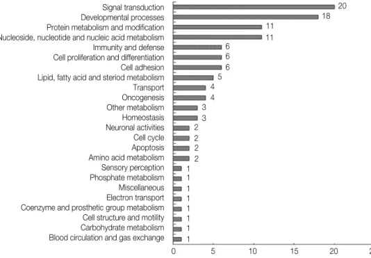
의 유전자를 생물학적 과정(biological process)에 따라 분 류하였다. wMTA에 의해 생긴 치수세포내의 유전자 발현 변화는 신호의 전달, 발생 과정, 단백질 대사, nucleoside, nucleotide and nucleic acid metabolism, 면역반응과 방 어기전, 세포의 증식과 분화, 세포의 부착에 관여하는 유전 자 순으로 많았다(Figure 1). 이에 비하여 Dycal에 의해서 생긴 치수세포내의 유전자 발현 변화는 nucleoside, nucleotide and nucleic acid metabolism, 신호의 전달, 발생 과정, 단백질 대사, 세포 주기(cell cycle), 세포의 구 조와 운동성(motility), 세포의 증식과 분화, 면역반응과 방 어기전에 관여하는 유전자의 순으로 많았다(Figure 2).
2. RT-PCR
RT-PCR은 wMTA와 Dycal을 6시간, 9시간, 24시간 적 용한 실험군에서 같은 시간대에 유전자 발현 정도의 비교가 가능한 bone morphogenetic protein 2 (BMP2), inter- leukin 11 (IL11), endothelin 1 (EDN1), CCAAT / enhancer-binding protein delta (CEBPD)와 각 시간대 에 특징적인 유전자 발현을 보이는 FBJ murine osteosar- coma viral oncogene homolog B (FOSB), tuftelin 1 (TUFT1), human homolog of the retroviral oncogene v-Fos (c-FOS), thrombospondin 1 (THBS1), decorin (DCN), bone morphogenetic protein 6 (BMP6), fibroblast growth factor 5 (FGF5)에 대하여 시행하였으 며 그 결과를 아래에 정리하였다(Table 3).
Table 2.Summary of expression profiles of genes differentially regulated by wMTA and Dycal Fold change
Number Gene symbol
wMTA Dycal
fold > 2 fold > 2 17 NUAK1, TRIB1, SGK, FZD8, NOX4, ABCA1, SLC35F2, BPGM, IL11, PMEPA1, ASNS, RCAN1, BMP2, PTGS2, PTHLH, STC1, HMOX1
fold < -2 1 EDN1
2 > fold > -2 25 LOH3CR2A, ANGPTL4, SH3BP5L, MMP10, DENR, DACT1, SCD, SLC46A3, UAP1L1, PDPN, FOSB, TUFT1, TNS1, ZNF365, ESM1, HBEGF, NKX3-1, MAZ, AK3L1, ETV4, MT1X, FOLR3, LRRC17, THBS1, COL10A1
Subtotal 43
fold < -2 fold > 2 7 KIAA1199, DHRS3, CEBPD, KYNU, SMAD6, ATOH8, SOCS2 fold < -2 5 PDE5A, HERC4, DDIT4L, VCAM1, SLC25A22
2 > fold > -2 13 TIMP2, FBN2, COL3A1, MFGE8, CNTNAP1, MFAD4, UBA7, PRRX2, VPS24, SIPA1, ASAM, PHF15, DCN
Subtotal 25
Figure 1.Classification of expressed genes by wMTA treatment. The genes which were up-or down-regulated by at least two- fold following a cellular treatment with wMTA at any time point were classified using the panther classification system (http://www.pantherdb.org).
Signal transduction Developmental processes Protein metabolism and modification Nucleoside, nucleotide and nucleic acid metabolism Immunity and defense Cell proliferation and differentiation Cell adhesion Lipid, fatty acid and steriod metabolism Transport Oncogenesis Other metabolism Homeostasis Neuronal activities Cell cycle Apoptosis Amino acid metabolism Sensory perception Phosphate metabolism Miscellaneous Electron transport Coenzyme and prosthetic group metabolism Cell structure and motility Carbohydrate metabolism Blood circulation and gas exchange
0 5 10 15 20 25 20 18 11
11 6
6 6 5 4 4 3 3 2 2 2 2 1 1 1 1 1 1 1 1
Figure 2. Classification of expressed genes by Dycal treatment. The genes which were up-or down-regulated by at least two- fold following a cellular treatment with Dycal at any time point were classified using the panther classification system.
Nucleoside, nucleotide and nucleic and metabolism Signal transduction Developmental processes Protein metabolism and modification Cell cycle Cell structure and motility Cell proliferation and differentiation Immunity and defense Transport Intracellular protein traffic Carbohydrate metabolism Lipid, fatty acid and steriod metabolism Other metabolism Oncogenesis Apoptosis Neuronal activities Amino acid metabolism Protein targeting and localization Muscle contraction Cell adhesion Sensory perception Miscellaneous Blood circulation and gas exchange Electron transport Sulfur metabolism Non-vertebrate process Coenzyme and prosthetic group metabolism
0 5 10 15 20 25 164 117
95 89 81 71 50 45 41 35 25 21 19 19 16 12
10 9 8 7 4 4 2 1
12 10 10
1 1
총괄 및 고찰
오랫동안 치수복조술에 사용된 수산화칼슘은 미분화된 치 수세포를 조상아세포로 분화시키고 치수가 노출된 곳에서 경조직을 형성한다고 알려져 있다. 그러나 수산화칼슘에 의 한 수복상아질의 형성은 재료자체의 고유한 능력에 의한 것 보다는 수산화칼슘의 자극에 대한 방어기전에 의한 결과로 알려져 왔다.24,25 최근 들어 생체적합성이 뛰어난 MTA가 치수복조술에 사용되었으며 우수한 결과들이 보고되었다.
그러나 유전자 수준에서 MTA와 수산화칼슘이 치수세포에 어떤 영향을 주는지에 대한 전반적인 정보는 많이 부족한 상태이다. 본 연구는 wMTA와 수산화칼슘을 적용한 치수 세포에서, 치수세포의 분화와 증식, 신생혈관형성, 석회화 과정 그리고 염증에 관련된 일련의 유전자들의 변화를 microarray로 보여준 최초의 연구였으며 연구결과에서는 치수복조제재로서 wMTA와 수산화칼슘이 치수세포에 서 로 다른 기전으로 작용을 한다는 점을 시사하였다.
손상 받은 치수의 상부에서 수복상아질이 생성되기까지는 몇 단계의 과정을 거친다.26우선 치수에 존재하는 줄기세포 나 전구세포가 손상 받은 부위로 모이게 된다. 이 세포들이 증식한 후 조상아세포양 세포(odontoblast-like cell)이나 조골세포양 세포(osteoblast-like cell)로 분화한다. 마지막 단계로 이런 세포들이 세포외 기질을 생성하고 석회화 과정 을 거치게 된다. 이번 연구에서도 wMTA와 Dycal을 적용 한 치수세포 내에 조상아세포양 세포 혹은 조골세포양 세포 로의 분화와 증식을 유도하는 유전자들의 변화가 확인 되었 다. BMP는 골 형성과 연골 형성에 중요한 역할을 하는 것
으로 알려져 있으며, 특히 BMP2는 수용체와 함께 인간 치 수세포에 많이 발현된다고 보고 되었다.27,28 절단된 치수에 recombinant human BMP2를 적용하였을 때 수복상아질 이 형성되는 결과가 관찰되었으며29 또한 인간치수세포가 조상아세포로 분화되는 과정이 BMP2에 의해서 촉진되는 현상이 보고되었다.30 본 실험에서는 wMTA를 6시간, 9시 간 적용한 실험군과 Dycal을 6시간, 9시간, 24시간 적용한 실험군에서 BMP2가 대조군보다 3배 이상 높게 발현되었 다. 이러한 사실을 종합해 볼 때 wMTA뿐만 아니라 Dycal 도 치수세포가 조상아세포로 분화하는데 관여하는 유전자 변화에 영향을 미치는 것으로 추측할 수 있다. 이러한 현상 은 기존의 연구에서 보고된 결과와 일치 하였다.31
BMP2와는 다르게 BMP6는 Dycal을 9시간, 24시간 적 용한 실험군에서만 2배 이상 증가하고 wMTA를 적용한 실 험군에서는 변화가 없었다는 것은 흥미로운 사실이다.
BMP6는 골 형성의 결정적 조절인자로 알려져 있으며 골 형성의 과정과 유사한 상아질 형성과정에서도 중요한 역할 을 하는 것으로 알려져 있다.32,33인위적인 치수세포의 분화 와 석회화를 유도한 실험에서 BMP6가 많이 발현되었다는 보고는 BMP6가 치수세포의 분화와 석회화에 중요한 역할 을 함을 시사한다.34이와 같은 사실을 근거로 볼 때 wMTA 뿐만 아니라 Dycal도 치수세포의 분화와 석회화에 관여하 는 유전자의 변화에 영향을 미치는 것으로 사료된다.
Drosophila protein인 mothers against decapenta- plegic (Mad)과 C. elegans (Sma)의 homolog인 SMAD 단백질은 세포의 신호전달에 작용을 하며 특히 BMP와 같 은 TGF-βfamily의 하위 신호전달인자로 작용을 한다.35 Table 3.The relative mRNA expression levels of genes analyzed by RT-PCR
Symbol 6hr 9hr 24hr
wMTA Dycal wMTA Dycal wMTA Dycal
BMP2 1.88 2.31
IL11 1.99 2.38
EDN1 1.41 0.26
CEBPD 1.16 2.07
FOSB 1.28
TUFT1 1.57
FOS 1.77
THBS1 1.89
DCN 0.58
BMP6 11.93
FGF5 0.25
The relative level of gene expression was normalized against GAPDH messenger RNA, and the control was set as 1.0.
Optical density values represent the mean (n=3).
포유류에서 확인된 SMAD 단백질은 8가지이며 이중 SMAD6는 Co-SMAD인 SMAD4와 경쟁하는 inhibitory SMAD로 작용하는 것으로 알려져 있다.36 본 실험에서 wMTA를 6시간 적용한 실험군에서 대조군보다 SMAD6가 50% 이하로 감소하며 Dycal을 9시간 적용한 실험군에서 는 대조군보다 2배 이상 증가하는 양상을 보였다. TGF-β family가 치수세포의 분화와 증식에 중요한 영향을 미친다 고 알려진 것을 고려해 볼 때 wMTA와 Dycal의 SMAD6 에 대한 상반된 결과는 흥미로운 현상이라 할 수 있다.
THBS1은 부착성 당단백질의 일종으로 세포간 상호작용 이나 세포와 기질의 상호작용을 매개하는 기능과 TGF-β에 결합하여 이를 활성화시키는 기능을 하는 것으로 알려져 있
다.37,38 THBS1은 치수조직이나 치은조직에서는 관찰되지
않으며 전상아질(predentin)에서만 분포하며 특히 조상아 세포에서 주로 관찰되었으며 이러한 현상을 종합해 볼 때 THBS1이 치수세포에 의해 발생하는 석회화 과정에서 중 요한 역할을 할 가능성이 제기되었다.39 본 실험에서 THBS1이 wMTA를 24시간 적용한 실험군에서 대조군보 다 2배 이상 증가하였다. 이러한 증가는 wMTA가 적용되 었을 때 치수세포 혹은 전구세포가 조상아세포로 분화하는 데 관여하는 유전자들의 변화가 발생하는 것이라 추측할 수 있다.
최근의 연구에서 EDN 수용체가 발육중인 치아 조직에 다양하게 분포하는 것을 확인한 후 EDN1이 치아의 발생 과정에 다양한 기능을 할 가능성을 제시하였다.40이전 연구 에서 일 차 세포 배양한 인간치수세포에 EDN1을 적용한 경우 치수세포가 증식되며 유사분열 기전이 활성화되며 collagen type I의 합성이 증가하는 양상을 관찰하였으며 이를 근거로 EDN1이 치수의 치유와 치수세포의 분화에 중 요한 역할을 할 것이라고 추론하였다.41본 실험에서 EDN1 이 wMTA를 6시간, 9시간, 24시간 적용한 실험군에서 대 조군보다 2배 이상 증가하는 양상을 보였고 Dycal을 6시 간, 9시간, 24시간 적용한 실험군에서 대조군보다 50% 이 하로 감소하는 양상을 보였다.
IL11은 골수성세포(myeloid cell)의 성장을 증가시키며, 지방생성(adipogenesis)을 억제시키며 조골세포생성 (osteoblastogenesis)을 촉진시키는 기능을 하는 것으로 알려져 있다.42 IL11을 정상보다 많이 발현하는 형질전환 쥐(transgenic mice)에서 더 많은 골괴(bone mass)가 형 성된다는 사실이 확인되었다.43 본 실험에서는 IL11이 wMTA를 6시간, 9시간, 24시간 적용한 실험군에서 대조군 보다 2배 이상 발현되었으며 특히 9시간에서는 대조군과 5 배 이상의 차이를 보였다. Dycal을 적용한 실험군에서는 9 시간에서만 2배 이상의 변화를 보였다.
세포의 분화, 증식, 그리고 세포사멸에 관여하는 유전자 발현을 조절하는 FOS family의 하나인 c-FOS는 골과 연
골의 세포분화에 영향을 미치는 것으로 알려져 있다.44이전 연구에서 c-FOS 유전자 결핍 모형(knockout model)에서 는 파골세포의 생성이 완전히 억제되고 골화석증 (osteopetrosis)이 발생하는 현상을 확인한 후 파골세포의 발생과 골의 remodeling에서 c-FOS의 중요성이 제시되었 다.45반대로 c-FOS가 과잉으로 발현되면 조골세포의 변형 에 의해 골육종을 발생시키는 것으로 알려져 있다.46본 실 험에서는 Dycal을 9시간 적용한 실험군에서 c-FOS가 2배 이상 발현되었다.
FOSB는 본 실험에서 wMTA를 3시간, 9시간 적용한 실 험군에서 2배 이상 발현되었다. 이전 연구에서 FOSB 유전 자 결핍 쥐(knockout mice)에서는 골 형성에는 별다른 이 상이 없었다는 결과가 발표 되었다.47그러나 세포의 분화와 증식에 관여하는 FOS family의 특성상 FOSB가 조상아세 포와 조골세포에 미치는 영향에 관해서도 계속적인 연구가 필요하리라 사료된다.
BMP, SMAD, THBS1, EDN1, IL11, c-FOS 그리고 FOS 유전자의 발현 변화를 보이는 결과를 볼 때 wMTA가 수산화칼슘보다 치수세포의 분화와 증식에 관여하는 유전 자의 변화에 더 많은 영향을 주는 것으로 사료된다. 또한 이 러한 분화와 증식에 관여하는 유전자의 발현 변화가 비교적 이른 시기에 시작됨을 알 수 있다.
성장 인자는 세포 외 기질에 존재하는 chemical signal- ing polypeptide의 일종으로 세포의 분화, 증식 그리고 생 존을 조절하는 기능을 한다.48치수조직이 손상을 받아 수복 상아질을 형성하는 과정에서 손상받은 부위로 건강한 혈관 계가 형성되는 과정이 필수적이라는 점에 착안하여 신생혈 관형성 유도물질(angiogenic inducer)에 대한 많은 연구가 진행되었다.
Vascular endothelial growth factor (VEGF)는 VEGF-A, -B, -C, -D, -E, 그리고 platelet-derived growth factor (PDGF)의 6가지 종류로 구성된다. 이중 VEGFA와 VEGFB가 신생혈관형성과 연관이 있는 것으로 알려져 있다.49 또한 VEGF는 배아의 형성(embryonic development) 과정 중이나 손상을 받은 조직에서 새로운 혈관을 생성하는 역할을 한다. 이전 연구에서 VEGF가 치 수세포에 자가 분비성(autocrine pattern)으로 직접 작용 하여 화학주성(chemotaxis)과 세포의 증식 및 분화를 촉진 시키며 알칼리성 인산분해효소(alkaline phosphatase)를 증가시킨다는 결과를 근거로 치수세포가 조상아세포로 분 화하는데 VEGF가 관여할 가능성을 제시 하였다.50다른 연 구에서 VEGF가 포함된 신생혈관 유도성 성장 인자 (angiogenic growth factor)가 상아치수 복합체 (dentinopulpal complex)의 치유 과정에 기여한다고 보고 되었다.51 본 실험에서 wMTA를 적용한 실험군에서는 VEGF에 대한 별다른 변화가 관찰되지 않았다. 그러나
Dycal을 적용한 실험군에서 VEGFA가 9시간에서 2배 이 상, 24시간에서 4배 이상의 변화를 보였다. 수산화칼슘이 작용하는 기전을 고려해 볼 때 이러한 현상이 단순히 손상 에 대응하는 치유과정을 표현하는지 아니면 조상아세포로 분화하는 과정에 관련이 있는 유전자의 변화를 표현하는지 에 대한 추가적인 분석이 필요하리라 사료된다.
Placental growth factor (PlGF)는 VEGF의 sub-fam- ily의 한 종류로 신생혈관형성에 중요한 역할을 한다. PlGF 는 VEGF와 이합체를 형성할 수 있으며 직접적인 유사분열 효과는 거의 없으나 낮은 농도의 VEGF를 정상보다 더 많 이 활성화 시키는 능력을 가지는 것으로 알려져 있다.52 본 실험에서는 Dycal을 24시간 적용한 실험군에서 2배 이상 증가하는 양상을 보였다.
FGF family는 배아의 형성, 형태형성(morphogenesis), 세포의 성장, 조직의 치유, 암의 성장과 침습에 관여하며, 이중 FGF5는 신생혈관 유도성 성장 인자의 한 종류로 알 려져 있다. 본 실험에서 다른 성장 인자와는 다르게 Dycal 을 적용한 실험군에서 6시간, 9시간, 24시간 적용한 경우에 FGF5가 50% 이상 감소하는 경향을 보였으며 이런 경향은 시간이 지날수록 더 명확해지는 양상을 보였다.
VEGFA와 같은 신생혈관형성 인자들은 기질을 분해하는 단백질 분해효소(proteinase)의 발현을 자극하여 세포 외 기질의 remodeling을 촉진하고 새로운 혈관의 침습(inva- sion)을 촉진하게 된다.53그러므로 이러한 단백질 분해효소 의 비활성화는 신생혈관형성을 방해하는 역할을 하게 된다.
Tissue inhibitor of metalloproteinases 2 (TIMP2)는 metalloproteinase의 활성을 감소시키며 인간 내피세포의 증식을 직접적으로 억제하는 기능을 가지고 있다.54,55 결과 적으로 TIMP2는 신생혈관형성을 방해하는 역할을 하며 본 실험에서는 wMTA를 24시간 적용한 실험군에서 TIMP2 의 발현이 50% 이상 감소하는 양상을 보였다. 신생혈관형 성을 유도하는 성장 인자의 유전자 발현은 wMTA보다 Dycal에 의해서 명확한 변화를 보이는 것으로 확인되었다.
이러한 현상이 과도한 염증에 대한 보상작용인지 수산화칼 슘이 wMTA와 다른 기전에 의한 효과인지에 대한 추가적 인 연구가 필요하리라 사료된다.
수복상아질이 형성되는 과정 중의 한 단계인 석회화를 조 절하는 유전자의 변화 역시 본 연구결과에서 확인할 수 있 었다. 본 실험에서는 collagen fibrillogenesis와 성숙(mat- uration)에 관련이 있는 DCN이 wMTA를 24시간 적용한 실험군에서 50% 이상 감소하였다. Collagen 기질의 석회 화 과정 중 DCN 유전자의 발현과 석회화의 반비례 관계가 여러 연구에서 보고 되었다.56 본 실험에서 wMTA를 적용 한 후 나타나는 DCN의 급격한 감소가 wMTA가 치수복조 제재로서 가지는 중요한 상아질 형성능력을 유전자 수준에 서 뚜렷하게 반영하는 결과라고 사료된다. Collagen type
X alpha 1 (COL10A1)은 연골내 골화(endochondral ossification)가 일어나는 동안 발생하는 비후성 연골세포 (hypertrophic chondrocyte)의 산물로 알려져 있다.57 최 근의 연구에서 발생중인 인간의 법랑질 organ과 법랑모세 포에 Collagen type X 이 다양하게 존재하는 것을 확인 하 였다.58본 실험에서는 wMTA를 24시간 적용한 실험군에서 COL10A1이 급격하게 증가하는 양상을 보였다. 그러나 col- lagen type I과 collagen type III는 각각 Dycal과 wMTA를 24시간 적용한 실험군에서 감소하는 경향을 보였다.
TUFT는 치아의 법랑질에서 발견되는 인산화된 당단백질 중 하나이다. TUFT는 주로 법랑질이 형성되는 시기 중 짧 은 기간에만 나타나기 때문에 법랑질의 석회화를 시작시키 는 물질로 제시 되었다.59 TUFT는 법랑질 형성의 초기에 분비성 법랑모세포(secretory ameloblast)에서 주로 관찰 되며 특히 Tomes’process에서 주로 발견된다. 또한 법랑 질이 형성되는 상아법랑질 경계 부위에서 집중되는 현상을 보인다.60 본 실험에서는 wMTA를 9시간 적용한 실험군에 서 TUFT1이 2배 이상 증가하는 양상을 보였다. wMTA를 적용한 치수세포에서 법랑질 형성과 관계되는 것으로 추측 되는 COL10A1과 함께 TUFT의 유전자가 발현되는 것은 재미있는 현상이라고 할 수 있다.
이외에도 염증에 의해 발생하는 유전자들의 변화도 다수 관찰되었다. 치수의 치유과정에서 줄기세포가 손상 받은 부 위로 이동하는데 필요한 조건이 적당한 염증이라면 감당할 수 있는 역치 내의 적당한 염증은 오히려 긍정적으로 해석 할 수도 있다. 또한 과도한 염증은 치수세포를 죽게 만들 수도 있기 때문에 치수복조제재가 염증을 야기하는 성질은 치수복 조제재의 선택에 있어서 신중히 고려해야 하는 항목이다.
Heme oxygenase의 3가지 isoform중 하나인 heme oxygenase 1 (HMOX1)은 다양한 자극에 의해 생겨나는 스트레스에 반응하는 단백질이다.61 HMOX1의 기능이 완 전히 알려지지는 않았지만 세포의 조절기전과 방어기전에 관여하는 것으로 알려져 있다.62 최근의 연구에서 전염증성 cytokine이나 과산화수소에 의한 치수세포내의 HMOX1의 발현을 확인한 후 HMOX1이 치수세포의 방어기전에 관여 할 가능성을 제시하였다.63,64본 실험에서 wMTA를 적용한 실험군에서는 24시간에서 2배 이상의 변화를 보이며 Dycal을 적용한 실험군에서는 3시간, 6시간, 9시간, 24시 간에서 모두 2배 이상의 변화를 보이며 특히 24시간에서는 대조군과 비교하여 14배나 많은 변화를 보였다. Dycal에서 HOMX1의 변화가 더 많이 발생하는 현상은 수산화칼슘이 wMTA보다 치수조직에 초기 염증을 더 많이 발생시키는 작용기전을 잘 반영하고 있다고 사료된다.
8개로 구성된 suppressors of cytokine signaling (SOCS) family는 Janus kinases (JAKs)와 Signal Transducers and Activators of Transcription (STATs)
의 pathway를 억제하는 역할을 한다.65 또한 SOCS는 multiple cytokine을 억제하는 기능을 가지고 있으며 특히 SOCS2는 아스피린에 의해 유도되는 lipoxin의 항염증성을 매개하는 것으로 알려져 있다.66 건강한 조직에서는 SOCS 의 발현이 드물지만 여러 조직에서 SOCS가 발현되는 정도 가 다양한 것은 염증반응의 결과로 추측된다.67최근의 연구 에서 치근단 육아종에 정상조직보다 SOCS1, SOCS2, SOCS3가 많이 발현된다는 사실을 보고하면서 이 물질을 치료의 대상으로 사용가능성을 제시하였다.68 본 실험에서 는 wMTA를 24시간 적용한 실험군에서 SOCS2가 50%
이하로 감소하는 양상을 보이며 Dycal을 24시간 적용한 실 험군에서는 반대로 2배 이상 발현되는 양상을 보였다.
다양한 종류의 세포분화와 염증의 조절에 영향을 미치는 것으로 알려져 있는 CEBPD는 염증 반응과 면역 반응에 관 계된 유전자의 조절에 중요한 역할을 한다.69,70또한 대식세 포의 분화와 활성화에 관여하는 유전자의 조절에 중요한 역 할을 하는 것으로 알려져 있다.71 본 실험에서는 wMTA를 24시간 적용한 실험군에서 CEBPD의 발현이 50% 이하로 감소하였고 반대로 Dycal을 9시간, 24시간 적용한 실험군 에서 CEBPD의 발현이 2배 이상 증가하였다. CEBPD가 염증반응에 작용하는 과정을 고려해볼 때 Dycal이 wMTA 보다 더 심한 염증 반응과 이물반응을 보이는 것으로 사료 된다.
본 실험에서 wMTA와 수산화칼슘 제재는 기전이 유사한 부분도 있는 반면 상반된 결과를 보이는 부분이 많이 존재 하는 것을 관찰하였으며 두 가지 제재 모두 치수복조제재로 서 치수세포의 변화를 유도하는 능력을 가지고 있는 것을 확인하였다. 향후 치수복조제재를 좀더 긴 시간 적용하여 실제로 단백질이 형성되는 것을 확인하는 실험이나 임상적 인 환경을 재현한 동물실험을 통한 추가적인 확인이 필요하 리라 사료된다.
결 론
wMTA를 치수세포에 적용한 경우 세포의 분화와 증식에 관여하는 유전자들(BMP2, SMAD6, THBS1, EDN1, IL11, FOSB)과 석회화에 관여하는 유전자들(DCN, COL10A1, TUFT1)의 유의한 발현 변화가 관찰되었다.
수산화칼슘 제재를 치수세포에 적용한 경우 세포의 분화와 증식에 관여하는 유전자들(BMP2, BMP6, IL11, FOS)과 신생혈관형성에 관여하는 유전자들(VEGFA, PlGF, FGF)의 유의한 발현 변화가 관찰되었다. 수산화칼슘 제재 를 치수세포에 적용한 경우 wMTA에 비하여 염증에 관여 하는 유전자들(HMOX1, SOCS2, CEBPD, KIAA1199) 이 더 많이 발현되는 양상을 보였다.
Conflict of Interest: No potential conflict of interest relevant to this article was reported.
REFERENCES
1. American Association of Endodontists. Glossary of endodontic terms, 7th ed. pp 40 2003.
2. Cox CF, Su¨bay RK, Ostro E, Suzuki S, Suzuki SH.
Tunnel defects in dentin bridges: their formation fol- lowing direct pulp capping. Oper Dent 1996;21:4-11.
3. Cox CF, Hafez AA, Akimoto N, Otsuki M, Suzuki S, Tarim B. Biocompatibility of primer, adhesive and resin composite systems on non-exposed and exposed pulps of non-human primate teeth. Am J Dent 1998;
11(Supplement):55-63.
4. Cox CF, Tarim B, Kopel H, Gu¨rel G, Hafez A. Technique sensitivity: biological factors contributing to clinical success with various restorative materials. Adv Dent Res 2001;15:85-90.
5. Yun YR, Yang IS, Hwang YC, Hwang IN, Choi HR, Yoon SJ, Kim SH, Oh WM. Pulp response of mineral trioxide aggregate, calcium sulfate or calcium hydrox- ide. J Kor Acad Cons Dent 2007;32:95-101.
6. Bae JH, Kim YG, Yoon PY, Cho BH, Choi YH. Pulp response of beagle dog to direct pulp capping materi- als: histological study. J Kor Acad Cons Dent 2010;35:
5-12.
7. Dominguez MS, Witherspoon DE, Gutmann JL, Opperman LA. Histological and scanning electron microscopy assessment of various vital pulp-therapy materials. J Endod 2003;29:324-333.
8. Ford TR, Torabinejad M, Abedi HR, Bakland LK, Kariyawasam SP. Using mineral trioxide aggregate as a pulp-capping material. J Am Dent Assoc 1996;127:
1491-1494.
9. Faraco IM Jr, Holland R. Response of the pulp of dogs to capping with mineral trioxide aggregate or a calcium hydroxide cement. Dent Traumatol 2001;17:163-166.
10. Hauman CH, Love RM. Biocompatibility of dental materials used in contemporary endodontic therapy: a review. Part 2. Root canal-filling materials. Int Endod J 2003;36:147-160.
11. Aeinehchi M, Eslami B, Ghanbariha M, Saffar AS.
Mineral trioxide aggregate (MTA) and calcium hydrox- ide as pulp-capping agents in human teeth: a prelimi- nary report. Int Endod J 2003;36:225-231.
12. Accorinte Mde L, Holland R, Reis A, Bortoluzzi MC, Murata SS, Dezan E Jr, Souza V, Alessandro LD.
Evaluation of mineral trioxide aggregate and calcium hydroxide cement as pulp-capping agents in human teeth. J Endod 2008;34:1-6.
13. Iwamoto CE, Adachi E, Pameijer CH, Barnes D, Romberg EE, Jeffries S. Clinical and histological evalu- ation of white ProRoot MTA in direct pulp capping. Am J Dent 2006;19:85-90.
14. Yasuda Y, Ogawa M, Arakawa T, Kadowaki T, Saito T. The effect of mineral trioxide aggregate on the min- eralization ability of rat dental pulp cells: an in vitro study. J Endod 2008;34:1057-1060.
15. Andelin WE, Shabahang S, Wright K, Torabinejad M.
Identification of hard tissue after experimental pulp capping using dentin sialoprotein (DSP) as a marker.J Endod 2003;29:646-650.
16. Kuratate M, Yoshiba K, Shigetani Y, Yoshiba N, Ohshima H, Okiji T. Immunohistochemical analysis of nestin, osteopontin, and proliferating cells in the reparative process of exposed dental pulp capped with mineral trioxide aggregate. J Endod 2008;34:970-974.
17. Min KS, Yang SH, Kim EC. The combined effect of mineral trioxide aggregate and enamel matrix deriva- tive on odontoblastic differentiation in human dental pulp cells. J Endod 2009;35:847-851.
18. Shi S, Robey PG, Gronthos S. Comparison of human dental pulp and bone marrow stromal stem cells by cDNA microarray analysis. Bone 2001;29:532-539.
19. McLachlan JL, Smith AJ, Bujalska IJ, Cooper PR.
Gene expression profiling of pulpal tissue reveals the molecular complexity of dental caries. Biochim Biophys Acta 2005;1741:271-281.
20. Syudo M, Yamada S, Yanagiguchi K, Matsunaga T, Hayashi Y. Early gene expression analyzed by a genome microarray and real-time PCR in osteoblasts cultured with a 4-META/MMA-TBB adhesive resin sealer. Oral Surg Oral Med Oral Pathol Oral Radiol Endod 2009;107:e77-81.
21. Martinez ZR, Naruishi K, Yamashiro K, Myokai F, Yamada T, Matsuura K, Namba N, Arai H, Sasaki J, Abiko Y, Takashiba S. Gene profiles during root canal treatment in experimental rat periapical lesions. J Endod 2007;33:936-943.
22. Yokose S, Kadokura H, Tajima Y, Fujieda K, Katayama I, Matsuoka T, Katayama T. Establishment and char- acterization of a culture system for enzymatically released rat dental pulp cells. Calcif Tissue Int 2000;
66:139-144.
23. Kim YB, Shon WJ, Lee WC, Kum KY, Baek SH, Bae KS. Gene Expression Profiling in Human Dental Pulp Cells Treated with Mineral Trioxide Aggregate. J Kor Acad Cons Dent 35:152-163, 2010.
24. Goldberg M, Six N, Decup F, Lasfargues JJ, Salih E, Tompkins K, Veis A. Bioactive molecules and the future of pulp therapy. Am J Dent 2003:16;66-76.
25. Almushayt A, Narayanan K, Zaki AE, George A.
Dentin matrix protein 1 induces cytodifferentiation of dental pulp stem cells into odontoblasts. Gene Ther 2006;13:611-620.
26. Goldberg M, Farges JC, Lacerda-Pinheiro S, Six N, Jegat N, Decup F, Septier D, Carrouel F, Durand S, Chaussain-Miller C, Denbesten P, Veis A, Poliard A.
Inflammatory and immunological aspects of dental pulp repair. Pharmacol Res 2008;58:137-147.
27. Reddi AH. Bone morphogenetic proteins: an unconven- tional approach to isolation of first mammalian mor- phogens. Cytokine Growth Factor Rev 1997;8:11-20.
28. Gu K, Smoke RH, Rutherford RB. Expression of genes for bone morphogenetic proteins and receptors in human dental pulp. Arch Oral Biol 1996;41:919-923.
29. Nakashima M, Nagasawa H, Yamada Y, Reddi AH.
Regulatory role of transforming growth factor-β, bone morphogenetic protein-2, and protein-4 on gene expression of extracellular matrix proteins and differ- entiation of dental pulp cells. Dev Biol 1994;162:18- 28.
30. Saito T, Ogawa M, Hata Y, Bessho K. Acceleration effect of human recombinant bone morphogenetic pro- tein-2 on differentiation of human pulp cells into odon- toblasts. J Endod 2004;30:205-208.
31. Yasuda Y, Ogawa M, Arakawa T, Kadowaki T, Saito
T. The effect of mineral trioxide aggregate on the min- eralization ability of rat dental pulp cells: an in vitro study. J Endod 2008;34:1057-1060.
32. Wozney JM, Rosen V. Bone morphogenetic protein and bone morphogenetic protein gene family in bone forma- tion and repair. Clin Orthop Relat Res 1998;346:26- 37.
33. Takeda K, Oida S, Goseki M, Iimura T, Maruoka Y, Amagasa T, Sasaki S. Expression of bone morpho- genetic protein genes in the human dental pulp cells.
Bone 1994;15:467-470.
34. Zhang Q, Wang X, Chen Z, Liu G, Chen Z. Semi-quan- titative RT-PCR analysis of LIM mineralization protein 1 and its associated molecules in cultured human den- tal pulp cells. Arch Oral Biol 2007;52:720-726.
35. Matsui S, Takeuchi H, Tsujimoto Y, Matsushima K.
Effects of Smads and BMPs induced by Ga-Al-As laser irradiation on calcification ability of human dental pulp cells. J Oral Sci 2008;50:75-81.
36. Imamura T, Takase M, Nishihara A, Oeda E, Hanai J, Kawabata M, Miyazono K. Smad6 inhibits signaling by the TGF-βsuperfamily. Nature 1997;389:622-626.
37. Lawler J. The functions of thrombospondin-1 and-2.
Curr Opin Cell Biol 2000;12:634-640.
38. Murphy-Ullrich JE, Schultz-Cherry S, Ho¨o¨k M.
Transforming growth factor-beta complexes with thrombospondin. Mol Biol Cell 1992;3:181-188.
39. Ueno A, Yamashita K, Nagata T, Tsurumi C, Miwa Y, Kitamura S, Inoue H. cDNA cloning of bovine throm- bospondin 1 and its expression in odontoblasts and predentin. Biochim Biophys Acta 1998;1382:17-22.
40. Neuhaus SJ, Byers MR. Endothelin receptors and endothelin-1 in developing rat teeth. Arch Oral Biol 2007;52:655-662.
41. Yan Y, Liu Z, Zhang WG. In vitro study of the effects of endothelin-1 on human dental pulp cells. Chin J Dent Res 1999;2:5-13.
42. Suga K, Saitoh M, Fukushima S, Takahashi K, Nara H, Yasuda S, Miyata K. Interleukin-11 induces osteoblast differentiation and acts synergistically with bone morphogenetic protein-2 in C3H10T1/2 cells. J Interferon Cytokine Res 2001;21:695-707.
43. Takeuchi Y, Watanabe S, Ishii G, Takeda S, Nakayama K, Fukumoto S, Kaneta Y, Inoue D, Matsumoto T, Harigaya K, Fujita T. Interleukin-11 as a stimulatory factor for bone formation prevents bone loss with advancing age in mice. J Biol Chem 2002;277:49011- 49018.
44. Takayanaqi H, Kim S, Koqa T, Nishina H, Isshiki M, Yoshida H, Saiura A, Isobe M, Yokochi T, Inoue J, Wagner EF, Mak TW, Kodama T, Taniguchi T.
Induction and activation of the transcription factor NFATc1 (NFAT2) integrate RANKL signaling in termi- nal differentiation of osteoclast. Dev Cell 2002;3:889- 901.
45. Grigoriadis AE, Wang ZQ, Cecchini MG, Hofstetter W, Felix R, Fleisch HA, Wagner EF. C-Fos: a key regula- tor of osteoclast-macrophage lineage determination and bone remodeling. Science 1994;266:443-448.
46. Grigoriadis AE, Schellander K, Wang ZQ, Wagner EF.
Osteoblasts are target cells for transformation in c-fos transgenic mice. J Cell Biol 1993;122:685-701.
47. Gruda MC, van Amsterdam J, Rizzo CA, Durham SK, Lira S, Bravo R. Expression of FosB during mouse development: normal development of FosB knockout
mice. Oncogene 1996;12:2177-2185.
48. Smith AJ, Murray PE, Lumley PJ. Preserving the vital pulp in operative dentistry: 1. a biological approach.
Dent Update 2002;29:64-69.
49. Keyt BA, Nguyen HV, Berleau LT, Duarte CM, Park J, Chen H, Ferrara N. Identification of vascular endothe- lial growth factor determinants for binding KDR and Flt-1 receptors: generation of receptorselective VEGF variants by site-directed mutagenesis. J Biol Chem 1996;271:5638-5646.
50. Matsushita K, Motani R, Sakuta T, Yamaguchi N, Koga T, Matsuo K, Nagaoka S, Abeyama K, Maruyama I, Torii M. The role of vascular endothelial growth fac- tor in human dental pulp cells: induction of chemo- taxis, proliferation, and differentiation and activation of the AP-1-dependent signaling pathway. J Dent Res 2000;79:1596-1603.
51. Roberts-Clark DJ, Smith AJ. Angiogenic growth factors in human dentine matrix. Arch Oral Biol 2000;45:
1013-1016.
52. Ribatti D. The crucial role of vascular permeability fac- tor/vascular endothelial growth factor in angiogenesis:
a historical review. Br J Haematol 2005;128:303-309.
53. Heissig B, Hattori K, Friedrich M, Rafii S, Werb Z.
Angiogenesis: vascular remodeling of the extracellular matrix involves metalloproteinases. Curr Opin Hematol 2003;10:136-141.
54. Seo DW, Li H, Guedez L, Wingfield PT, Diaz T, Salloum R, Wei BY, Stetler-Stevenson WG. TIMP-2 mediated inhibition of angiogenesis: an MMP-indepen- dent mechanism. Cell 2003;114:171-180.
55. Murphy AN, Unsworth EJ, Stetler-Stevenson WG.
Tissue inhibitor of metalloproteinases-2 inhibits bFGF- induced human microvascular endothelial cell prolifera- tion. J Cell Physiol 1993;157:351-358.
56. Mochida Y, Duarte WR, Tanzawa H, Paschalis EP, Yamauchi M. Decorin modulates matrix mineralization in vitro. Biochem Biophys Res Commun 2003;305:6-9.
57. Alini M, Marriott A, Chen T, Abe S, Poole AR. A novel angiogenic molecule produced at the time of chondro- cyte hypertrophy during endochondral bone formation.
Dev Biol 1996;176:124-132.
58. Felszeghy S, Hollo′K, Mo′dis L, Lammi MJ. Type X col- lagen in human enamel development: a possible role in mineralization. Acta Odontol Scand 2000;58:171-176.
59. Deutsch D, Palmon A, Fisher LW, Kolodny N, Termine JD, Young MF. Sequencing of bovine enamelin (“tuftelin”) a novel acidic enamel protein. J Biol Chem 1991;266:16021-10628.
60. Paine CT, Paine ML, Luo W, Okamoto CT, Lyngstadaas SP, Snead ML. A tuftelin-interacting pro- tein (TIP39) localizes to the apical secretory pole of
mouse ameloblasts. J Biol Chem 2000;275:22284- 22292.
61. Otterbein LE, Choi AE. Heme oxygenase: colors of defense against cellular stress. Am J Physiol Lung Cell Mol Physiol 2000;279:L1029-1037.
62. Foresti R, Motterlini R. The heme oxygenase pathway and its interaction with nitric oxide in the control of cellular homeostasis. Free Radic Res 1999;31:459- 475.
63. Min KS, Lee HJ, Kim SH, Lee SK, Kim HR, Pae HO, Chung HT, Shin HI, Lee SK, Kim EC. Hydrogen per- oxide induces heme oxygenase-1 and dentin sialophos- phoprotein mRNA in human pulp cells. J Endod 2008;34:983-989
64. Min KS, Kwon YY, Lee HJ, Lee SK, Kang KH, Lee SK, Kim EC. Effects of proinflammatory cytokines on the expression of mineralization markers and heme oxyge- nase-1 in human pulp cells. J Endod 2006;32:39-43.
65. Krebs DL, Hilton DJ. SOCS proteins: negative regula- tors of cytokine signaling. Stem Cells 2001;19:378- 387.
66. Machado FS, Johndrow JE, Esper L, Dias A, Bafica A, Serhan CN, Aliberti J. Anti-inflammatory actions of lipoxin A(4) and aspirin-triggered lipoxin are SOCS-2 dependent. Nat Med 2006;12:330-334.
67. Alexander WS, Hilton DJ. The role of suppressors of cytokine signaling (SOCS) proteins in regulation of the immune response. Annu Rev Immunol 2004;22:503- 529.
68. Menezes R, Garlet TP, Trombone AP, Repeke CE, Letra A, Granjeiro JM, Campanelli AP, Garlet GP. The potential role of suppressors of cytokine signaling in the attenuation of inflammatory reaction and alveolar bone loss associated with apical periodontitis. J Endod 2008;34:1480-1484.
69. Schrem H, Klempnauer J, Borlak J. Liver-enriched transcription factors in liver fuction and development.
Part II: The C/EBPs and D site binding protein in cell cycle control, carcinogenesis, circadian gene regulation, liver regulation, apoptosis, and liver-specific gene reg- ulation. Pharmacol Rev 2004;56:291-330.
70. Liu YW, Chen CC, Wang JM, Chang WC, Huang YC, Chung SY, Chen BK, Hung JJ. Role of transcriptional factors Sp1, c-Rel, and c-Jun in LPS-induced C/EBPdelta gene expression of mouse macrophages.
Cell Mol Life Sci 2007;64:3282-3294.
71. Caivano M, Gorgoni B, Cohen P, Poli V. The induction of cyclooxygenage-2 mRNA in macrophages is biphasic and requires both CCAAT/enhancer-binding protein beta (C/EBP beta) and C/EBP delta transcription fac- tors. J Biol Chem 2001; 276:48693-48701.
국문초록
인간치수세포에 Mineral Trioxide Aggregate와 수산화칼슘 제재 적용 시 유전자 발현 양상 비교
김용범∙손원준∙이우철∙금기연∙백승호∙배광식*
서울대학교 치의학대학원 치과보존학교실
연구목적: 이 연구에서는 mineral trioxide aggregate 제재인 white ProRoot MTA (wMTA)와 수산화칼슘 제재인 Dycal 을 인간치수세포에 적용한 후 치수세포의 분화와 증식, 석회화, 신생혈관형성(angiogenesis) 그리고 염증에 관여하는 유전 자들의 발현 변화를 비교하였다.
연구 재료 및 방법: 실험군은 wMTA와 Dycal을 테플론 튜브(내경 10 mm, 길이 1 mm)에 담아 4시간 경화시킨 후 일차세 포배양한 인간치수세포에 적용하였고, 대조군은 빈 튜브만을 적용하였다. 3시간, 6시간, 9시간, 24시간 후 total RNA를 추 출하고 oligonucleotide microarray 방법을 통하여 유전자 발현 양상을 분석하였다. 위의 결과를 역전사 중합효소 연쇄반응 (reverse transcriptase polymerase chain reaction)으로 재확인하였다.
결과: wMTA를 적용한 실험군에서 24,546개의 유전자 중 43개 유전자의 발현이 2배 이상 증가하였으며(예. BMP2, FOSB, THBS1, EDN1, IL11, COL10A1, TUFT1, HMOX1) 25개 유전자의 발현이 50% 이하로 감소하였다(예.
SMAD6, TIMP2, DCN, SOCS2, CEBPD, KIAA1199). Dycal을 적용한 실험군에서 239개 유전자의 발현이 2배 이상 증가하였으며(예. BMP2, BMP6, SMAD6, IL11, FOS, VEGFA, PlGF, HMOX1, SOCS2, CEBPD, KIAA1199) 358 개 유전자의 발현이 50% 이하로 감소하였다(예. EDN1, FGF).
결론: wMTA를 적용한 치수세포에서는 분화와 증식 그리고 석회화에 관여하는 유전자들의 변화가 관찰되었다. Dycal을 적 용한 치수세포에서는 분화와 증식 그리고 신생혈관형성에 관여하는 유전자들의 변화가 관찰되었다. 또 Dycal이 염증에 관여 하는 유전자들을 더 많이 발현시키는 양상을 보였다.
주요단어: 수산화칼슘, 역전사 중합효소 연쇄반응, 치수복조, 치수세포, Microarray, Mineral trioxide aggregate