© 2013 Korean Breast Cancer Society. All rights reserved. http://ejbc.kr | pISSN 1738-6756
INTRODUCTION
For patients with locally advanced breast cancer (LABC), even after mastectomy and systemic therapy, the possibility of occult disease cannot be excluded. Postmastectomy radio-
therapy (PMRT) is performed to improve locoregional con- trol (LRC) and survival, a strategy supported by the findings of a number of randomized trials [1-3].
Axillary lymph node (ALN) status is an important prognos- tic factor for LRC and survival in patients with breast cancer, and the 7th American Joint Committee on Cancer (AJCC) staging system for breast cancer is based on the absolute num- ber of pathologically positive ALNs [4]. Recently, several stud- ies have reported that the nodal ratio (NR), the proportion of involved ALNs amongst all excised ALNs, is of equal prognos- tic importance [5-10]. In addition, both gross pathologic and biomolecular parameters can be useful prognostic factors for breast cancer. In this regard, hormone receptor (HR) status and c-erbB-2/HER2 status are markers of specific intrinsic subtypes of breast cancer. The Ki-67 index, a marker of cell
Prognostic Value of the Nodal Ratio and Ki-67 Expression in Breast Cancer Patients Treated with Postmastectomy Radiotherapy
Tae Ryool Koo, Keun Yong Eom, Eun Young Kang1, Yu Jung Kim1, Sung Won Kim1, Jee Hyun Kim1, Jae Sung Kim, In Ah Kim
Department of Radiation Oncology, Seoul National University College of Medicine, Seoul; 1Breast Care Center, Seoul National University Bundang Hospital, Seongnam, Korea
ORIGINAL ARTICLE
Purpose: This pilot study aimed to evaluate prognostic factors of postmastectomy radiotherapy (PMRT) for breast cancer patients undergoing systemic therapy in either preoperative or postopera- tive setting. Methods: Between 2003 and 2009, 113 patients re- ceived PMRT: 61 underwent preoperative systemic therapy (PST subgroup) and 52 received postoperative systemic therapy (non- PST subgroup). Results: The median follow-up time was 72.3 months (range, 34.0-109.4 months) for surviving patients. In uni- variate analysis of all patients, disease-free survival (DFS) was as- sociated with age, nodal ratio (NR), and Ki-67 expression; overall survival (OS) was associated with NR and Ki-67 expression.
Pathologic N stage and HER2 expression were marginally asso- ciated with DFS and OS. In the non-PST subgroup, DFS was as- sociated with age, NR, venous invasion, and Ki-67 expression;
OS was associated with age. In the PST subgroup, DFS was as- sociated with ypN stage and NR; OS was associated with ypN, histologic grade, HER2 expression, and p53 expression. In multi- variate analysis of all patients, DFS and OS were significantly as- sociated with NR (p=0.003 and p=0.019, respectively) and Ki-67
expression (p=0.002 and p=0.015, respectively). Patients were classified into low-risk (NR ≤0.2 and Ki-67 ≤20%; n=34), inter- mediate-risk (NR >0.2 or Ki-67 >20%; n=63), and high-risk (NR
>0.2 and Ki-67 >20%; n=16) subgroups. All low-risk patients were alive at the time of analysis. High-risk (p<0.001 and p=0.001, respectively) and intermediate-risk (p=0.022 and p=
0.008, respectively) patients had significantly shorter DFS and OS than low-risk patients. This prognostic model was statistically significant for DFS when applied to the PST (p=0.001) and non- PST (p=0.016) subgroups separ ately. Conclusion: For breast can- cer patients undergoing PMRT, NR and Ki-67 are potential prog- nostic factors. A model using these factors might help predict a poor prognosis. Whether NR and Ki-67 are also prognostic for different setting of systemic therapy, preoperative or postopera- tive, warrants further study.
Key Words: Breast neoplasms, Ki-67 antigen, Lymph nodes, Mastectomy, Radiotherapy
Correspondence to: In Ah Kim
Department of Radiation Oncology, Seoul National University Bundang Hospital, Seoul National University College of Medicine, 182 Gumi-ro 173beon-gil, Bundang-gu, Seongnam 463-707, Korea
Tel: +82-31-787-7651, Fax: +82-31-787-4019 E-mail: [email protected]
This work was supported by the grant (No. 0820010) for Cancer Control Program from Korean Ministry of Health & Welfare and for BAERI (No.
2007-2001198) from National Research Foundation.
Received: March 22, 2013 Accepted: July 21, 2013
Cancer
proliferation, is likewise a marker of a specific intrinsic sub- type [11,12] and is also associated with breast cancer recur- rence and death [13-16].
Conventionally, PMRT was performed following postoper- ative systemic therapy in LABC patients. Recently however, preoperative systemic therapy (PST) following PMRT has been widely used in order to facilitate conservation of breast tissue. Here, we report the results of a pilot study designed to identify prognostic or predictive factors for patients with LABC who undergo PMRT in either preoperative or postop- erative setting of systemic therapy.
METHODS
With the approval of the Institutional Review Board of
Seoul National University Bundang Hospital (B-1205/153- 107), we retrospectively reviewed the medical records of 113 patients with LABC who underwent mastectomy followed by PMRT between March 2003 and December 2009 (Figure 1).
Patients who had synchronous metastases at diagnosis, a his- tory of malignancy, or incomplete radiotherapy (RT) were ex- cluded from the present study. The pathologic stage was grad-
Table 1. Patient characteristics
Variable No. (%)
Age (yr)* 47 (27-77)
Excised lymph nodes* 22 (1-55) Menopausal status
Pre 74 (65)
Post 39 (35)
Clinical T†
cT1 3 (5)
cT2 11 (18)
cT3 29 (48)
cT4 18 (30)
Clinical N†
cN0 4 (7)
cN1 31 (51)
cN2 18 (30)
cN3 8 (13)
Clinical stage†
II 11 (18)
III 50 (82)
Pathologic T
ypT0 9 (8)
ypT1 32 (28)
ypT2 52 (46)
ypT3 16 (14)
ypT4 4 (4)
Pathologic N
ypN0 24 (21)
ypN1 25 (22)
ypN2 34 (30)
ypN3 30 (27)
Pathologic stage
0 7 (6)
I 8 (7)
II 29 (26)
III 69 (61)
*Median (range); †The patients with preoperative systemic therapy are includ- ed only.
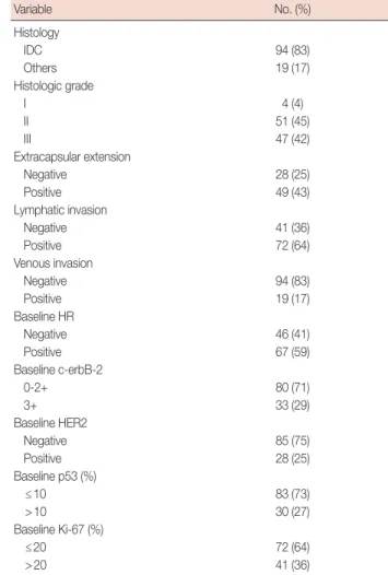
Table 2. Tumor characteristics
Variable No. (%)
Histology
IDC 94 (83)
Others 19 (17)
Histologic grade
I 4 (4)
II 51 (45)
III 47 (42)
Extracapsular extension
Negative 28 (25)
Positive 49 (43)
Lymphatic invasion
Negative 41 (36)
Positive 72 (64)
Venous invasion
Negative 94 (83)
Positive 19 (17)
Baseline HR
Negative 46 (41)
Positive 67 (59)
Baseline c-erbB-2
0-2+ 80 (71)
3+ 33 (29)
Baseline HER2
Negative 85 (75)
Positive 28 (25)
Baseline p53 (%)
≤10 83 (73)
>10 30 (27)
Baseline Ki-67 (%)
≤20 72 (64)
>20 41 (36)
IDC=infiltrating ductal carcinoma; HR=hormone receptor.
Figure 1. A flow sheet on treatment of breast cancer: preoperative sys- temic therapy (PST) was considered in patients with advanced clinical T stage or axillary lymph node involvement.
*Chemotherapy was administered before and after mastectomy; or ad- ditional chemotherapy was given in patients with adverse pathologic features.
PST subgroup (n=61)
Maste * ctomy
Mastectomy
Chemo therapy
Hormonal therapy Chemo
therapy
Chemotherapy Non-PST subgroup
(n=52)
ed according to the seventh edition of the AJCC cancer stag- ing system [4]. Patient and tumor characteristics are listed in Tables 1 and 2.
Surgery
All the patients underwent mastectomy. ALN dissection (level I and II) was performed in 110 cases (97%), with senti- nel lymph node biopsy alone performed in the remaining 3 (3%). Of the patients undergoing ALN dissection, 70 under- went ALN dissection alone and 40 underwent sentinel lymph node biopsy followed by ALN dissection (Table 3).
Chemotherapy
The most common PST regimen was DA (docetaxel and doxorubicin) followed by ACT (doxorubicin, cyclophospha- mide, and paclitaxel). After completion of PST, the PST sub- group patients underwent mastectomy with ALN dissection.
ACT was the most common adjuvant chemotherapy regimen.
Adjuvant hormonal therapy was administered in patients with a positive HR status and consisted of 5 years of tamoxifen for premenopausal women and initial aromatase inhibitor ther- apy or a switch from tamoxifen to aromatase inhibitor therapy for postmenopausal women. Trastuzumab was recommended for all patients with a tumor exhibiting c-erbB-2 overexpres-
sion (3+) or HER2 gene amplification (Table 3).
Radiotherapy
For RT, the chest wall and supraclavicular fossa were irradi- ated with up to 50.4 Gy at 1.8 Gy per fraction with 5 fractions per week; for a scar boost, 9 Gy at 1.8 Gy per fraction with electrons was administered. Two opposing tangential and one anterior photon beam were used for chest wall and supracla- vicular fossa RT, respectively (Table 3). PMRT was started after the completion of adjuvant chemotherapy. When capecitabine was used as the adjuvant chemotherapeutic agent, the patient received PMRT concurrently (n=4).
Biomarkers
We reviewed the following histopathologic parameters: es- trogen receptor (ER) status; progesterone receptor (PR) status;
and the expression of c-erbB-2, p53, Ki-67, and COX-2. Base- line histopathologic parameters were evaluated by immuno- histochemical (IHC) analysis using pre-PST biopsy specimens (PST subgroup) or surgical specimens (non-PST subgroup).
IHC staining was performed using a BenchMark XT auto- stainer (Ventana Medical Systems, Tucson, USA) and an i-View detection kit (Ventana Medical Systems) as previously described [17]. The positive cut-off values were IHC staining in ≥1% for ER/PR [18], in >10% for p53, and a 3+ staining score for COX-2 and c-erbB-2. The NR was defined as the number of ALNs with cancer involvement divided by the total number of excised ALNs. Lymph node (LN) status was evalu- ated by hematoxylin and eosin staining. Fluorescence in situ hybridization was performed for the analysis of HER2 gene amplification as reported previously [17].
Follow-up
The base follow-up duration was defined from the date when the first treatment was initiated. In cases of treatment failure, we analyzed the first site of relapse. Locoregional re- currence (LRR) included recurrences in the ipsilateral chest wall or ipsilateral regional LNs (axillary, supra/infraclavicular, and internal mammary). Relapses in the contralateral chest wall, axillary LNs, supra/infraclavicular LNs, internal mam- mary LNs, cervical LNs, or other organs were defined as dis- tant metastases (DM).
Statistics
Using the Kaplan-Meier method and the log-rank test, sur- vival curves and differences between subgroups were esti- mated. For multivariate analysis, the Cox proportional haz- ards method was used. To compare proportions between sub- groups, Pearson chi-square and Fisher exact test were used.
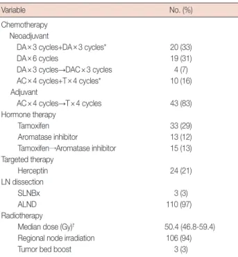
Table 3. Treatment regimens
Variable No. (%)
Chemotherapy Neoadjuvant
DA×3 cycles+DA×3 cycles* 20 (33)
DA×6 cycles 19 (31)
DA×3 cycles→DAC×3 cycles 4 (7) AC×4 cycles+T×4 cycles* 10 (16) Adjuvant
AC×4 cycles→T×4 cycles 43 (83) Hormone therapy
Tamoxifen 33 (29)
Aromatase inhibitor 13 (12)
Tamoxifen→Aromatase inhibitor 15 (13) Targeted therapy
Herceptin 24 (21)
LN dissection
SLNBx 3 (3)
ALND 110 (97)
Radiotherapy
Median dose (Gy)† 50.4 (46.8-59.4) Regional node irradiation 106 (94)
Tumor bed boost 3 (3)
DA=docetaxel and doxorubicin; AC=doxorubicin and cyclophosphamide;
T =paclitaxel; LN =lymph node; SLNBx =sentinel lymph node biopsy;
ALND=axillary lymph node dissection.
*Chemotherapy was performed before and after surgery; †Median (range).
SPSS version 18.0 (SPSS, Chicago, USA) was used for statisti- cal analyses. A p-value less than 0.05 was deemed to be statis- tically significant.
Generally, a value above 10% to 20% of the Ki-67 index was defined as a high level [12-14,16]. We compared survival curves using 3 hypothetical cut-off values, 10%, 15%, and 20%
of the baseline Ki-67 index, and found that the latter gave the most significant differences.
The NR cut-off value used in previous studies varied from 0.15 to 0.25 [5,7-10]. We used 6 candidates for the cut-off val- ue of the NR, ranging from 0.05 to 0.3 with intervals of 0.05.
The maximal chi-square method in the R program version 2.13.0 (R Development Core Team, Vienna, Austria; available from http://www.R-project.org) was used to obtain the opti- mal cut-off value of the NR, which was 0.2.
RESULTS
A total of 61 patients with an advanced clinical T stage tu- mor (T3 and T4) or ALN involvement received PST. In the PST subgroup, 7 patients received additional chemotherapy because of adverse pathologic features such as advanced stage
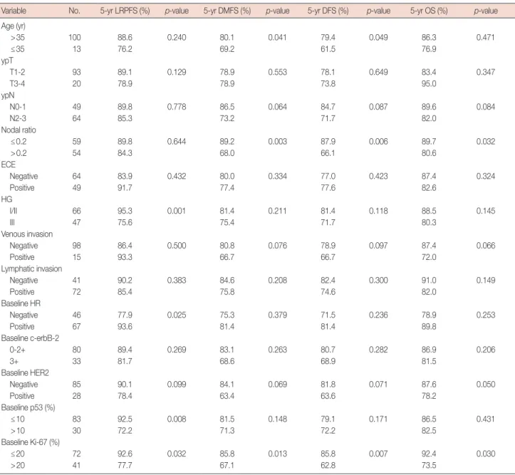
Table 4. Univariate analysis for entire patients
Variable No. 5-yr LRPFS (%) p-value 5-yr DMFS (%) p-value 5-yr DFS (%) p-value 5-yr OS (%) p-value Age (yr)
>35 100 88.6 0.240 80.1 0.041 79.4 0.049 86.3 0.471
≤35 13 76.2 69.2 61.5 76.9
ypT
T1-2 93 89.1 0.129 78.9 0.553 78.1 0.649 83.4 0.347
T3-4 20 78.9 78.9 73.8 95.0
ypN
N0-1 49 89.8 0.778 86.5 0.064 84.7 0.087 89.6 0.084
N2-3 64 85.3 73.2 71.7 82.0
Nodal ratio
≤0.2 59 89.8 0.644 89.2 0.003 87.9 0.006 89.7 0.032
>0.2 54 84.3 68.0 66.1 80.6
ECE
Negative 64 83.9 0.432 80.0 0.334 77.0 0.423 87.4 0.324
Positive 49 91.7 77.4 77.6 82.6
HG
I/II 66 95.3 0.001 81.4 0.211 81.4 0.118 88.5 0.145
III 47 75.6 75.4 71.7 80.3
Venous invasion
Negative 98 86.4 0.500 80.8 0.076 78.9 0.097 87.4 0.066
Positive 15 93.3 66.7 66.7 72.0
Lymphatic invasion
Negative 41 90.2 0.383 84.6 0.208 82.4 0.300 91.0 0.149
Positive 72 85.4 75.8 74.6 82.0
Baseline HR
Negative 46 77.9 0.025 75.3 0.379 71.5 0.236 78.9 0.253
Positive 67 93.6 81.4 81.4 89.8
Baseline c-erbB-2
0-2+ 80 89.4 0.269 83.1 0.263 80.7 0.282 86.9 0.206
3+ 33 81.7 68.6 68.9 81.5
Baseline HER2
Negative 85 90.1 0.099 84.1 0.069 81.8 0.071 87.6 0.050
Positive 28 78.4 63.4 63.6 78.2
Baseline p53 (%)
≤10 83 92.5 0.008 81.5 0.148 79.1 0.171 86.5 0.431
>10 30 72.2 71.3 72.2 82.5
Baseline Ki-67 (%)
≤20 72 92.6 0.032 85.8 0.013 85.8 0.007 92.4 0.030
>20 41 77.7 67.1 62.8 73.5
LRPFS=locoregional progression-free survival; DMFS=distant metastasis-free survival; DFS=disease-free survival; OS=overall survival; ECE=extracapsular ex- tension; HG=histologic grade; HR=hormone receptor.
or negative HR status. The other 52 patients received adjuvant systemic therapy. Chest wall and supraclavicular fossa irradia- tion was administered in 106 patients, and chest wall irradia- tion only in 7 patients. A total of 3 patients received a scar boost. The median number of excised ALNs was 22 (range,
1-55) in the whole cohort and 23 (range, 1-55) and 21 (range, 5-50) in the non-PST and PST subgroups, respectively. The median NR was 0.19 (range, 0-1) in the whole cohort, includ- ing patients with pathologically noninvolved ALNs (pN0), and 0.26 (range, 0.03-1.0) in patients with pathologically in-
p=0.644
Nodal ratio ≤0.2 Nodal ratio >0.2
Probability (%)
100
80
60
40
20
00 2 4 6 8 10
Year A
p=0.032
Ki-67 >20%
Ki-67 ≤20%
Probability (%)
100
80
60
40
20
00 2 4 6 8 10
Year B
p=0.007
Ki-67 >20%
Ki-67 ≤20%
Probability (%)
100
80
60
40
20
00 2 4 6 8 10
Year D
Nodal ratio ≤0.2
Nodal ratio >0.2
0 2 4 6 8 10
Probability (%)
100
80
60
40
20
0
p=0.006
Year C
Nodal ratio ≤0.2
Nodal ratio >0.2
0 2 4 6 8 10
Probability (%)
100
80
60
40
20
0
p=0.032
Year E
Figure 2. Survival curves in the patients with breast cancer having postmastectomy radiotherapy: locoregional progression-free survival according to the nodal ratio (A) and the baseline Ki-67 (B); disease-free survival according to the nodal ratio (C) and the baseline Ki-67 (D); overall survival according to the nodal ratio (E) and the baseline Ki-67 (F).
p=0.030
Ki-67 >20%
Ki-67 ≤20%
Probability (%)
100
80
60
40
20
00 2 4 6 8 10
Year F
With respect to the type of initial disease relapse, LRR oc- curred in 4 patients (PST subgroup, 4), DM in 14 patients (non-PST subgroup, 7; PST subgroup, 7), and both LRR and DM in 10 patients (non-PST subgroup, 3; PST subgroup, 7).
One of the patients with initial LRR underwent resection and the other 3 underwent systemic therapy. Of those patients with initial LRR and DM, 1 patient underwent resection and systemic therapy, 1 patient underwent chemotherapy and whole brain irradiation, and 6 patients were treated using sys- temic therapy only.
volved ALNs (pN+). We used the NR of 0.2 as a cut-off value to classify patients into high and low NR groups.
Follow-up and failure analysis
The median follow-up duration was 72.3 months (range, 34.0-109.4 months) for surviving patients. In the entire co- hort, the 5-year survival rates were 87.2%, 78.9%, 77.3%, and 85.3% for locoregional progression-free survival (LRPFS), dis- tant metastasis-free survival (DMFS), disease-free survival (DFS), and overall survival (OS), respectively.
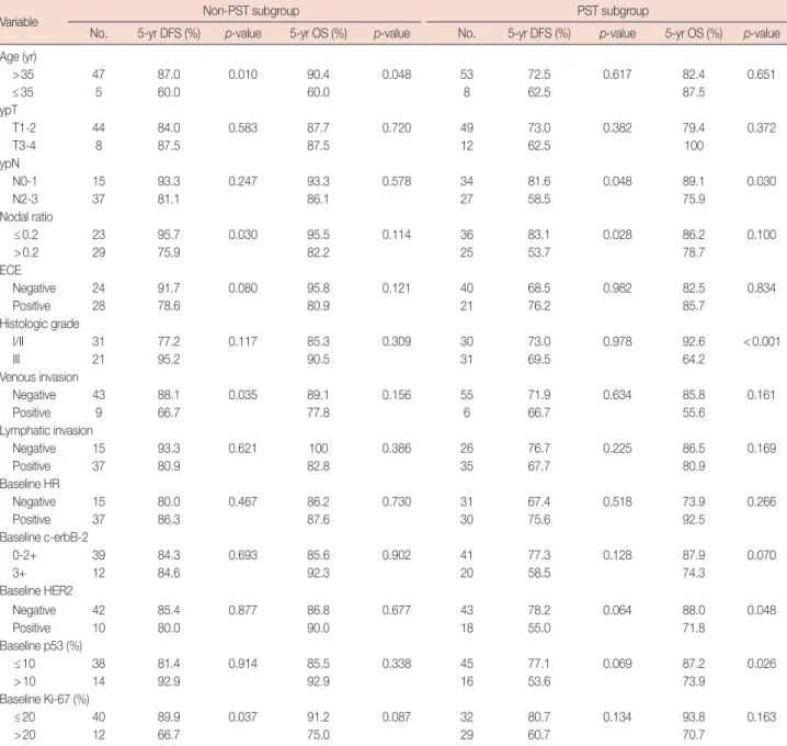
Table 5. Univariate analysis according to sequence of systemic therapy
Variable Non-PST subgroup PST subgroup
No. 5-yr DFS (%) p-value 5-yr OS (%) p-value No. 5-yr DFS (%) p-value 5-yr OS (%) p-value Age (yr)
>35 47 87.0 0.010 90.4 0.048 53 72.5 0.617 82.4 0.651
≤35 5 60.0 60.0 8 62.5 87.5
ypT
T1-2 44 84.0 0.583 87.7 0.720 49 73.0 0.382 79.4 0.372
T3-4 8 87.5 87.5 12 62.5 100
ypN
N0-1 15 93.3 0.247 93.3 0.578 34 81.6 0.048 89.1 0.030
N2-3 37 81.1 86.1 27 58.5 75.9
Nodal ratio
≤0.2 23 95.7 0.030 95.5 0.114 36 83.1 0.028 86.2 0.100
>0.2 29 75.9 82.2 25 53.7 78.7
ECE
Negative 24 91.7 0.080 95.8 0.121 40 68.5 0.982 82.5 0.834
Positive 28 78.6 80.9 21 76.2 85.7
Histologic grade
I/II 31 77.2 0.117 85.3 0.309 30 73.0 0.978 92.6 <0.001
III 21 95.2 90.5 31 69.5 64.2
Venous invasion
Negative 43 88.1 0.035 89.1 0.156 55 71.9 0.634 85.8 0.161
Positive 9 66.7 77.8 6 66.7 55.6
Lymphatic invasion
Negative 15 93.3 0.621 100 0.386 26 76.7 0.225 86.5 0.169
Positive 37 80.9 82.8 35 67.7 80.9
Baseline HR
Negative 15 80.0 0.467 86.2 0.730 31 67.4 0.518 73.9 0.266
Positive 37 86.3 87.6 30 75.6 92.5
Baseline c-erbB-2
0-2+ 39 84.3 0.693 85.6 0.902 41 77.3 0.128 87.9 0.070
3+ 12 84.6 92.3 20 58.5 74.3
Baseline HER2
Negative 42 85.4 0.877 86.8 0.677 43 78.2 0.064 88.0 0.048
Positive 10 80.0 90.0 18 55.0 71.8
Baseline p53 (%)
≤10 38 81.4 0.914 85.5 0.338 45 77.1 0.069 87.2 0.026
>10 14 92.9 92.9 16 53.6 73.9
Baseline Ki-67 (%)
≤20 40 89.9 0.037 91.2 0.087 32 80.7 0.134 93.8 0.163
>20 12 66.7 75.0 29 60.7 70.7
PST=preoperative systemic therapy; DFS=disease-free survival; OS=overall survival; ECE=extracapsular extension; HR=hormone receptor.
Univariate analysis
Univariate analysis revealed that patients with a NR of >0.2 had a significantly lower DMFS (p=0.003), DFS (p=0.006), and OS (p=0.032) than those with a NR of ≤0.2. Patients with a baseline Ki-67 index of >20% had a significantly lower LRPFS (p=0.032), DMFS (p=0.013), DFS (p=0.007), and OS (p=0.030) than those with a baseline Ki-67 index of
Table 6. Multivariate analysis
Variable LRPFS DMFS DFS OS
p-value RR (95% CI) p-value RR (95% CI) p-value RR (95% CI) p-value RR (95% CI)
Young age (≤35 yr) - - - -
Histologic grade (III) 0.004 6.308 (1.778-22.373) - - -
High nodal ratio (>0.2) - 0.002 4.063 (1.701-9.701) 0.003 3.589 (1.567- 8.220) 0.019 3.444 (1.227- 9.669) Baseline Ki-67 (>20%) - 0.004 3.125 (1.450-6.731) 0.002 3.274 (1.536- 6.979) 0.015 3.133 (1.249- 7.856)
Baseline HR (+) - - - -
Baseline p53 (>10%) - - - -
LRPFS=locoregional progression-free survival; DMFS=distant metastasis-free survival; DFS=disease-free survival; OS=overall survival; RR=relative risk;
CI=confidence interval; HR=hormone receptor.
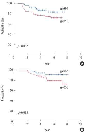
≤20%. The baseline HR level was associated with LRPFS (p=0.025) but not with DMFS (p=0.379), DFS (p=0.236), and OS (p=0.253). The pathologic nodal stage was marginal- ly associated with DMFS (p=0.064), DFS (p=0.087), and OS (p=0.084). These results are detailed in Table 4, Figures 2 and 3.
We also performed subgroup analysis for the non-PST and PST subgroups. In the former, age (p=0.010), NR (p=0.030), venous invasion (p=0.035), and the baseline Ki-67 index (p=
0.037) were associated with DFS, although only age (p=0.048) was associated with OS. In the PST subgroup, ypN stage (p=
0.048) and NR (p=0.028) were associated with DFS and ypN (p=0.030), histologic grade (p<0.001), baseline HER2 ex- pression (p=0.048), and baseline p53 expression (p=0.026) were associated with OS (Table 5).
Multivariate analysis
We performed multivariate analysis incorporating the NR, baseline Ki-67 index, age, histologic grade, and baseline p53 expression, all of which were found to be significantly associ- ated with DFS or OS in univariate analysis of the entire cohort.
A high NR was associated with poor DMFS (relative risk [RR], 4.063; 95% confidence interval [CI], 1.701-9.701; p=0.002), DFS (RR, 3.589; 95% CI, 1.567-8.220; p=0.003), and OS (RR, 3.444; 95% CI, 1.227-9.669; p=0.019). A high baseline Ki-67 index was associated with poor DMFS (RR, 3.125; 95% CI, 1.450-6.731; p=0.004), DFS (RR, 3.274; 95% CI, 1.536-6.979;
p=0.002), and OS (RR, 3.133; 95% CI, 1.249-7.856; p=0.015).
Results of the multivariate analysis are detailed in Table 6.
Prognostic model
We devised a prognostic model using the NR and baseline Ki-67 index, with a score of zero points for a NR of ≤0.2 or a baseline Ki-67 index of ≤20% and 1 point for a NR of >0.2 or a baseline Ki-67 index of >20%. Patients were classified into 3 subgroups according to their total score: low risk (0 point, n=34), intermediate risk (1 point, n=63), and high risk (2 points, n=16). No deaths occurred in the low-risk group, whereas 13 patients in the intermediate-risk group and Figure 3. Survival curves in the patients with breast cancer having post-
mastectomy radiotherapy according to pathologic nodal stage: (A) dis- ease-free survival and (B) overall survival.
Probability (%)
0 2 4 6 8 10
0 2 4 6 8 10 Year
100
80
60
40
20
0
Probability (%)
Year 100
80
60
40
20
0
p=0.084
ypNO-1 ypNO-1
ypN2-3 ypN2-3
p=0.087
A
B
6 patients in the high-risk group had died at the time of the last follow-up. When comparing the high- and low-risk pa- tients, a significant difference was found in LRPFS (p=0.040), DMFS (p<0.001), DFS (p<0.001), and OS (p<0.001). A sig- nificant difference was also observed between the intermedi-
ate- and low-risk groups with respect to DMFS (p=0.031), DFS (p=0.022), and OS (p=0.008), but not LRPFS (p=0.204) (Figure 4).
We used the Cox proportional hazards method in order to evaluate the RR among the different risk groups. For LRPFS, the high- and intermediate-risk groups demonstrated RRs of 4.898 (95% CI, 0.897-26.753; p=0.067) and 2.599 (95% CI, 0.562-12.032; p=0.222), respectively, and for DMFS, the RRs of the high- and intermediate-risk groups were 14.110 (95%
CI, 3.089-64.448; p=0.001) and 4.400 (95% CI, 1.006-19.241;
p=0.049), respectively. With respect to DFS, the high- and in- termediate-risk patients showed RRs of 14.264 (95% CI, 3.122-65.165; p=0.001) and 4.785 (95% CI, 1.100-20.814; p=
0.037), respectively.
We applied this prognostic model to the non-PST and PST subgroups. There was a significant difference in DMFS (p=
0.016 and p<0.001) and DFS (p=0.016 and p<0.001) in the non-PST and PST subgroups, respectively. There was no sig- nificant difference in the LRPFS in the non-PST (p=0.364) and PST (p=0.224) subgroups, although there was a signifi- cant difference with respect to OS in the PST subgroup (p=
0.045) but not in the non-PST subgroup (p=0.074).
DISCUSSION
Several large, randomized studies have shown that PMRT improves LRC and survival in breast cancer patients, particu- larly those with more than 3 involved ALNs [1-3]. To date though, the role of PMRT in breast cancer patients with fewer than 4 metastatic ALNs has not been evaluated. Overgaard et al. [19] conducted a reanalysis of Danish trials and found that PMRT benefited patients with 1 to 3 positive ALNs. Recently, the number of excised ALNs was shown to be as important as the number of involved ALNs, suggesting that the NR is an important prognostic factor [5-10]. In a study by the Surveil- lance, Epidemiology and End Results program, the NR was found to be better at predicting disease-specific survival than the number of involved ALNs [7]. Truong et al. [8] reported that an NR of 0.25 was associated with a poor prognosis with respect to LRR, DM, and OS in patients with 1 to 3 involved ALNs. Ahn et al. [10] analyzed a nationwide registry of pN+
patients and concluded that the NR was a better prognostic factor than pN stage, particularly in patients with high-risk factors such as young age, a HER2/neu-enriched tumor, or a triple-negative tumor.
In the study we report here, the pN stage showed only a borderline association with recurrence or survival in univari- ate analysis. A possible reason for this finding may be the het- erogeneity in the sequence of systemic therapy. After PST,
p=0.204 p=0.040
Year
Low risk Intermdiate risk
Low vs. Intermediate Low vs. High
High risk
Probability (%)
100
80
60
40
20
00 2 4 6 8 10
A
Year B
p=0.022 p<0.001 Low vs. Intermediate
Low vs. High
Low risk Intermdiate risk
High risk
0 2 4 6 8 10
Probability (%)
100
80
60
40
20
0
Figure 4. Survival curves in the patients with breast cancer having post- mastectomy radiotherapy according to the risk group: (A) locoregional progression-free survival, (B) disease-free survival, and (C) overall sur- vival.
C p=0.008 p<0.001 Low vs. Intermediate
Low vs. High
Low risk Intermdiate risk
High risk
0 2 4 6 8 10
Probability (%)
Year 100
80
60
40
20
0
more patients could have a lower N stage as a result of chemo- therapy. This finding might also be explained by the relatively small size and short follow-up period of our study. As shown in Figure 2, the DFS and OS curves differed between patients with pN0-1 and pN2-3, and it is possible that with a greater number of patients and a longer follow-up period, a statisti- cally significant relationship might be found between survival and pN stage.
Although this pilot study included patients with different pN stages, the NR (cut-off value of 0.2) was associated with a high risk of metastasis and short survival in LABC patients.
Because the NR reflects the absolute number of excised ALNs, it might have a higher prognostic value than pN stage [20]. In the current cancer staging system [4], the usefulness of the absolute number of involved nodes for predicting disease bur- den in the axilla is confounded by the number of nodes re- moved [21]. When additional ALNs are excised, less residual occult disease may be expected. In Canada, ALN dissection, including all level I and II ALNs, is recommended for accurate staging and reducing the risk of recurrence in the axilla [22].
Although several studies have reported a possible prognos- tic role for the NR in LRC [8,23,24], we could not establish a relationship between the NR and LRC in this study. This may have been because of the relatively short follow-up duration (approximately 6 years). Improved LRC as a result of regional RT [25,26] might also account for the lack of any significant difference in LRPFS between the high NR and low NR patient groups. We suggest therefore that a randomized controlled study focusing specifically on the prognostic role of NR be conducted.
In addition to the NR, biomolecular markers might also have prognostic value for LABC patients. It is generally ac- cepted that biomolecular markers of cell proliferation, such as the baseline Ki-67 index used in our study, are associated with the response to systemic therapy [27]. Our study showed that a high baseline Ki-67 index was associated with a high risk of mortality. Furthermore, a relationship between a high Ki-67 index and other indicators of a poor prognosis has been pre- viously reported [28]. This negative relationship would ex- plain the prognostic value of Ki-67 index. In the present study, a Ki-67 index in excess of 20% was associated with baseline negative HR expression (p<0.001).
Consistent with our findings, the Ki-67 index has been shown to be a possible prognostic marker in several other studies. Cheang et al. [12] classified invasive breast cancer into luminal A, luminal B, and HER2-positive intrinsic subtypes on the basis of hormone receptor status, HER2 status, and the Ki-67 index, as determined using IHC. The Ki-67 index was used to distinguish luminal B from luminal A, using a cut-off
value of 14%. The luminal B and luminal HER2 subtypes were found to have a poor prognosis with respect to breast cancer recurrence-free and disease-specific survival. The 10-year breast cancer-specific survival rates were 92%, 79%, and 78%
in luminal A, luminal B, and HER2 positive cancer, respec- tively (p<0.001). In a meta-analysis study of early breast can- cer, Ki-67 positivity (cut-off points were defined by the au- thors of the studies being included) was associated with in- creased relapse (RR, 1.93; 95% CI, 1.74-2.14; p<0.001) and shorter survival (RR, 1.95; 95% CI, 1.70-2.24; p<0.001) in all patients. The authors of that study suggested that Ki-67 posi- tivity was a prognostic marker in patients with early breast cancer [14].
Despite these studies, in general, the association between specific biomolecular markers and LRC remains unclear. Two previous studies found that the Ki-67 index was a possible prognostic factor of LRC [13,16]. Voduc et al. [13] defined the luminal B subtype as being ER(+) or PR(+) and HER2(-) and having a Ki-67 index of ≥14% in patients who had undergone mastectomy. The luminal B subgroup was associated with a high risk of local and regional recurrences. Selz et al. [16] re- ported that a Ki-67 index of >20% was prognostic for LRFPS (RR, 4.18; 95% CI, 1.11-15.77; p=0.0215) in breast cancer pa- tients with pN0 after modified radical mastectomy.
In addition to HER2 status, the Ki-67 index, representing tumor aggressiveness, may also be a means of identifying high-risk groups among breast cancer patients. However, con- troversy still exists regarding the optimal cut-off point for Ki- 67; a level of Ki-67 above 10% to 20% has been suggested to define a high-risk group in several studies [12-14,16]. In our study, the baseline Ki-67 index was used to determine the risk groups, using a cut-off point of 20%. The 2011 St. Gallen Con- sensus [11] recommended a Ki-67 labeling index of 14% as the cut-off point to classify the intrinsic subtype of breast can- cer; however, these guidelines have not been clarified. It there- fore remains necessary to develop a standardized approach to using the Ki-67 index, including a single cut-off value and a reproducible way of determining the index.
Here, we propose a prognostic model using 2 parameters, the NR and baseline Ki-67 index, both of which are signifi- cantly associated with disease relapse, reflecting the probabili- ty of residual tumor on a macroscopic scale and the possibility of disease relapse on a microscopic scale, respectively. Our prognostic model is simple to apply and can identify the poor prognostic group amongst a heterogeneous population with disparate pN stages or sequences of systemic therapy. Using our prognostic model, patients with a high risk of disease re- lapse can be identified, and intensified adjuvant treatment can be considered to improve their survival. With respect to LRC,
however, the high-risk group tended to have a worse progno- sis than the low-risk group (p=0.067), and the intermediate- risk group showed no association. We expect that this prog- nostic model would be more useful to identify the high-risk group among LABC patients with an increased long-term fol- low-up period.
Our study has several limitations. The patients needed to be analyzed independently according to the use of PST because the NR has a prognostic value in patients with PST [6]. How- ever, subgroup multivariate analysis was not performed be- cause of an insufficient number of patients. In addition, the relatively short follow-up duration was a hindrance to com- paring OS. These limitations may have made it more difficult to identify a relationship between treatment outcomes and well-known prognostic factors, such as T/N stage and HR sta- tus. Furthermore, the study included patients with a range of different N stages and 21% of patients had pN0 tumors (21%), whereas the other studies on the prognostic value of NR dis- cussed here only involved node-positive patients. Therefore, a further study is needed with a more homogenous patient group with respect to the sequence of systemic therapy and pN stage. Additionally, for a more precise prognostic model, the change in biomarker status before and after PST [15,29]
should be considered.
In conclusion, we found that the NR and baseline Ki-67 in- dex were potential prognostic markers in LABC patients who underwent PMRT. Our prognostic model, using these 2 fac- tors, might be able to identify patients at high risk of disease relapse. Improved prognostic models will help to individual- ize treatment regimens for breast cancer patients.
CONFLICT OF INTEREST
The authors declare that they have no competing interests.
REFERENCES
1. Overgaard M, Hansen PS, Overgaard J, Rose C, Andersson M, Bach F, et al. Postoperative radiotherapy in high-risk premenopausal women with breast cancer who receive adjuvant chemotherapy. Danish Breast Cancer Cooperative Group 82b Trial. N Engl J Med 1997;337:949-55.
2. Overgaard M, Jensen MB, Overgaard J, Hansen PS, Rose C, Andersson M, et al. Postoperative radiotherapy in high-risk postmenopausal breast-cancer patients given adjuvant tamoxifen: Danish Breast Cancer Cooperative Group DBCG 82c randomised trial. Lancet 1999;353:
1641-8.
3. Ragaz J, Olivotto IA, Spinelli JJ, Phillips N, Jackson SM, Wilson KS, et al.
Locoregional radiation therapy in patients with high-risk breast cancer receiving adjuvant chemotherapy: 20-year results of the British Colum- bia randomized trial. J Natl Cancer Inst 2005;97:116-26.
4. Edge SB, Byrd DR, Compton CC, Fritz AG, Greene FL, Trotti A; Amer-
ican Joint Committee on Cancer. AJCC Cancer Staging Manual. 7th ed.
New York: Springer; 2010.
5. Han TJ, Kang EY, Jeon W, Kim SW, Kim JH, Kim YJ, et al. The prognos- tic value of the nodal ratio in N1 breast cancer. Radiat Oncol 2011;
6:131.
6. Keam B, Im SA, Kim HJ, Oh DY, Kim JH, Lee SH, et al. Clinical signifi- cance of axillary nodal ratio in stage II/III breast cancer treated with neoadjuvant chemotherapy. Breast Cancer Res Treat 2009;116:153-60.
7. Vinh-Hung V, Verkooijen HM, Fioretta G, Neyroud-Caspar I, Rapiti E, Vlastos G, et al. Lymph node ratio as an alternative to pN staging in node-positive breast cancer. J Clin Oncol 2009;27:1062-8.
8. Truong PT, Berthelet E, Lee J, Kader HA, Olivotto IA. The prognostic significance of the percentage of positive/dissected axillary lymph nodes in breast cancer recurrence and survival in patients with one to three positive axillary lymph nodes. Cancer 2005;103:2006-14.
9. Truong PT, Woodward WA, Thames HD, Ragaz J, Olivotto IA, Buch- holz TA. The ratio of positive to excised nodes identifies high-risk sub- sets and reduces inter-institutional differences in locoregional recur- rence risk estimates in breast cancer patients with 1-3 positive nodes: an analysis of prospective data from British Columbia and the M. D. An- derson Cancer Center. Int J Radiat Oncol Biol Phys 2007;68:59-65.
10. Ahn SH, Kim HJ, Lee JW, Gong GY, Noh DY, Yang JH, et al. Lymph node ratio and pN staging in patients with node-positive breast cancer:
a report from the Korean Breast Cancer Society. Breast Cancer Res Treat 2011;130:507-15.
11. Goldhirsch A, Wood WC, Coates AS, Gelber RD, Thürlimann B, Senn HJ; Panel members. Strategies for subtypes--dealing with the diversity of breast cancer: highlights of the St. Gallen International Expert Con- sensus on the Primary Therapy of Early Breast Cancer 2011. Ann On- col 2011;22:1736-47.
12. Cheang MC, Chia SK, Voduc D, Gao D, Leung S, Snider J, et al. Ki67 index, HER2 status, and prognosis of patients with luminal B breast cancer. J Natl Cancer Inst 2009;101:736-50.
13. Voduc KD, Cheang MC, Tyldesley S, Gelmon K, Nielsen TO, Kennecke H. Breast cancer subtypes and the risk of local and regional relapse. J Clin Oncol 2010;28:1684-91.
14. de Azambuja E, Cardoso F, de Castro G Jr, Colozza M, Mano MS, Dur- becq V, et al. Ki-67 as prognostic marker in early breast cancer: a meta- analysis of published studies involving 12,155 patients. Br J Cancer 2007;96:1504-13.
15. Jones RL, Salter J, A’Hern R, Nerurkar A, Parton M, Reis-Filho JS, et al.
The prognostic significance of Ki67 before and after neoadjuvant che- motherapy in breast cancer. Breast Cancer Res Treat 2009;116:53-68.
16. Selz J, Stevens D, Jouanneau L, Labib A, Le Scodan R. Prognostic value of molecular subtypes, Ki67 expression and impact of postmastectomy radiation therapy in breast cancer patients with negative lymph nodes after mastectomy. Int J Radiat Oncol Biol Phys 2012;84:1123-32.
17. Jang MH, Kim EJ, Choi Y, Lee HE, Kim YJ, Kim JH, et al. FGFR1 is am- plified during the progression of in situ to invasive breast carcinoma.
Breast Cancer Res 2012;14:R115.
18. Hammond ME, Hayes DF, Wolff AC. Clinical Notice for American So- ciety of Clinical Oncology-College of American Pathologists guideline recommendations on ER/PgR and HER2 testing in breast cancer. J Clin Oncol 2011;29:e458.
19. Overgaard M, Nielsen HM, Overgaard J. Is the benefit of postmastecto-
my irradiation limited to patients with four or more positive nodes, as recommended in international consensus reports? A subgroup analysis of the DBCG 82 b&c randomized trials. Radiother Oncol 2007;82:247- 53.
20. Schmoor C, Sauerbrei W, Bastert G, Bojar H, Schumacher M; German Breast Cancer Study Group. Long-term prognosis of breast cancer pa- tients with 10 or more positive lymph nodes treated with CMF. Eur J Cancer 2001;37:1123-31.
21. Woodward WA, Vinh-Hung V, Ueno NT, Cheng YC, Royce M, Tai P, et al. Prognostic value of nodal ratios in node-positive breast cancer. J Clin Oncol 2006;24:2910-6.
22. Canadian Association of Radiation Oncologists; The Steering Commit- tee on Clinical Practice Guidelines for the Care and Treatment of Breast Cancer. Axillary dissection. CMAJ 1998;158(Suppl 3):S22-6.
23. Katz A, Buchholz TA, Thames H, Smith CD, McNeese MD, Theriault R, et al. Recursive partitioning analysis of locoregional recurrence patterns following mastectomy: implications for adjuvant irradiation. Int J Radi- at Oncol Biol Phys 2001;50:397-403.
24. Grills IS, Kestin LL, Goldstein N, Mitchell C, Martinez A, Ingold J, et al.
Risk factors for regional nodal failure after breast-conserving therapy:
regional nodal irradiation reduces rate of axillary failure in patients with four or more positive lymph nodes. Int J Radiat Oncol Biol Phys 2003;
56:658-70.
25. Wai ES, Lesperance M, Speers CH, Truong PT, Jones S, Tyldesley S, et al.
Increased use of regional radiotherapy is associated with improved out- come in a population-based cohort of women with breast cancer with 1-3 positive nodes. Radiother Oncol 2010;97:301-6.
26. Truong PT, Jones SO, Kader HA, Wai ES, Speers CH, Alexander AS, et al. Patients with t1 to t2 breast cancer with one to three positive nodes have higher local and regional recurrence risks compared with node- negative patients after breast-conserving surgery and whole-breast ra- diotherapy. Int J Radiat Oncol Biol Phys 2009;73:357-64.
27. Paik S, Shak S, Tang G, Kim C, Baker J, Cronin M, et al. A multigene as- say to predict recurrence of tamoxifen-treated, node-negative breast cancer. N Engl J Med 2004;351:2817-26.
28. Viale G, Giobbie-Hurder A, Regan MM, Coates AS, Mastropasqua MG, Dell’Orto P, et al. Prognostic and predictive value of centrally re- viewed Ki-67 labeling index in postmenopausal women with endo- crine-responsive breast cancer: results from Breast International Group Trial 1-98 comparing adjuvant tamoxifen with letrozole. J Clin Oncol 2008;26:5569-75.
29. Yerushalmi R, Woods R, Ravdin PM, Hayes MM, Gelmon KA. Ki67 in breast cancer: prognostic and predictive potential. Lancet Oncol 2010;
11:174-83.