ABSTRACT
Objective: The aim of this study was to confirm the incidence and implications of a lymphatic spread pattern involving para-aortic lymph node (PAN) metastasis in the absence of pelvic lymph node (PLN) metastasis in patients with endometrial cancer.
Methods: We carried out a retrospective chart review of 380 patients with endometrial cancer treated by surgery including PLN dissection and PAN dissection at Hokkaido Cancer Center between 2003 and 2016. We determined the probability of PAN metastasis in patients without PLN metastasis and investigated survival outcomes of PLN−PAN+ patients.
Results: The median numbers of PLN and PAN removed at surgery were 41 (range: 11–107) and 16 (range: 1–65), respectively. Sixty-four patients (16.8%) had lymph node metastasis, including 39 (10.3%) with PAN metastasis. The most frequent lymphatic spread pattern was PLN+PAN+ (7.9%), followed by PLN+PAN− (6.6%), and PLN−PAN+ (2.4%). The probability of PAN metastasis in patients without PLN metastasis was 2.8% (9/325). The 5-year overall survival rates were 96.5% in PLN−PAN−, 77.6% in PLN+PAN−, 63.4% in PLN+PAN+, and 53.6% in PLN−PAN+ patients.
Conclusion: The likelihood of PAN metastasis in endometrial cancer patients without PLN metastasis is not negligible, and the prognosis of PLN−PAN+ is likely to be poor.
The implications of a PLN−PAN+ lymphatic spread pattern should thus be taken into consideration when determining patient management strategies.
Keywords: Endometrial Neoplasms; Lymphatic Metastasis; Lymph nodes; Prognosis
INTRODUCTION
Endometrial cancer is the most common malignancy of the female genital tract in the United States, with an estimated number of 61,380 new cases in 2017 [1]. The annual death rate of 6,000 in 1997 [2] had almost doubled to 10,920 by 2017 [1]. Surgery is the principal treatment for endometrial cancer, including hysterectomy, bilateral salpingo-oophorectomy, and establishment of lymph node status. However, there is currently no consensus on the therapeutic value of lymphadenectomy, and this issue remains a topic for debate.
Original Article
Received: Mar 31, 2017 Revised: Apr 21, 2017 Accepted: May 4, 2017 Correspondence to Yukiharu Todo
Division of Gynecologic Oncology, National Hospital Organization, Hokkaido Cancer Center, 4-2 Kikusui, Shiroishi-ku, Sapporo 003-0804, Japan.
E-mail: [email protected] Copyright © 2017. Asian Society of Gynecologic Oncology, Korean Society of Gynecologic Oncology
This is an Open Access article distributed under the terms of the Creative Commons Attribution Non-Commercial License (https://
creativecommons.org/licenses/by-nc/4.0/) which permits unrestricted non-commercial use, distribution, and reproduction in any medium, provided the original work is properly cited.
ORCID iDs Yukiharu Todo
https://orcid.org/0000-0001-8692-4789 Sho Takeshita
https://orcid.org/0000-0002-5484-7309 Kazuhira Okamoto
https://orcid.org/0000-0003-3678-6669 Katsushige Yamashiro
https://orcid.org/0000-0001-6020-9496 Hidenori Kato
https://orcid.org/0000-0002-2153-5843 Conflict of Interest
No potential conflict of interest relevant to this article was reported.
Yukiharu Todo ,1 Sho Takeshita ,1 Kazuhira Okamoto ,1 Katsushige Yamashiro ,2 Hidenori Kato 1
1Division of Gynecologic Oncology, National Hospital Organization, Hokkaido Cancer Center, Sapporo, Japan
2Division of Pathology, National Hospital Organization, Hokkaido Cancer Center, Sapporo, Japan
Implications of para-aortic lymph
node metastasis in patients with
endometrial cancer without pelvic
lymph node metastasis
Sentinel lymph node (SLN) mapping is expected to offer a trade-off between systematic lymphadenectomy and no dissection at all in clinical stage I patients. However, while SLN mapping in patients with endometrial cancer is increasingly credible in Western countries [3,4], Japan lags behind in this research, because of some plausible concerns. The first concern involves the tracer-injection site for SLN mapping. There are 3 possible injection sites for SLN mapping in endometrial cancer: the endometrium, the subserosa of the uterine corpus, and the uterine cervix. The former 2 are technically demanding, while the latter is technically simple and has been used favorably in Western countries. The second issues concern the possibility of false-negative results, i.e., reduced sensitivity for lymph node metastasis (LNM) because of failure to detect SLNs. Unfortunately, the ability of cervical injection to detect para-aortic SLNs is poor, and if a patient has no pelvic lymph node (PLN) metastasis, but does have para-aortic lymph node (PAN) metastasis, SLN mapping by cervical injection may result in the opportunity to establish PAN status being missed. The possibility of such an underdiagnosis of PLN−PAN+
patients is a matter of great concern. Indeed, a third of false-negative results following cervical injection were attributable to para-aortic SLN detection failure [5].
The aim of the present study was to clarify the clinical implications of this underdiagnosis.
The importance of PLN−PAN+ should be assessed in terms of both its probability and prognostic risk. Taking the possible outcome of SLN mapping by cervical injection into account, the probability of PAN metastasis in patients without PLN metastasis is a more appropriate endpoint of this study than the probability of PAN metastasis in all patients (patients with and without PLN metastasis). We investigated both the probability of PAN metastasis in patients without PLN metastasis and biological nature of PLN−PAN+ in terms of its aggressivity. Finally, we consider the implications of failing to search for PAN in patients without PLN metastasis.
MATERIALS AND METHODS
1. Patients and assessment of LNM for the present cohort study A total of 880 patients with uterine corpus malignancy were treated at the National Hospital Organization, Hokkaido Cancer Center between January 2003 and December 2016. We excluded 85 who had carcinosarcoma/sarcoma, and 41 who did not undergo surgical treatment. A total of 754 patients with endometrial cancer thus underwent surgical treatment. Of these 754 patients, 199 (26.4%) did not undergo lymph node dissection, 175 (23.2%) underwent PLN dissection (PLND) alone, and 380 (50.4%) underwent both PLNDs and PAN dissections (PANDs). Information concerning age, body mass index (BMI), the International Federation of Gynecology and Obstetrics (FIGO) stage, extent of surgery, and final pathological reports was collected by review of the relevant medical records. Lymph node sites were classified into PLN and PAN. PAN was further classified into low PAN and high PAN, indicating the region between the bifurcation of the aorta and the inferior mesenteric artery, and the region between the inferior mesenteric artery and the renal vessels, respectively. The survival outcome measure was overall survival (OS), defined as the time from the starting date of initial treatment to death. Patients known to still be alive or lost to follow-up at the time of analysis were censored at their last follow-up. Survival rates were estimated by the Kaplan-Meier method. Unpaired numerical data were compared using Student's unpaired t-tests. Variables were compared between groups using Fisher's exact test, χ2 test, or Mann-Whitney U tests. The significance level was set at 0.05. Statistical analyses were performed using StatView J-5. 0 PPC (SAS Institute, Cary, NC, USA).
Author Contributions
Conceptualization: T.Y.; Data curation: T.S., O.K.; Formal analysis: T.Y.; Investigation: Y.K.;
Methodology: T.Y.; Project administration: T.Y.;
Software: T.Y.; Supervision: T.Y.; Validation:
O.K., K.H.; Writing - original draft: T.Y.; Writing - review & editing: O.K., K.H.
2. Study selection and data extraction for a pooled analysis
Using the keywords “endometrial cancer,” “uterine cancer,” “para-aortic lymphadenectomy,”
and “para-aortic lymph node metastasis,” a PubMed search for English language publications published from 1983 to 2016 was conducted. Research published only in abstract format was not included. Publications were selected for initial review if the study showed its proportion of PAN metastasis. If 2 or more reports overlapping study periods from the same institution were identified, only 1 was selected in the analysis to avoid duplication of cases. If the PAN metastasis rate was calculated based on background data with an unknown probability of patients with PAND in the study population, that study was excluded from the present analysis. In a nutshell, published studies with information on all the following were included in the analysis: 1) number of patients negative for PLN and PAN metastases (PLN−PAN−);
2) number of patients negative for PLN metastasis but positive for PAN metastasis (PLN−
PAN+); 3) number of patients positive for PLN metastasis but negative for PAN metastasis (PLN+PAN−); and 4) number of patients positive for PLN and PAN metastases (PLN+PAN+).
These 4 values must have been obtained based exclusively on data for patients who
underwent both PLNDs and PANDs. Twenty-five eligible reports were finally identified from the pooled data [6-30].
RESULTS
The clinical and pathological characteristics of the 754 patients treated by surgery are shown in Table 1. The median age was 59 years (range: 20–93 years). Grouping according to the extent of surgery showed that 32.7% of patients aged >70 years were in the no- lymphadenectomy group, 22.9% in the PLND group, and 6.6% in the PLND+PAND group (p<0.001). The mean BMI of all patients was 24.7 kg/m2 (standard deviation: 5.65). The proportions of patients with a BMI >30 kg/m2 were 20.6% in the no-lymphadenectomy group, 17.1% in the PLND group, and 12.4% in the PLND+PAND group (p=0.029). In terms of histological variants, there were 413 (54.8%) grade 1 endometrioid adenocarcinomas (G1), 149 (19.8%) grade 2 endometrioid adenocarcinomas (G2), and 89 (11.8%) grade 3 endometrioid adenocarcinomas (G3), and 103 (13.6%) non-endometrioid carcinomas.
Table 1. Characteristics of 754 patients with endometrial cancer who underwent surgical treatment
Characteristic LND (−) (n=199) PLND alone (n=175) PLND+PAND (n=380) p-value
Age (yr) 59.0 (20–93) 60.0 (33–83) 58.5 (28–76)
≥70 65 (32.7) 40 (22.9) 25 (6.6) <0.001
≥75 40 (20.1) 19 (10.9) 1 (0.3) <0.001
BMI (kg/m2) 23.6 (15.5–51.8) 23.1 (15.5–46.6) 23.8 (13.4–44.9)
>30 41 (20.6) 30 (17.1) 47 (12.4) 0.029
>35 17 (8.5) 9 (5.1) 14 (3.7) 0.046
Final pathology <0.001
Endometrioid grade 1 129 (64.8) 120 (68.6) 164 (43.2)
Endometrioid grade 2 28 (14.1) 30 (17.1) 91 (23.9)
Endometrioid grade 3 16 (8.0) 8 (4.6) 65 (17.1)
Other 26 (13.1) 17 (9.7) 60 (15.8)
Postoperative stage <0.001
IA 132 (66.3) 121 (69.1) 188 (49.5)
IB 34 (17.1) 27 (15.4) 79 (20.8)
II 9 (4.5) 7 (4.0) 21 (5.5)
III 7 (3.5) 11 (6.3) 82 (21.6)
IV 17 (8.5) 9 (5.1) 10 (2.6)
Values are presented as median (range) or number (%).
BMI, body mass index; LND, lymph node dissection; PAND, para-aortic lymph node dissection; PLND, pelvic lymph node dissection.
The incidence rates of grade 3/non-endometrioid carcinomas were 21.1% in the no- lymphadenectomy group, 14.3% in the PLND group, and 32.9% in the PLND+PAND group (p<0.001). The overall proportions of FIGO stage were 581 (77.0%) in stage I, 37 (4.9%) in stage II, 100 (13.3%) in stage III, and 36 (4.8%) in stage IV. The incidence rates of patients with stage IA disease were 66.3% in the no-lymphadenectomy group, 69.1% in the PLND group, and 49.5% in the PLND+PAND group, while the rates of stage III/IV disease were 12.0% in the no-lymphadenectomy group, 11.4% in the PLND group, and 24.2% in the PLND+PAND group (p<0.001).
The surgical results for the 380 patients who underwent both PLNDs and PANDs are shown in Table 2. Removal of PAN up to the renal vein was performed in 375 (98.7%) patients, while the remaining 5 (1.3%) patients (1 PLN−PAN−, 1 PLN−PAN+, 3 PLN+PAN+) did not undergo removal of PAN above the inferior mesenteric artery. The median numbers of PLN and PAN removed were 41 (range: 11–107) and 16 (range: 1–65), respectively. The overall LNM rate was 16.9% (64/380) and the PAN metastasis rate was 10.3% (39/380). The most frequent lymphatic spread pattern was PLN+PAN+ (7.9%), followed by PLN+PAN− (6.6%), and PLN−
PAN+ (2.4%). The proportion of PAN metastasis in patients without PLN metastasis were 2.8% (9/325). Of the 9 patients with PLN−PAN+, 3 (33.3%) had peritoneal disease. Complete surgery, including removal of peritoneal disease, was performed in all 3 cases, and one patient achieved long-term disease-free survival (Table 3).
Table 2. Surgical results of 380 patients who underwent PLND and PAND
Characteristic Value (n=380)
Type of PAND
Low PAN alone (below the IMA) 5
Low PAN and high PAN 375
No. of lymph nodes removed
PLN 41 (11–107)
PAN 16 (1–65)
Total 56.5 (18–131)
Lymphatic spread pattern
PLN−PAN− 316 (83.1)
PLN−PAN+ 9 (2.4)
PLN+PAN− 25 (6.6)
PLN+PAN+ 30 (7.9)
Values are presented as median (range) or number (%).
IMA, inferior mesenteric artery; PAN, para-aortic lymph node; PAND, para-aortic lymph node dissection; PLN, pelvic lymph node; PLND, pelvic lymph node dissection.
Table 3. Profile of the 9 patients with PLN−PAN+
Age (yr) Peritoneal
disease Final histological
type Myometrial
invasion Cervical
involvement LVSI Adnexal
metastasisPeritoneal
washing Preoperative
histological type Myometrial
invasion (MRI) Radiological signs of
extrauterine disease Outcome OS (M)
46 + G2 <1/2 + + + + G2 >1/2 − NED 75
70 + S >1/2 + + − + S >1/2 + DOD 8
63 + G2 >1/2 − + + + G1 >1/2 + NED 7
67 − G2 >1/2 + + + + G1 >1/2 + DOD 15
70 − Mixed <1/2 − + + + S >1/2 − NED 9
40 − G2 >1/2 − + − + G1 >1/2 − DOD 43
55 − G1 >1/2 − + − − G1 >1/2 − NED 63
63 − G1 >1/2 + + − − G1 >1/2 − NED 61
61 − G1 >1/2 − + − − G2 >1/2 − NED 22
DOD, died of disease; G1, grade 1 endometrioid adenocarcinoma; G2, grade 2 endometrioid adenocarcinoma; LVSI, lympho-vascular space invasion; M, month;
Mixed, mixed epithelial carcinoma; MRI, magnetic resonance imaging; NED, no evidence of disease; OS, overall survival; PAN, para-aortic lymph node; PLN, pelvic lymph node; S, serous adenocarcinoma.
The lymphatic spread patterns in the 60 patients who underwent PAND up to the renal vein and were diagnosed with LNM are shown in Fig. 1. Thirty-five (58.3%) had PAN metastasis. Of these 35 patients, 28 (80.0%) had high PAN metastasis.
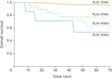
Kaplan-Meier OS curves by lymphatic spread pattern are shown in Fig. 2. The overall median follow-up period was 58 months. The 5-year OS rates were 96.5% in PLN−PAN− patients, 77.6% in PLN+PAN−, 63.4% in PLN+PAN+, and 53.6% in PLN−PAN+ patients. There was no significant difference in survival between the PLN−PAN+ and the PLN+PAN+ groups (log- rank test, p=0.41), and no significant difference in survival between the PLN−PAN+ and PLN+PAN− groups (log-rank test, p=0.40).
The results of the pooled analysis are shown in Table 4. A total of 6,532 patients with endometrial cancer who underwent both PLNDs and PANDs were identified from the pooled data [6-30]. The overall LNM rate was 16.7% (1,092/6,532) and the PAN metastasis rate was 9.7% (634/6,532). The PAN metastasis rates were 8.5% (174/2,056) among studies with the number of PAN removed <10 and 12.7% (280/2,197) among studies with the number of PAN High PAN+
Low PAN−
PLN−
n=4 (6.7%)
High PAN−
Low PAN+
PLN−
n=3 (5.0%)
High PAN+
Low PAN+
PLN−
n=1 (1.7%)
High PAN−
Low PAN−
PLN+
n=25 (41.7%)
High PAN+
Low PAN−
PLN+
n=8 (13.3%)
High PAN−
Low PAN+
PLN+
n=4 (6.7%)
High PAN+
Low PAN+
PLN+
n=15 (25.0%)
Fig. 1. Lymphatic spread pattern in 60 patients who underwent PAND up to the renal vein and were diagnosed with LNM.
LNM, lymph node metastasis; PAN, para-aortic lymph node; PAND, para-aortic lymph node dissection; PLN, pelvic lymph node.
Time (mo)
0.6
0.4
0.2
0 20 30 50 70
Overall survival
0.8 1.0
40 60
10
PLN−PAN−
PLN+PAN−
PLN+PAN+
PLN−PAN+
Fig. 2. Kaplan-Meier OS curves according to lymphatic spread pattern.
OS, overall survival; PAN, para-aortic lymph node; PLN, pelvic lymph node.
removed >10. The proportion of PAN metastasis in patients without PLN metastasis was 2.7% (149/5,599). The PAN metastasis rates in patients without PLN metastasis were 1.6%
(29/1,781) among studies with the number of PAN removed <10 and 3.5% (64/1,819) among studies with the number of PAN removed >10.
DISCUSSION
In this study, we focused on the probability of PAN metastasis in endometrial cancer patients without PLN metastasis, in light of the increasing use of SLN mapping by cervical tracer injection in Western countries. As noted above, the ability of cervical injection to assess PAN status is poor. Before discussing a significance of the probability, we considered our results in light of previous studies in patients at risk of LNM. In our study, PAND was implemented at the discretion of the attending surgeons and was performed in 50.4% of patients who underwent surgical treatment. The overall LNM rate was 16.9% among patients who underwent both PLNDs and PANDs, which was in close agreement with the rate of 16.7% derived from the pooled analysis. During the last decade, the Mayo criteria have been recognized as the standard decision-making model for implementing lymphadenectomy in Table 4. Results of pooled analysis: lymphatic spread pattern in endometrial cancer according to number of PANs removed
Author Year No. FIGO stage
III/IV (%) NE histology
(%) No. of PLNs
removed* No. of PANs
removed* A B C D B/A+B
PLN−/PAN− PLN−/PAN+ PLN+/PAN− PLN+/PAN+ (%) No. of PAN removed: <10
Larson et al. [6] 1993 50 28 0 13 5 50 0 2 8 0.0
Fanning et al. [7] 1996 60 8 0 21 7 55 0 5 0 0.0
Yokoyama et al. [8] 1997 63 13 3 14 6 45 4 6 8 8.2
Lee et al. [9] 2009 349 NA 0 (22.8) (9.5) 277 7 26 39 2.5
Abu-Rustum et al. [10] 2009 847 NA NA 16 5 722 12 52 61 1.6
Chiang et al. [11] 2011 171 22 6 17 5 154 2 12 3 1.3
Solmaz et al. [12] 2015 516 NA 0 22 8.5 449 4 37 26 0.9
Subtotal 2,056 1,752 29 140 145 1.6
No. of PAN removed: not available
Chen and Lee [13] 1983 74 NA 11 NA NA 63 3 3 5 4.5
Creasman et al. [14] 1987 621 22 4 NA NA 551 12 36 22 3.9
Ayhan et al. [15] 1995 209 NA NA NA NA 173 6 17 13 3.4
Hirahatake et al. [16] 1997 200 42 4 NA NA 158 2 24 16 1.3
Milam et al. [17] 2012 582 11 0 NA NA 520 12 31 19 2.3
Sueoka et al. [18] 2015 502 17 18 NA NA 422 15 27 38 3.4
Mahdi et al. [19] 2015 91 NA NA NA NA 56 6 18 11 9.7
Subtotal 2,279 1,943 56 156 124 2.8
No. of PAN removed: >10
Onda et al. [20] 1997 173 24% 1 (37.9) (28.7) 143 2 10 18 1.4
Matsumoto et al. [21] 2002 106 NA 5 (36.8) (30.5) 79 2 7 18 2.5
Mariani et al. [22] 2008 281 NA NA 35 17 218 10 24 29 4.4
Fujimoto et al. [23] 2009 355 25 0 42 19 306 7 20 22 2.2
Dogan et al. [24] 2012 161 21 21 (49.5) (19.0) 143 2 11 5 1.4
Odagiri et al. [25] 2014 266 NA 17 62.5 20 224 7 16 19 3.0
Altay et al. [26] 2015 173 NA 27 26 12 135 7 12 19 4.9
Tomisato et al. [27] 2014 260 46 17 50 22 169 9 34 48 5.1
Fotopoulou et al. [28] 2015 128 15 24 29 21.5 101 4 8 15 3.9
Sautua et al. [29] 2015 90 NA NA (11.9) (10.7) 77 6 3 4 7.2
Alay et al. [30] 2015 204 26 23 (44.1) (24.9) 160 8 17 19 4.8
Subtotal 2,197 1,755 64 162 216 3.5
Total 6,532 5,450 149 458 485 2.7
FIGO, International Federation of Gynecology and Obstetrics; NA, not available; NE, non-endometrioid; PAN, para-aortic lymph node; PLN, pelvic lymph node.
*Values are presented as median (mean).
patients with endometrial cancer. These criteria divide patients into “not at-risk for lymph node metastasis” and “at-risk for lymph node metastasis” groups. The former includes cases of G1/G2, <50% myoinvasion, and tumor diameter <2 cm, while the latter includes all other cases [22,31]. According to their protocol, lymphadenectomy is not recommended for the
“not at-risk” group, but both pelvic and para-aortic lymphadenectomies are recommended for the “at-risk” group. Kumar et al. [31] found prevalence of PLN and PAN metastases of 17% and 12%, respectively, among patients at risk of LNM determined by the Mayo criteria, compared with 14.5% and 10.3%, respectively, in our cohort, suggesting that our results do not overestimate the true probability of PAN metastasis in patients without PLN metastasis.
The incidence of PAN metastasis in patients without PLN metastasis was 2.8% in our cohort, which was in close agreement with 2.7% in the pooled analysis. Previous studies [20-30] with sufficient numbers of PAN removed (>10) found incidences up to 3.5%. Overall, these results suggest that the probability of PAN metastasis in the absence of PLN metastasis is remote, but not improbable, in patients at risk of LNM. SLN mapping by cervical tracer injection should thus be performed cautiously in such a population. Our results also demonstrated the significance of the upper para-aortic region. To the best of our knowledge, SLNs in the upper para-aortic region have not been detected by cervical injection, and cervical injection lacks the ability to detect para-aortic SLNs above the inferior mesenteric artery, which represents a major disadvantage of this procedure.
SLN mapping by cervical injection could be considered safe if the implications of PLN−PAN+
were negligible, and the relevance of PLN−PAN+ should thus be assessed in light of not only its probability, but also its prognostic risk. There was no difference in survival between the PLN−PAN+ and PLN+PAN+ groups in the present study. Tomisato et al. [27] also showed a 5-year progression-free survival rate of 44.4% for PLN−PAN+ (compared with 87.1% for PLN−
PAN−, 67.5% for PLN+PAN−, and 33.2% for PAN+PAN+), with no significant difference in survival between the PLN−PAN+ and PLN+PAN+ groups, despite there being few PLN−PAN+
cases. These results were consistent with ours, and suggest that the prognosis of patients with PLN−PAN+ status is poor. A group of patients with endometrial cancer at risk of LNM thus consists of a minority (around 3%) of PLN−PAN+ cases at risk of a poor prognosis.
In light of the increasing attention given to SLN mapping using cervical tracer injection, it may be necessary to create a novel patient category, i.e., “at-risk but not at-risk for PAN metastasis.”
We suggest that this type of SLN mapping should only be applied in patients at negligible risk of PAN metastasis [32]. The probability of isolated PAN metastasis may be deemed low enough to forego the need to determine PAN status. However, this could be considered to be a utilitarian approach with an emphasis on economic efficiency, at the potential expense of a minority of patients with a poor prognosis, in case of PLN−PAN+. The utilitarian concept threatens medical evolution by reducing treatment for patients at risk of a poor prognosis. Regarding the treatment strategy for PLN−PAN+ patients, we wish to question the emphasis placed on the low prevalence of PLN−PAN+ and ask if its prognostic risk has been fairly assessed. We suggest that gynecologic oncologists should consider establishing treatment strategies aimed at the specific care of minorities, such as patients with PLN−PAN+ status.
The current study had some limitations. The number of patients was too small to produce conclusive results. Furthermore, the study was inevitably subject to selection bias because of its retrospective, single-institution nature. It should be noted that the proportion of PLN−PAN+ in this study does not represent the corresponding one in the general
population which include both “not at-risk” and “at-risk” groups. However, the issue of PAN metastasis might not be studied in a general population that includes “not at-risk” patients because the probability of PAN metastasis is greatly reduced in this population, and the implication of PLN−PAN+ is subsequently undervalued. In addition, there were no strict rules for applying PLNDs and PANDs in our patients. However, we performed a systematic review to overcome this weakness. Our patient group with PLND and PAND is likely to resemble the “at-risk” groups identified in other studies. In conclusion, PAN metastasis may occur in patients without PLN metastasis, with a non-negligible effect on survival.
REFERENCES
1. Siegel RL, Miller KD, Jemal A. Cancer statistics, 2017. CA Cancer J Clin 2017;67:7-30.
PUBMED | CROSSREF
2. Parker SL, Tong T, Bolden S, Wingo PA. Cancer statistics, 1997. CA Cancer J Clin 1997;47:5-27.
PUBMED | CROSSREF
3. Abu-Rustum NR. The increasing credibility of sentinel lymph node mapping in endometrial cancer. Ann Surg Oncol 2013;20:353-4.
PUBMED | CROSSREF
4. Cormier B, Rozenholc AT, Gotlieb W, Plante M, Giede CCommunities of Practice (CoP) Group of Society of Gynecologic Oncology of Canada (GOC). Sentinel lymph node procedure in endometrial cancer: a systematic review and proposal for standardization of future research. Gynecol Oncol 2015;138:478-85.
PUBMED | CROSSREF
5. Ballester M, Dubernard G, Lécuru F, Heitz D, Mathevet P, Marret H, et al. Detection rate and diagnostic accuracy of sentinel-node biopsy in early stage endometrial cancer: a prospective multicentre study (SENTI-ENDO). Lancet Oncol 2011;12:469-76.
PUBMED | CROSSREF
6. Larson DM, Johnson KK. Pelvic and para-aortic lymphadenectomy for surgical staging of high-risk endometrioid adenocarcinoma of the endometrium. Gynecol Oncol 1993;51:345-8.
PUBMED | CROSSREF
7. Fanning J, Nanavati PJ, Hilgers RD. Surgical staging and high dose rate brachytherapy for endometrial cancer: limiting external radiotherapy to node-positive tumors. Obstet Gynecol 1996;87:1041-4.
PUBMED | CROSSREF
8. Yokoyama Y, Maruyama H, Sato S, Saito Y. Indispensability of pelvic and paraaortic lymphadenectomy in endometrial cancers. Gynecol Oncol 1997;64:411-7.
PUBMED | CROSSREF
9. Lee KB, Ki KD, Lee JM, Lee JK, Kim JW, Cho CH, et al. The risk of lymph node metastasis based on myometrial invasion and tumor grade in endometrioid uterine cancers: a multicenter, retrospective Korean study. Ann Surg Oncol 2009;16:2882-7.
PUBMED | CROSSREF
10. Abu-Rustum NR, Khoury-Collado F, Pandit-Taskar N, Soslow RA, Dao F, Sonoda Y, et al. Sentinel lymph node mapping for grade 1 endometrial cancer: is it the answer to the surgical staging dilemma? Gynecol Oncol 2009;113:163-9.
PUBMED | CROSSREF
11. Chiang AJ, Yu KJ, Chao KC, Teng NN. The incidence of isolated para-aortic nodal metastasis in completely staged endometrial cancer patients. Gynecol Oncol 2011;121:122-5.
PUBMED | CROSSREF
12. Solmaz U, Mat E, Dereli ML, Turan V, Tosun G, Dogan A, et al. Lymphovascular space invasion and positive pelvic lymph nodes are independent risk factors for para-aortic nodal metastasis in endometrioid endometrial cancer. Eur J Obstet Gynecol Reprod Biol 2015;186:63-7.
PUBMED | CROSSREF
13. Chen SS, Lee L. Retroperitoneal lymph node metastases in Stage I carcinoma of the endometrium:
correlation with risk factors. Gynecol Oncol 1983;16:319-25.
PUBMED | CROSSREF
14. Creasman WT, Morrow CP, Bundy BN, Homesley HD, Graham JE, Heller PB. Surgical pathologic spread patterns of endometrial cancer. A Gynecologic Oncology Group Study. Cancer 1987;60:2035-41.
PUBMED | CROSSREF
15. Ayhan A, Tuncer ZS, Tuncer R, Yüce K, Küçükali T. Tumor status of lymph nodes in early endometrial cancer in relation to lymph node size. Eur J Obstet Gynecol Reprod Biol 1995;60:61-3.
PUBMED | CROSSREF
16. Hirahatake K, Hareyama H, Sakuragi N, Nishiya M, Makinoda S, Fujimoto S. A clinical and pathologic study on para-aortic lymph node metastasis in endometrial carcinoma. J Surg Oncol 1997;65:82-7.
PUBMED | CROSSREF
17. Milam MR, Java J, Walker JL, Metzinger DS, Parker LP, Coleman RL, et al. Nodal metastasis risk in endometrioid endometrial cancer. Obstet Gynecol 2012;119:286-92.
PUBMED | CROSSREF
18. Sueoka K, Umayahara K, Abe A, Usami T, Yamamoto A, Nomura H, et al. Prognosis for endometrial cancer patients treated with systematic pelvic and para-aortic lymphadenectomy followed by platinum- based chemotherapy. Int J Gynecol Cancer 2015;25:81-6.
PUBMED | CROSSREF
19. Mahdi H, Jernigan A, Nutter B, Michener C, Rose PG. Lymph node metastasis and pattern of recurrence in clinically early stage endometrial cancer with positive lymphovascular space invasion. J Gynecol Oncol 2015;26:208-13.
PUBMED | CROSSREF
20. Onda T, Yoshikawa H, Mizutani K, Mishima M, Yokota H, Nagano H, et al. Treatment of node-positive endometrial cancer with complete node dissection, chemotherapy and radiation therapy. Br J Cancer 1997;75:1836-41.
PUBMED | CROSSREF
21. Matsumoto K, Yoshikawa H, Yasugi T, Onda T, Nakagawa S, Yamada M, et al. Distinct lymphatic spread of endometrial carcinoma in comparison with cervical and ovarian carcinomas. Cancer Lett 2002;180:83-9.
PUBMED | CROSSREF
22. Mariani A, Dowdy SC, Cliby WA, Gostout BS, Jones MB, Wilson TO, et al. Prospective assessment of lymphatic dissemination in endometrial cancer: a paradigm shift in surgical staging. Gynecol Oncol 2008;109:11-8.
PUBMED | CROSSREF
23. Fujimoto T, Nanjyo H, Fukuda J, Nakamura A, Mizunuma H, Yaegashi N, et al. Endometrioid uterine cancer: histopathological risk factors of local and distant recurrence. Gynecol Oncol 2009;112:342-7.
PUBMED | CROSSREF
24. Dogan NU, Gungor T, Karsli F, Ozgu E, Besli M. To what extent should para-aortic lymphadenectomy be carried out for surgically staged endometrial cancer? Int J Gynecol Cancer 2012;22:607-10.
PUBMED | CROSSREF
25. Odagiri T, Watari H, Kato T, Mitamura T, Hosaka M, Sudo S, et al. Distribution of lymph node metastasis sites in endometrial cancer undergoing systematic pelvic and para-aortic lymphadenectomy: a proposal of optimal lymphadenectomy for future clinical trials. Ann Surg Oncol 2014;21:2755-61.
PUBMED | CROSSREF
26. Altay A, Toptas T, Dogan S, Simsek T, Pestereli E. Analysis of metastatic regional lymph node locations and predictors of para-aortic lymph node involvement in endometrial cancer patients at risk for lymphatic dissemination. Int J Gynecol Cancer 2015;25:657-64.
PUBMED | CROSSREF
27. Tomisato S, Yamagami W, Susumu N, Kuwahata M, Takigawa A, Nomura H, et al. Clinicopathological study on para-aortic lymph node metastasis without pelvic lymph node metastasis in endometrial cancer.
J Obstet Gynaecol Res 2014;40:1733-9.
PUBMED | CROSSREF
28. Fotopoulou C, El-Balat A, du Bois A, Sehouli J, Harter P, Muallem MZ, et al. Systematic pelvic and paraaortic lymphadenectomy in early high-risk or advanced endometrial cancer. Arch Gynecol Obstet 2015;292:1321-7.
PUBMED | CROSSREF
29. Sautua RR, Goiri K, Calle MA, Marin IJ, Artola AL. Incidence of nodal metastasis and isolated aortic metastases in patients with surgically staged endometrioid endometrial cancer. Int J Gynecol Cancer 2015;25:875-8.
PUBMED | CROSSREF
30. Alay I, Turan T, Ureyen I, Karalok A, Tasci T, Ozfuttu A, et al. Lymphadenectomy should be performed up to the renal vein in patients with intermediate-high risk endometrial cancer. Pathol Oncol Res 2015;21:803-10.
PUBMED | CROSSREF
31. Kumar S, Podratz KC, Bakkum-Gamez JN, Dowdy SC, Weaver AL, McGree ME, et al. Prospective assessment of the prevalence of pelvic, paraaortic and high paraaortic lymph node metastasis in endometrial cancer. Gynecol Oncol 2014;132:38-43.
PUBMED | CROSSREF
32. Todo Y, Okamoto K, Takeshita S, Sudo S, Kato H. A patient group at negligible risk of para-aortic lymph node metastasis in endometrial cancer. Gynecol Oncol 2016;141:155-9.
PUBMED | CROSSREF