Inhibitory Effect of Bee Venom Toxin on the Growth of Cervix Cancer C33A Cells via Death Receptor
Expression and Apoptosis
Seong Cheol Ko and Ho Sueb Song*
Department of Acupuncture & Moxibustion Medicine, College of Orinetal Medicine, Gachon University
[Abstract]
Objectives : We investigated whether bee venom(BV) inhibit cell growth through enhancement of death receptor expressions in the human cervix cancer C33A cells.
Methods : BV(1~5 ㎍/㎖) inhibited the growth of cervix cancer C33A cells by the induction of apoptotic cell death in a dose dependent manner.
Results : Consistent with apoptotic cell death, expression of Fas, death receptor(DR) 3, 4, 5 and 6 was increased concentration dependently in the cells. Moreover, Fas, DR3 and DR6 revealed more sensitivity to BV. Thus, We reconfirmed whether they actually play a critical role in anti-proliferation of cervix cancer C33A cells. Consecutively, expression of DR downstream pro-apoptotic proteins including caspase-8, -3, -9 was upregulated and Bax was concomitantly overwhelmed the expression of Bcl-2. NF-κB were also inhibited by treatment with BV in C33A cells.
Conclusions : These results suggest that BV could exert anti-tumor effect through induction of apoptotic cell death in human cervix cancer C33A cells via enhancement of death receptor expression, and that BV could be a promising agent for preventing and treating cervix cancer.
Key words : Bee venom;
Cervix cancer;
C33A;
Death receptor;
Apoptosis
Received : 2014. 05. 14.
Revised : 2014. 05.23.
Accepted : 2014. 05.28.
On-line : 2014. 06.20.
✱ Corresponding author : Department of Acupuncture & Moxibustion Medicine, Gil Oriental Medicine Hospital of Gachon Univercity, 12, Dokjeom-ro 29beon-gil, Namdong-gu, Incheon, 405-760, Republic of Korea
Tel : +82-70-7120-5012 Email : [email protected]
This is an Open-Access article distributed under the terms of the Creative Commons Attribution Non-Commercial License (http://creativecommons.org/licenses/by-nc/3.0) which permits unrestricted non-commercial use, distribution, and reproduction in any medium, provided the original work is properly cited.
The Acupuncture is the Journal of Korean Acupuncture & Moxibustion Medicine Society. (http://www.TheAcupuncture.org) Copyright © 2014 KAMMS. Korean Acupuncture & Moxibustion Medicine Society. All rights reserved.
Ⅰ. Introduction
Carcinoma of the uterine cervix is the second common prevalent malignancy in women worldwide, which contributes to the leading cancer-induced mortality in the developing countries1,2).
The treatment of the malignancy consists of surgery, radiation therapy and chemotherapy, which mostly depends on its stage3).
A combination of therapeutic approaches was thought to be beneficial to increase the 5-year survival rate in women with locally advanced cervix cancers4-8). A randomized controlled trials substantiated that weekly administration of the cisplatin was the ideal chemotherapy regimen4). Consequently, the role of concurrent cisplatin(CDDP) chemotherapy was established as an adjuvant to radiation therapy until now5,9). However, a controversy remains due to the risk of normal tissue injury and its various toxicity related complications such as anemia, febrile neutropenia, nausea, vomiting and neuropathy. Such complications can reduce lifespan and impair the quality of life in the patients7). Thus, the identification and the development of natural novel agents which enhances cytotoxicity and diminishes complications of CDDP is necessary.
Resveratrol, a biologically active natural polyphenol substance commonly found in grape, giant knotweed rhizome, inhibits the growth of cervix cell lines C33A, SiHa and HeLa via induction of cell apoptosis in a time- and dose-dependent manner10,11). Furthermore, the addition of luteolin, a kind of flavonoid to tumor necrosis factor(TNF) related apoptosis inducing ligand(TRAIL) overcomes the TRAIL resistance in HeLa cells by increasing the death receptor(DR) expression11).
Bee venom(BV) contains various different peptides including melittin, phospholipase A2, apamin, adolapin, and mast cell-degranulating peptide(MCDP)12,13) and it has been used in Korean medicine as a pharm- acopuncture to treat back pain, rheumatism, and many skin diseases due to its antibacterial, antiviral, and anti-inflammatory effects14,15). Moreover, several ex- perimental studies reported that BV and/or melittin have anti-cancer effects in breast, cervix, renal14),
prostate15), liver16) cancer cells.
However, the experiments demonstrating the anti- cancer effects and molecular mechanisms of BV in cervix cancer cells have not been reported. Therefore, in this study, I have investigated anti-proliferative effects of BV on cervix cancer C33A cells and explored the mechanism associated with the DR-mediated apoptosis.
Ⅱ. Materials and methods
A. Materials
BV was purchased from You-Miel BV Ltd(Hwasoon, Jeonnam, Korea). The composition of the BV was as follows: 45-50 % melittin, 2.5~3 % mast cell degranulating peptide, 12 % phospholipase A2, 1 % lysophospholipase A, 1~1.5 % histidine, 4~5 % 6-pentyl a-pyrone lipids, 0.5 % secarpin, 0.1 % tertiapin, 0.1 % procamine, 1.5~2 % hyaluronidase, 2~3 % amine, 4~5 % carbohydrate, and 19~27 % of others, including protease inhibitor, glucosidase, invertase, acid phosphomonoesterase, dopamine, norepinephrine, and unknown amino acids, with 99.5 % purity. All of the secondary antibodies such as Bax, Bcl-2, caspase-3, -8, -9, cleaved caspase-3, -8, -9, TNF‐like weak inducer of apoptosis(TWEAK), Fas L, Fas, DR 3, DR 4, DR 5 and DR 6 used in western blot analysis were purchased from Santa Cruz Biotechnology(Santa Cruz, CA, USA). T4 polynucleotide kinase was obtained from Promega(Madison, WI). Poly(dI-dC), horseradish per- oxidase-labeled donkey anti-rabbit secondary antibody, and ECL detection reagent were obtained from Amersham Pharmacia Biotech(Piscataway, NJ, USA).
Reagents for sodium dodecyl sulfate(SDS)–polyacryla- mide gel electrophoresis were purchased from Bio-Rad (Hercules, CA, USA).
B. Cell culture
The cervix cancer C33A cells lines were purchased from the American Type Culture Collection(Manassas,
VA, USA). C33A cancer cells were cultured in RPMI- 1640 medium supplemented with 10 % fetal bovine serum(FBS) and penicillin/streptomycin(100 U/㎖). Cell cultures were then maintained at 37 ℃ in a humidified atmosphere with 5 % CO2.
C. Cell viability assay
To determine the cell number, cervix cancer C33A cells were plated in 24-well plates(5×10⁴ cells/well), and subconfluent cells were subsequently treated with BV(1, 2 and 5 ㎍/㎖) for 24 hrs. After treatment, cells were trypsinized and pelleted by centrifugation for 5 min at 1,500 rpm, resuspended in 5 ㎖ of phosphate- buffered saline(PBS), and 0.1 ㎖ of 0.2 % trypan blue was added to the cancer cell suspension in each of the solutions(0.9 ㎖ each). Subsequently, a drop of suspension was placed into a Neubauer chamber and the living cancer cells were counted. Cells that showed signs of staining were considered to be dead, whereas those that excluded trypan blue were considered viable.
Each assay was carried out in triplicate.
D. Western blot analysis
Cells were homogenized with lysis buffer(50 mM Tris, pH 8.0, 150 mM NaCl, 0.02 % NaN3, 0.2 % SDS, 1 mM phenylmethylsulfonyl fluoride, 10 ㎕/㎖ aprotinin, 1 % igapel 630(Sigma–-Aldrich, St Louis, MO, USA), 10 mM NaF, 0.5 mM EDTA, 0.1 mM EGTA, and 0.5 % sodium deoxycholate) and centrifuged at 23,000 g for 1 hr.
Equal amounts of proteins(80 ㎍) were separated on SDS 12 % polyacrylamide gels and then transferred to a nitrocellulose membrane(Hybond ECL; Amersham Pharmacia Biotech, NJ, USA). Blots were blocked for 2 hrs at room temperature with 5 %(w/v) nonfat dried milk in Tris buffered saline(10 mM Tris, pH 8.0, 150 mM NaCl) containing 0.05 % Tween 20. The membrane was incubated for 5 hrs at room temperature with specific antibodies: rabbit polyclonal for caspase-3, cleaved caspase-3, caspase-8, cleaved caspase-8, caspase-9, cleaved caspase-9, TWEAK, Fas L, Fas, DR3, DR4,
DR5, DR6(1:1,000 dilution, Cell Signaling Technology, Beverly, MA, USA), Bcl-2, Bax, goat polyclonal antibody to p50, p65(1:500 dilution, Santa Cruz Biotechnology, CA, USA) and phospho-IκBa(1:200, Santa Cruz Biotechnology, CA, USA). The blot was then incubated with the corresponding conjugated anti- rabbit and anti-mouse immunoglobulin G-horseradish peroxidase(1:2,000 dilutions, Santa Cruz Biotech- nology, CA, USA). Immunoreactive proteins were detected with the ECL western blotting detection system.
E. Transfection assay
Cervix cancer C33A cells(3×10⁴ cells/well) were plated in 24-well plates and transiently transfected with siRNA, using a mixture of siRNA and the WelFect-EXPLUS reagentin OPTI-MEN, according to the manufacturer’s specification(WelGENE, Seoul, Korea). The transfected cells were treated with 2 ㎍/㎖ BV for 24 hrs. Thereafter, cell viability assay was performed as described above.
F. Apoptosis evaluation
Cervix cancer C33A cells(2.5×10⁵ cells/well) were cultured on 8-chamber slides. The cells were treated with BV(1, 2 and 5 ㎍/㎖). The cells were washed twice with PBS and fixed by incubation in 4 % para- formaldehyde in PBS for 1 hr at room temperature.
Membrane was permeabilized by exposure to 0.1 % Triton X-100 in phosphate-buffered saline for 5 min at room temperature. TdT-mediated dUTP nick and labeling(TUNEL) assays were performed by using the in situ cell death detection kit(Roche Diagnostics GmbH, Mannheim, Germany) according to the manufacturer’s instructions. For 4'-6-Diamidino-2-phenyl indole(DAPI) staining, slides were incubated for 15 min at room temperature in the dark with mounting medium for fluorescence containing DAPI(Vector Laboratories Inc., Burlingame, CA). The cells were then observed through a fluorescence microscope(Leica Microsystems AG, Wetzlar, Germany).
G. Preparation of nuclear extracts and electromobility shift assays
It was performed according to the manufacturer's recommendations(Promega, Madison, WI, USA). Briefly, 1×10⁶ cells/㎖ was washed twice with 1× PBS, followed by the addition of 1 ㎖ of PBS, and the cells were scraped into a cold Eppendorf tube. Cells were spun down at 15,000 g for 1 min, and the resulting supernatant was removed. Solution A(50 mM HEPES, pH 7.4, 10 mM KCl, 1 mM EDTA, 1 mM EGTA, 1 mM dithiothreitol, 0.1 ㎍/㎖ phenylmethyl- sulfonyl fluoride, 1 ㎍/㎖ pepstatin A, 1 ㎍/㎖ leupeptin, 10 ㎍/㎖ soybean trypsin inhibitor, 10 ㎍/㎖ aprotinin, and 0.5 % Nonidet P-40) was added to the pellet in a 2:1 ratio(v/v) and allowed to incubate on ice for 10 min. Solution C(solution A + 10 % glycerol and 400 mM KCl) was added to the pellet in a 2:1 ratio(v/v) and vortexed on ice for 20 min. The cells were centrifuged at 15,000 g for 7 min, and the resulting nuclear extract supernatant was collected in a chilled Eppendorf tube.
Consensus oligonucleotides were end-labeled using T4 polynucleotide kinase and [g-32P] ATP for 10 min at 37 ℃. Gel shift reactions were assembled and allowed to incubate at room temperature for 10 min followed by the addition of 1 ㎕(50,000-200,000 cpm) of 32P-labeled oligonucleotide and another 20 min of incubation at room temperature. For supershift assays, nuclear extracts from cells treated with BV(1~5 ㎍/㎖) were incubated with specific antibodies against NF-κB for 1 hr before EMSA. For competition assays, nuclear extracts from cells treated with BV(1~5 ㎍) were incubated with unlabelled NF-κB oligonuclaotide(50×, 100× and 200×) or labeled SP-1(100X) and AP-1(100X) for 30 min before EMSA. Subsequently 1 ㎕ of gel loading buffer was added to each reaction and loaded onto a 6 % nondenaturing gel and electrophoresed until the dye was three-fourths of the way down the gel.
The gel was dried at 80 ℃ for 1 hr and exposed to film overnight at 70 ℃. The relative density of the DNA-protein binding bands was scanned by densi- tometry using MyImage(SLB, Seoul, Korea), and quantified by Labworks 4.0 software(UVP Inc, Upland, CA, USA).
H. Data analysis
The data were analyzed using the GraphPad Prism 4 ver. 4.03 software(GraphPad Software, La Jolla, CA, USA). Data are presented as mean ± SD. The differences in all data were assessed by one-way analysis of variance(ANOVA). When the p value in the ANOVA test indicated statistical significance, the differences were assessed by the Dunnett’s test. A value of p<0.05 was considered to be statistically significant.
Ⅲ. Results
A. Effect of bee venom on cell growth in cervix cancer C33A cells
To assess the inhibitory effect of BV on cell growth of cervix cancer C33A cells, we analyzed cell viability by direct cell counting. The cells were treated with several concentrations of BV(1, 2, and 5 ㎍/㎖) for 24 hrs. BV inhibited cell proliferation of cervix cancer C33A cells in a concentration-dependent manner with IC50 value of 5.5 ㎍/㎖(Fig. 1). Morphologic observation showed that the cells were gradually reduced in size and changed into a small round single cell shape dose-dependently in C33A cells(Fig. 1).
B. Apoptotic cell death by bee
venom in cervix cancer C33A cells
To determine the inhibition of cell growth by BV via apoptosis, we evaluated the changes in the chromatin morphology of cells by using DAPI staining followed by TUNEL staining assays, and then the double labeled cells were analyzed by fluorescence microscope. Con- versely well with cell growth inhibition, DAPI-stained TUNEL-positive cells were significantly increased in BV treated cells concentration dependently. The treatment of BV(5 ㎍/㎖) resulted in about 90 % induction of
Fig. 1. Effect of bee venom on cell viability in cervix cancer C33A cells
Concentration-dependent effect of BV was shown on the cell viability assay in cervix cancer C33A cells. After treatment of BV(1, 2 and 5 ㎍/㎖) for 24 hrs, the cells were harvested by trypsinization and stained with 0.2 % trypan blue. Relative cell survival rate was determined by counting live and dead cells. The results were expressed as a percentage of viable cells. Morphologic observation with the treatment of BV in C33A cells.
Data are shown means ± SE(con : control)
* : p<0.05, significantly different from untreated control cells.
apoptotic cell death in cervix cancer C33A cells(Fig. 2).
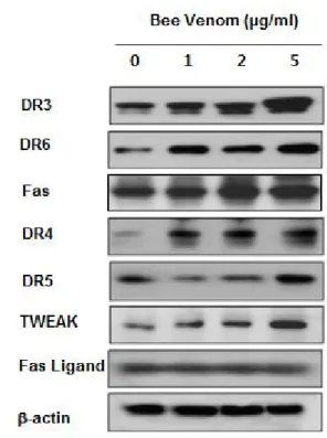
C. Expression of death receptors and related ligands in cervix cancer C33A cells by bee venom
Apoptosis can be induced by stimulation of death receptors and their ligands. Therefore, to investigate expression of them in cervix cancer C33A cells undergoing apoptotic cell death, we performed western bot analysis.
The results showed that BV treatment generally increased activities of Fas, DR 3, DR 4, DR 5 and DR 6 as well as TWEAK, a TNF superfamily, TNF week inducer of apoptosis dose dependently, in cervix cancer C33A cells, and it enhanced more sensitively enhanced the expression of Fas, DR 3 and DR 6(Fig. 3).
Fig. 2. Effect of bee venom on apoptotic cell death in cervix cancer C33A cells
The cervix cancer cells, C33A were treated with BV(1,2 and 5 ㎍/㎖) for 24 hrs, and then labeled with DAPI and TUNEL solution. Total number of cells in a given area was determined by using DAPI nuclear staining(fluorescent microscope). The green color in the fixed cells marks TUNEL-labeled cells. The apoptotic index was determined as the DAPI-stained TUNEL-positive cell number/total DAPI stained cell number(magnification, 200×).
Data are shown means ± SE(con : control)
Fig. 3. Effect of bee venom on expression of death recptors and their ligands in cervix cancer C33A cells
Cells were treated with BV(1, 2, and 5 ㎍/㎖) for 24 hrs, and examined for expressions of TWEAK, Fas L, Fas, DR-3, -4, -5, -6 by western blot analysis. β-actin was used as an control.
D. Effect of bee venom on the expression of apoptotic regulatory proteins in cervix cancer C33A cells
To investigate the relationship between the induction of apoptotic cell death and increase of DR activity in cervix cancer C33A cells treated by 1~5 ㎍/㎖ of BV, and the expression of related regulatory proteins of apoptotic cell death was investigated by western blots analysis. The expression of pro-apoptotic proteins overwhelmed anti-apoptotic protein: Bcl-2 was decreased. However, Bax, caspase-3, -8, -9, cleaved form of caspase-3, -8 and -9 was increased by treatment of BV in a concentration dependent manner(Fig. 4).
Fig. 4. Effect of bee venom on the expression of apoptosis regulatory proteins in cervix cancer C33A cells
Expression of apoptosis regulatory proteins was determined using western blot analysis. The cervix cancer C33A cells were treated with different concentrations of BV(1, 2, and 5 ㎍/㎖) for 24 hrs. Equal amounts of total proteins(50 ㎍/lane) were subjected to 12 % or 8 % SDS-PAGE. Expression of caspase-3, caspase-8, caspase-9, cleaved caspase-3,-8,-9, Bax, Bcl-2, and β-actin were detected by western blotting using specific antibodies. β-actin protein here was used as an internal control.
E. Reversed effects of DR siRNAs on bee venom-induced cell growth inhibition in cervix cancer C33A cells
To reconfirm whether BV inhibited cell growth of cervix cancer C33A cells through DR-mediated apoptosis, I transfected cervix cancer C33A cells with DR siRNA using a transfection agent. The cells were transfected with 100nM siRNA of DRs including Fas, DR 3 and DR 6 for 24 hrs, and then treated with BV(5
㎍/㎖) for 24 hrs. Thereafter, cancer cell growth was measured by direct counting after trypan blue staining.
As results, siRNA transfected Fas, DR 3 and DR 6 reversed BV inducing anti-proliferation through DR- mediated apoptosis in cervix cancer C33A cells(Fig. 5).
Fig. 5. Effects of siRNA of death receptors on bee venom-induced anti-proliferation in the cervix cancer C33A cells
The cervix cancer C33A cells were transfected with the DR siRNA(100 nM) for 24 hrs, the cells were then and treated with BV(5 ㎍/㎖) for another 24 hrs. Cell viability assay was performed to examine change of cell growth.
Data are shown means ± SE(con: control).
* : p<0.05, ** : p<0.01(significantly different from untreated control cells).
F. Inhibition of NF-κB and its signal molecules
NF-κB is known to inhibitory transcription factor of apoptosis. Whether to prevent anti-apoptotic ability of NF-κB is crucial for a agent causing cancer cells go
Fig. 6. Inhibition of NF-κB in cervix cancer C33A cells by bee venom
Activation of NF-κB was determined by electrophoretic mobility shift assay(EMSA), as described in materials and methods. Nuclear extracts from cervix cancer C33A cells with BV (1, 2, 5 ㎍/㎖) were incubated in binding reactions of 32P-labeled oligonucleotide containing the B sequence.
NF-κB DNA binding activity was determined by EMSA. Equal amounts of total proteins(50 ㎍/lane) were subjected to 12 % or 8 % SDS-PAGE. Expression of p50, p65, IκBα, p-IκBα and histone-1 were detected by western blotting using specific antibodies. Histone-1 protein here was used as an internal control.
apoptosis. To determine the effect of BV on the growth of cervix cancer C33A cells through apoptosis, I assessed NF-κB activity in the cells treated for different concentration with for 24 hrs BV by EMSA and observed NF-κB signal molecules in the cells by western blot analysis. NF-κB was highly activated in this cell.
However, the enhanced expression of NF-κB and its signal molecules were concentration- dependently decreased by the culture in the presence of BV in the cells(Fig. 6).
Ⅳ. Discussion
According to previous studies5-8), radiotherapy and chemotherapy were usually used combinationally and increased survival rate of patients with advanced
cervix cancer, of which radiotherapy was regarded to be more essential to ultimate recovery of these patients. Unexpectedly, some of them were not able to respond to radiation and concurrent CDDP due to the resistance or to tolerate fatal side effects resulting from combining two cytotoxic therapies such as normal tissue injury, leading to morbidity and even mortality.
In addition, a relatively increasing imbalance between proliferation and apoptosis was allegedly responsible for cervix carcinogensis17,18).
Thus, in order to resolve the above challenges and develop a novel agent of cervix cancer, more attention should be paid to explore promising agents restoring the balance through induction of DR mediated apoptosis than to strengthen the cytotoxicity of combined radiation and chemotherapy.
Apoptosis, a programmed cell death can play a decisive role in keeping up the balance related with carcinogenesis of cervix cancer, which is mainly induced by interplay of signaling the intrinsic mitochondria-mediated pathway or the extrinsic DR– mediated pathway19). DRs majorly involve in the both apoptotic pathways and trigger apoptosis20,21). They generally bind to their specific ligands or antibodies and induce trimerization of themselves22,23), and then they cluster at their own characteristic death domain(DD), recruiting intracellular adaptor proteins and procaspase-8 and forming DR inducing signal complex(DISC)24,25). Once the DISC is made, an intracellular cascade of caspases is activated, resulting in cleavage of so-called death-substrates PARP(poly (ADP)-ribose polymerase) and eventually go apoptosis22,23). Caspase-8 is considered as a key initiator caspase, which can activate other down-stream caspases such as caspase-326). Besides direct activation by caspase-8, caspase-3 can also become activated through induction of a mitochondrial intrinsic apoptosis resulting in caspase-9 activation and concomitantly caspase-3 activation following cytochrome C release by loosened mitochondrial membrane potential27-32). Cleavage of the Bcl-2 family member Bid by caspase-8 conveys the apoptotic signal to the mitochondria, initiating intrinsic mitochondrial apoptosis in a same way as the above33,34).
Previous studies35-37)demonstrated that all cervix
cancer cell lines are not always susceptible to apoptosis despite high levels of DR expression, suggesting intracellular apoptotic regulatory mechanisms play a major role in the determination of the susceptibility of cervix cancer cells to DR-mediated apoptosis.
Among them, Ryu et al35). confirmed increased TRAIL receptor expression in cervix cancer cells. In addition, Hougardy et al. treated human cervix cell lines including HeLa, CaSki, SiHa and C33A with Fas monoclonal antibody in combination with interferon and cisplatin and confirmed increase of caspases 3, 8 and 9 activities, indicating anti-Fas therapy could be used in a complementary way to cisplatin due to its property enhancing Fas mediated apoptosis38). Inter- estingly, from the previous reports substantiating that resveratrol from medicinal plants such as grape, giant knotweed rhizome exerts wide spectrum of anti- proliferative effect on various cancers such as leukemia, breast cancer, ovarian cancer, hepatoma39-44), Shen et al10) applied the results to cervix cancer cells and similarly found that it also inhibits the pro- liferation of cervix cell lines C33A, SiHa and HeLa through apoptosis. Moreover, according to Horinaka et al' report11), the role of luteolin was revealed as a helper to get over TRAIL resistance in HeLa cells through enhancement of DR expression11). Recently, several studies have demonstrated anti-cancer effects of BV and melittin. Of them, Park et al14) confirmed that BV and melittin significantly suppressed PMA- induced invasion by inhibiting MMP-9 expression in Caki-1 renal cells through blocking the PMA- stimulated activations of activator protein-1(AP-1) and nuclear factor-κB as well as their upstream factors including extracellular signal-regulated kinase(ERK) and c-Jun N-terminal kinase(JNK). Park et al15) found that BV and melittin inhibited cancer cell growth through induction of apoptotic cell death via suppression of constitutively activated NF-κB in LNCaP, DU145, and PC-3 human prostate cancer cells invitro as well as in nude mice implanted with PC-3 cells invivo, implying BV and melittin could inhibit prostate cancer in invitro and invivo by anti-apoptotic NF-κB and pro-apoptotic caspase signal mediated induction of apoptotic cell death. Liu et al16) reported
that melittin can inhibit cell motility drastically and prevent Hepatocellular carcinoma (HCC) metastasis invivo through the suppression of expression of the Rac1, a critical regulator for the metastasis of HCC, indicating melittin is a potential novel drug for HCC. Furthermore, Jang et al45) reported BV should exerted anti-tumor effect in human A549 lung cancer cells through induc- tion of apoptotic cell death in lung cancer cells via enhancing expression of DRs including TNF R1 (DR 1), Fas(DR 2), Apo-3(DR 3), suggesting that BV could be a promising agent for preventing and treating lung cancer.
Together with the above results, I hypothesized that BV could inhibit cancer growth of cervix cancer through DR-mediated apoptosis. Consequently, in this study, cell viability was decreased in the BV treated cervix cancer C33A cells dose dependently(Fig.1), in which charateristic morphologic change of apoptosis was observed(Fig. 1) and caspase active cells was increased concentration dependently, even reaching to 90 % by 5 ㎍/㎖ of BV(Fig. 2), implying that BV result in inhibition of cervix cancer C33A cell growth through vivid induction of apoptotic cell death in cervix cancer C33A cells. In the western blot analysis to elucidate the mechanism how BV trigger apoptosis, expression of Fas, DR 3, DR 4 DR 5 and DR 6 as well as TWEAK, a TNF superfamily, TNF week inducer of apoptosis was generally and dose-dependently enhanced in C33A cervix cancer(Fig. 3). Concomitantly, Bax overwhelmed Bcl-2 and pro-apoptotic caspase -3, -8 and -9 was increased dose-dependently by BV(Fig. 4). Moreover, Fas, DR 3 and DR 6 represented relative higher sensitivity to the cells than the other DRs and ligands(Fig. 3). In the transfection assay to reconfirm this, siFas, siDR 3, siDR 6 revealed the reverse effects that decreased cell viability results by BV was increased again following transfection of siRNA to Fas, DR 3 and DR 6(Fig. 5). Although previous studies mainly emphasized a role of enhanced Fas with Fas ligand, DR 4 and DR 5 with TRAIL in the induction of apoptosis of cervix cancer10,11,35,38), this study additionally demonstrated that of DR 3 and DR 6 in apoptosis of cervix cancer C33A cells, which is minorly different from Jang et al.'s result reporting enhancement of DR1, DR 2 and DR 3 in lung cancer A549 cells.
Meanwhile, regarding etiology, cervix cancer is caused by human papillomavirus(HPV) infection which has properties to induce increase of anti-apoptotic NF-κB activity46-48) which closely involves in pathogenesis of cervix cancer and ultimately results in aggravation of it including proliferation, metastasis, and other poor prognosis49). Therefore, the machinery of NF-κB activation also plays a major role in mediating apoptosis50).
In EMSA assay of present study to investigate the effect of BV on activity of anti apoptotic NF-κB, BV consecutively suppressed the activity of NF-κB and its signal molecules such as p50, p65 and IκB(Fig. 6).
Considering all these results, it is suggested that BV should exert anti-proliferative influence upon cervix cancer C33A cells through induction of broad spectrum of DR mediated apoptosis involving regulation of representative pro- or anti- apoptotic proteins in the intrinsic as well as extrinsic apoptotic pathway.
Consequently, although further studies are needed to substantiate my investigation concretely and expand to animal experiments invivo, these present data provide that BV could be available as a promising agent alternative or complementary to CDDP due to not only enhancing anti-proliferation of C33A, cervix cancer cells through but overcoming the resistance of them via initiation of DR-mediated apoptosis.
V. Conclusion
These results suggest that BV could exert anti- tumor effect through induction of apoptotic cell death in human cervix cancer C33A cells via enhancement of death receptor expression, and that BV could be a promising agent for preventing and treating cervix cancer.
Ⅴ. References
1. Parkin DM, Pisani P, Ferlay J. Estimates of the
worldwide incidence of eighteen major cancers in 1985. Int J Cancer. 1993 ; 54(4) : 594-606.
2. Parkin DM, Bray F, Ferlay J, Pisani P. Global cancer statistics, 2002. CA Cancer J Clin. 2005 ; 55(2) : 74–108.
3. Ryu HS. Concurrent chemoradiotherapy in cervical cancer(a new paradigm in cervical cancer treat- ment). Yonsei Med J. 2002 ; 43(3) : 749-53.
4. Koh WRP. Locally advanced cervical cancer. In:
Gershenson D MW, editor. Gynecologic cancer:
controversies in management, Vol. Philadelphia : Elsevier Churchill-Livingstone. 2004 : 175–86.
5. Rose PG, Bundy BN, Watkins ER et al. Concurrent cisplatin-based radiotherapy and chemotherapy for locally advanced cervical cancer. N Engl J Med. 1999 ; 340(15) : 1144–53.
6. Whitney CW, Sause W, Bundy BN et al. Randomized comparison of fluorouracil plus cisplatin versus hydroxyurea as an adjunct to radiation therapy in stages IIB-IVA carcinoma of the cervix with negative para-aortic lymph nodes. A Gynecologic Oncology Group and Southwest Oncology Group study. J Clin Oncol. 1999 ; 17(5) : 1339-48.
7. Morris M, Eifel PJ, Lu J et al. Pelvic radiation with concurrent chemotherapy versus pelvic and para-aortic radiation for high-risk cervical cancer.
A Randomized Radiation Therapy Oncology Group clinical trial. N Engl J Med. 1999 ; 340(5) : 1137-43.
8. PetersⅢ WA, Lui PY et al. Concurrent chemo- therapy and pelvic radiation therapy compared with pelvic radiation therapy alone as adjuvant therapy after radical surgery in high-risk early- stage cancer of the cervix. J Clin Oncol. 2000 ; 18(8) : 1606-13.
9. Keys HM, Bundy BN, Stehman FB et al. Cisplatin, radiation, and adjuvant hysterectomy compared with radiation and adjuvant hysterectomy for bulky stage in cervical carcinoma. N Engl J Med.
1999 ; 340(15) : 1154–61.
10. Shen Xin, Lv Shulan, Zhang Jing, Li Shengnan, Gao Jiyong, Pan Cheng’'en. Effects of res on proliferation and apoptosis of human cervical carcinoma cell lines C33A, SiHa and HeLa. J of
Medical Colleges of PLA. 2009 ; 24(3) : 148–54 11. Horinaka M, Yoshida T, Shiraishi T et al. Luteolin
induces apoptosis via death receptor 5 upregulation in human malignant tumor cells.
Oncogene. 2005 ; 24(48) : 7180–9.
12. Park HJ, Lee SH, Son DJ et al. Antiarthritic effect of bee venom: inhibition of inflammation mediator generation by suppression of NF-κB through interaction with the p50 subunit. Arthritis Rheum.
2004 ; 50(11) : 3504–15.
13. Wang C, Chen T, Zhang N et al. Melittin, a major component of bee venom, sensitizes human hepatocellular carcinoma cells to tumor necrosis factor-related apoptosis-inducing ligand(TRAIL)- induced apoptosis by activating CaMKII-TAK1- JNK/p38 and inhibiting IκBα kinase-NFκB. J Biol Chem. 2009 ; 284(6) : 3804–13.
14. Park JH, Jeong YJ, Park KK et al. Melittin suppresses PMA-induced tumor cell invasion by inhibiting NF-kappaB and AP-1-dependent MMP-9 expression. Mol Cells. 2010a ; 29(2) : 209–15.
15. Park MH, Choi MS, Kwak DH et al. Anti-cancer effect of bee venom in prostate cancer cells through activation of caspase pathway via inactivation of NF-ΚB. Prostate. 2011 ; 71(8) : 801–12.
16. Liu S, Yu M, He Y et al. Melittin prevents liver cancer cell metastasis through inhibition of the Rac1-dependent pathway. Hepatology. 2008 ; 47(6) : 1964–73.
17. Sheets EE, Yeh J. The role of apoptosis in gynaecological malignancies. Ann Med. 1997 ; 29(2) : 121-6.
18. Isacson C, Kessis TD, Hedrick L, Cho KR. Both cell proliferation and apoptosis increase with lesion grade in cervical neoplasia but do not correlate with human papillomavirus type. Cancer Res. 1996 ; 56(4) : 669-74.
19. Ghobrial IM, Witzig TE, Adjei AA. Targeting apoptosis pathways in cancer therapy. CA-Cancer J Clin.
2005 ; 55(3) : 178-94.
20. Costantini P, Jacotot E, Decaudin D, Kroemer G.
Mitochondrion as a novel target of anticancer chemotherapy. J Natl Cancer Inst. 2000 ; 92(13) :
1042-53.
21. Wallach D, Varfolomeev EE, Malinin NL, Goltsev YV, Kovalenko AV, Boldin MP. Tumor necrosis factor receptor and Fas signaling mechanisms.
Annu Rev Immunol. 1999 ; 17(1) : 331–7.
22. Ashkenazi A. Targeting death and decoy receptors of the tumor necrosis factor superfamily. Nat Rev, Cancer. 2002 ; 2(6) : 420-30.
23. Timmer T, de Vries EG, de Jong S. Fas receptor- mediated apoptosis. A clinical application. J Pathol. 2002 ; 196(2) : 125-34.
24. Kischkel FC, Hellbardt S, Behrmann I et al.
Cytotoxicity-dependent APO-1(Fas/CD95)-associated proteins form a death-inducing signaling complex (DISC) with the receptor. EMBO J. 1995 ; 14(22) : 5579-88.
25. Peter ME, Krammer PH. The CD95(APO-1/Fas) DISC and beyond. Cell Death Differ. 2003 ; 10(1) : 26-35.
26. Enari M, Talanian RV, Wong WW, Nagata S.
Sequential activation of ICE-like and CPP32-like proteases during Fas-mediated apoptosis.
Nature. 1996 ; 380(6576) : 723-6.
27. Scaffidi C, Fulda S, Srinivasan A et al. Two CD95(APO-1/Fas) signaling pathways. EMBO J.
1998 ; 17(6) : 1675–87.
28. Green DR, Reed JC. Mitochondria and apoptosis.
Science. 1998 ; 281(5381) : 1309–12.
29. Hengartner MO. The biochemistry of apoptosis.
Nature. 2000 ; 407(6805) : 770-6.
30. Reed JC. Apoptosis-regulating proteins as targets for drug discovery. Trends Mol. Med. 2001 ; 7(7) : 314–9.
31. Li JY, Xu ZJ, Tan MY, Su WK, Gong XG. 3- (4-(Benzo[d]thiazol-2-yl) -1-phenyl-1Hpyrazol-3-yl) phenyl acetate induced HepG2 cell apoptosis through a ROS-mediated pathway. Chem Biol Interact. 2010 ; 183(3) : 341–8.
32. Cain K, Brown DG, Langlais C, Cohen GM. Caspase activation involves the formation of the aposome, a large(approximately 700 kDa) caspaseactivating complex. J Biol Chem. 1999 ; 274(32) : 22686–92.
33. Luo X, Budihardjo I, Zou H, Slaughter C, Wang X.
Bid, a Bcl2 interacting protein, mediates cytochrome c
release from mitochondria in response to acti- vation of cell surface death receptors. Cell. 1998 ; 94(4) : 481-90.
34. Li H, Zhu H, Xu CJ, Yuan J. Cleavage of BID by caspase 8 mediates the mitochondrial damage in the Fas pathway of apoptosis. Cell. 1998 ; 94(4) : 491- 501.
35. Ryu HS, Chang KH, Chang SJ, Kim MS, Joo HJ, Oh KS. Expression of TRAIL(TNF-related apoptosis- inducing ligand) receptors in cervical cancer. Int J Gynecol Cancer. 2000 ; 10(5) : 417-24.
36. Horinaka M, Yoshida T, Shiraishi T et al. The com- bination of TRAIL and luteolin enhances apoptosis in human cervical cancer HeLa cells. Biochem Biophys Res Commun. 2005 ; 333(3) : 833-8.
37. Hougardy BM, Maduro JH, van der Zee AG, Willemse PH, de Jong S, de Vries EG. Clinical potential of inhibitors of survival pathways and activators of apoptotic pathways in treatment of cervical cancer: changing the apoptotic balance. Lancet Oncol. 2005 ; 6(8) : 589-98.
38. Hougardy BM, van der Zee AG, van den Heuvel FA, Timmer T, de Vries EG, de Jong S. Sensitivity to Fas-mediated apoptosis in high- risk HPVpositive human cervical cancer cells.
Relationship with Fas, caspase-8, and Bid.
Gynecol Oncol. 2005 ; 97(2) : 353–64.
39. Baatout S, Derradji H, Jacquet P et al. Increased radiation sensitivity of an eosinophilic cell line following treatment with epigallocatechingallate, resveratrol and curcuma. Int J Mol Med. 2005 ; 15(2) : 337–52.
40. Le Corre L, Chalabi N, Delort L et al. Resveratrol and breast cancer chemoprevention. Molecular mechanisms. Mol Nutr Food Res. 2005 ; 49(5) : 462-71.
41. Levi F, Pasche C, Lucchini F et al. Resveratrol and breast cancer risk. Eur J Cancer Preo. 2005 ; 14(2) : 139–42.
42. Pozo-Guisado E, Merino JM, Mulero-Navarro S et al. Resveratrol induced apoptosis in MCF-7 human breast cancer cells involves a caspase- independent mechanism with downregulation of Bcl-2 and NF-kappaB. Int J Cancer. 2005 ; 115(1) : 74–84.
43. Tyagi A, Singh RP, Agarwal C et al. Resveratrol causes Cdc2-try15 phosphorylation via ATM/
ATR-Chk1/2-Cdc25C pathway as a central mech- anism for S phase arrest in human ovarian carcinoma Ovcar-3 cell. Carcinogenesis. 2005 ; 26(11) : 1978–87.
44. Yu L, Sun ZJ, Wu SL et al. Effect of resveratrol on cell cycle proteins in murine transplantable liver cancer. J Gastroenterol. 2003 ; 9(10) : 2341-3.
45. Jang DM, Song HS. Inhibitory effects of bee venom on growth of A549 lung cancer cells via induction of death receptors. The Acupuncture.
2012 ; 30(1) : 57-70.
46. James MA, Lee JH, Klingelhutz AJ. Human papillomavirus type 16 E6 activates NF-κB, induces cIAP-2 expression, and protects against apoptosis in a PDZ binding motif-dependent manner. J Virol. 2006 ; 80(11) : 5301–7.
47. Kutuk O, Basaga H. Aspirin inhibits TNFalpha- and IL-1-induced NF-kappaB activation and sensitizes HeLa cells to apoptosis. Cytokine. 2004 ; 25 : 229–37.
48. Nair A, Venkatraman M, Maliekal TT, Nair B, Karunagaran D. NF-kappaB is constitutively acti- vated in high-grade squamous intraepithelial lesions and squamous cell carcinomas of the human uterine cervix. Oncogene. 2003 ; 22(5) : 50–8.
49. Li J, Jia H, Xie L et al. Association of cons- titutive nuclear factor-kappaB activation with aggressive aspects and poor prognosis in cervical cancer. Int J Gynecol Cancer. 2009 ; 19(8) : 1421–6.
50. Karin M. Nuclear factor-kappaB in cancer devel- opment and progression. Nature. 2006 ; 441(7092) : 431–6.