Granular Cell Tumor in the Sartorius Muscle
Jun-Bum Kim, M.D., Kwang-Min Choi, M.D., Sai-Won Kwon, M.D. , Jae-Hwi Nho, M.D., You-Sung Suh, M.D., and Jong-Seok Park, M.D.
Department of Orthopaedic Surgery, Soonchunhyang University Hospital Cheonan, Cheonan, Korea
Granular cell tumor, a soft tissue neoplasm that originates in the nervous system, is a very unusual tumor. Granular cell tumor appears as a solitary painless lesion, which can arise at virtually any body site, but is mainly found on the skin, oral cavity, respiratory tract or digestive tract. However, an intramuscular granular cell tumor is very rare. We report on a case of a granular cell tumor in the sartorius muscle in a 71-year-old male patient along with a review of the literature.
Key words: granular cell tumor, neoplasm, intramuscular tumor, malignancy
Granular cell tumor is a rare tumor that can originate from the neu- ral system, and it typically occurs in the tongue, breast, skin, and respiratory or digestive tract.1-3) Although most granular cell tumors have excellent outcomes after surgical resection, 0.5% to 2% may be malignant and have a poor prognosis with high potential of local recurrence and distant metastasis.3,4) The incidence of granular cell tumors in deep soft tissue of the extremities is very low, especially those of intramuscular origin.2,4,5) In recent researches, there are few reports of intramuscular granular cell tumor. We describe here a case of a granular cell tumor in the sartorius muscle that was successfully treated with surgical resection.
CASE REPORT
A 71-year-old man was referred to Soonchunhyang University Hospital Cheonan with a 10-month history of a non-tender, hard, growing mass in the anterior portion of his right thigh. He had no previous history of any significant underlying illness except for well- controlled hypertension. He had no problems with gait or squatting position. There was no evidence of infection including redness or heating sensation in the right thigh. Laboratory investigations on admission revealed the following: white blood cell count of 5,700,
erythrocyte sedimentation rate of 3 mm/h, and C-reactive protein level of 0.3 mg/dl. Abnormal findings were not observed in routine laboratory investigations.
On physical examination, there was an oval-shaped and child-fist sized mass that was located in the anterior aspect of the right thigh.
The overlying skin was normal. The mass appeared to be deeply located and arising from the sartorius muscle. The mass was mobile with no tenderness. The patient had no tingling sensation or tinnel sign around mass. A plain radiograph showed normal findings with no osseous abnormality or soft tissue calcification. Ultrasonography showed an ill-defined hypoechoic mass approximately 3.7×3.0 cm
Copyright © 2014 by The Korean Orthopaedic Association
“This is an Open Access article distributed under the terms of the Creative Commons Attribution Non-Commercial License (http://creativecommons.org/licenses/by-nc/3.0/) which permits unrestricted non-commercial use, distribution, and reproduction in any medium, provided the original work is properly cited.”
The Journal of the Korean Orthopaedic Association Volume 49 Number 1 2014 Received June 12, 2013 Revised July 29, 2013 Accepted October 23, 2013 Correspondence to: Sai-Won Kwon, M.D.
Department of Orthopaedic Surgery, Soonchunhyang University Hospital Cheonan, 31 Sunchunhyang 6-gil, Dongnam-gu, Cheonan 330-721, Korea
TEL: +82-41-570-2170 FAX: +82-41-572-7234 E-mail: [email protected]
Figure 1. Preoperative ultrasound image shows a low echoic mass measuring 6×4 cm in size with a central high echoic lesion in the sartorius muscle.
Jun-Bum Kim, et al
in size, which caused acoustic shadowing in the posterior section of the mass (Fig. 1).
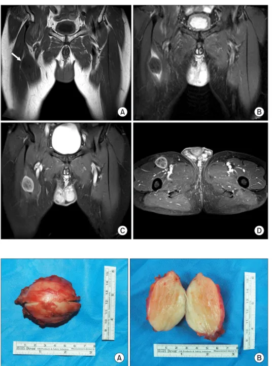
On the preoperative magnetic resonance imaging (MRI), the mass showed a hypointense signal relative to the surrounding muscle, and had an ill-defined margin on T1-weighted sequences. On T2- weighted sequences, the mass had a lower signal intensity than fat, but a slightly higher signal intensity than the muscle. The mass had heterogeneous signal intensity, an ill-defined margin, and a periph- eral rim enhancement. There was no bony involvement or abnormal intramedullary signal changes (Fig. 2).
The patient underwent a surgical excision of the tumor. The tu-
mor has more likely benign nature in preoperative MRI, so we per- formed marginal excision despite of the possibility of malignancy.
The mass was carefully dissected from the sartorius muscle using electrocautery. The excised mass was oval-shaped, 6×5×4 cm in size, and had a smooth boundary. On the cut section, the cut surface showed a yellow homogeneous appearance. There was no hemor- rhage or necrosis within the tumor. The mass extended to the resec- tion margins of the specimen at multiple points (Fig. 3).
The histological examination revealed that the tumor was com- posed of ill-defined cords and nests of oval epithelioid cells that had abundant polygonal cells with granular eosinophilic cytoplasm
Figure 2. (A) Coronal non-contrast T1- weighted image of the pelvis shows a low signal intensity mass within the mid portion of the sartorius muscle measuring 6×5×4 cm in size (arrow). (B) Coronal non-contrast T2-weighted image shows a low signal intensity mass with peripheral ovoid high signal intensity. (C) T1-weighted enhanced image shows a heterogeneous enhancing mass with a clear margin. (D) Axial T1- weighted enhanced image shows a mass in the mid portion of the sartorius muscle.
Figure 3. (A) The submitted specimen was comprised of a well demarcated ovoid mass measuring 4×5×6 cm in size. (B) On sectioning, a pale yellow surface with firm consistency was observed.
in haematoxylin and eosin stain. There were fibrous septae between the clusters, cells with vesicular nuclei with a prominent nucleolus, eosinophilic granular cytoplasm, and eosinophilic intracytoplasmic particles surrounded by a clear halo (Fig. 4). Histological diagnosis was confirmed with granular cell tumor by two pathologists.
The patient had an ordinary post-operative recovery within two weeks. Partial weight bearing was done with clutch postoperatively.
After total stitch out, he was allowed full weight bearing without as- sistive devices. At the 3-month, 6-month, and 1-year follow-up, the operation site was observed carefully, and there is no evidence of recurrence in physical examination and ultrasonography.
DISCUSSION
The characteristics of granular cell tumor are a solitary painless le- sion and unique cytoplasm containing eosinophilic granules.1,6,7) Granular cell tumor usually arises in the nervous system, and can originate in the skin, oral cavity and digestive tract.6-8) However, in- tramuscular granular cell tumor which is located in sartorius muscle is very rare.8) After performing an extensive search of the literature, we could not find any similar cases.
Conventional granular cell tumor is a benign neoplasm. However, malignancy occurs in less than 0.5% to 2% of patients, and they show a higher risk for local recurrence and metastasis with a poor prognosis.3) A large tumor size (>5 cm), older age, female gender, deep location (intramuscular), occurrence in the lower extremities, recent rapid tumor growth after an extended period, and local re-
currence are the known factors that increase the malignant potential of granular cell tumor.3,8) The distinction between a benign and ma- lignant granular cell tumor is difficult based on morphology alone, as the different grades are all histologically similar. The diagnosis of malignant granular cell tumor is based on a combination of histo- logical findings, including cellular pleomorphism and elevated mi- totic activity, and clinical manifestations.2,8) Because of the possibility of malignancy, wide resection around the tumor is recommended.2,3) Furthermore, Rosenthal et al.9) suggested the application of radiation therapy after complete excision. Although most granular cell tumors are managed successfully with surgical excision, radiation therapy has led to limb preservation and better cosmetic results.9)
In imaging studies, granular cell tumors are best evaluated with MRI. The preoperative MRI is helpful to differentiate this tumor from other soft tissue tumors based on some characteristic find- ings.4,5,10) Typically, the tumors are slightly hypointense or isoin- tense on T1-weighted sequences, and show homogeneous contrast enhancement after intravenous injection of gadolinium. On T2- weighted sequences, the tumor generally shows heterogeneous increased signal intensity. Peripheral high intensity seems highly specific to granular cell tumors, especially in those of intramuscular origin. This finding corresponds to the lymphocytic infiltration and inflammation noted at the tumor margin.2,10)
In this case, we could successfully treat the patient with complete surgical excision without radiation therapy. The radiation therapy was considered for this patient, but it was not initiated due to old age. After the operation, he had a satisfactory relief of discomfort in his right anterior thigh, and there was no recurrence at 1 year post- operative.
Hence, preoperative MRI should be performed in a patient having clinical manifestations suspicious for intramuscular granular cell tu- mor. The characteristic MRI findings can help establish the accurate diagnosis and planning resection of the tumor. After establishing the diagnosis, surgeons should remove the tumor completely due to the possibility of malignancy.
REFERENCES
1. Khansur T, Balducci L, Tavassoli M. Granular cell tumor.
Clinical spectrum of the benign and malignant entity. Cancer.
1987;60:220-2.
2. Arai E, Nishida Y, Tsukushi S, Sugiura H, Ishiguro N. Intra- muscular granular cell tumor in the lower extremities. Clin Orthop Relat Res. 2010;468:1384-9.
Figure 4. Histologic examination showed the characteristic appearance of nests of large polyhedral cells with abundant granular cytoplasm (H&E, ×400).
Jun-Bum Kim, et al
3. Fanburg-Smith JC, Meis-Kindblom JM, Fante R, Kindblom LG. Malignant granular cell tumor of soft tissue: diagnostic criteria and clinicopathologic correlation. Am J Surg Pathol.
1998;22:779-94.
4. Elkousy H, Harrelson J, Dodd L, Martinez S, Scully S. Granu- lar cell tumors of the extremities. Clin Orthop Relat Res.
2000;380:191-8.
5. Thacker MM, Humble SD, Mounasamy V, Temple HT, Scully SP. Case report. Granular cell tumors of extremities: compari- son of benign and malignant variants. Clin Orthop Relat Res.
2007;455:267-73.
6. Ordóñez NG. Granular cell tumor: a review and update. Adv Anat Pathol. 1999;6:186-203.
7. Lack EE, Worsham GF, Callihan MD, et al. Granular cell tu- mor: a clinicopathologic study of 110 patients. J Surg Oncol.
1980;13:301-16.
8. Tsuchida T, Okada K, Itoi E, Sato T, Sato K. Intramuscular malignant granular cell tumor. Skeletal Radiol. 1997;26:116- 21.
9. Rosenthal SA, Livolsi VA, Turrisi AT 3rd. Adjuvant radiother- apy for recurrent granular cell tumor. Cancer. 1990;65:897- 900.
10. Blacksin MF, White LM, Hameed M, Kandel R, Patterson FR, Benevenia J. Granular cell tumor of the extremity: magnetic resonance imaging characteristics with pathologic correlation.
Skeletal Radiol. 2005;34:625-31.
봉공근 내 발생한 과립세포종
김준범 • 최광민 • 권세원 • 노재휘 • 서유성 • 박종석
순천향대학교 천안병원 정형외과
과립 세포종은 신경계통에서 기원하는 연부 조직 종양이며 매우 드문 질환으로, 통증이 없는 단독 종괴로 나타난다. 과립 세포종은 인체 내 어떤 곳에서도 발생할 수 있지만 대부분은 피부, 구강 내, 호흡기, 소화기관에 발생하며 근육 내의 발생이 보고되는 경우는 매우 드물다. 저자들은 대퇴 전방부의 종물을 주소로 내원한 71세 남자 환자에서 발생한 봉공근 내부의 과립 세포종 1예를 경험하여 문헌 고찰과 함께 보고하는 바이다.
색인단어: 과립세포종, 신생물, 근육내 종물, 악성
접수일 2013년 6월 12일 수정일 2013년 7월 29일 게재확정일 2013년 10월 23일 책임저자 권세원
천안시 동남구 순천향6길 31, 순천향대학교 천안병원 정형외과
TEL 041-570-2170, FAX 041-572-7234, E-mail [email protected]
Copyright © 2014 by The Korean Orthopaedic Association
“This is an Open Access article distributed under the terms of the Creative Commons Attribution Non-Commercial License (http://creativecommons.org/licenses/by-nc/3.0/) which permits unrestricted non-commercial use, distribution, and reproduction in any medium, provided the original work is properly cited.”
대한정형외과학회지:제 49권 제 1호 2014