악교정 수술을 동반한 임플란트 보철물을 이용한 완전구강회복 증례
안혜림
1∙허지예
1∙김철훈
2∙황희성
1∙김복주
2* 동아대학교 의료원
1치과보철학교실,
2구강악안면외과학교실
c cc
2016 The Korean Academy of Prosthodontics
This is an Open Access article distributed under the terms of the Creative Commons Attribution Non-Commercial License (http://creativecommons.org/licens- es/by-nc/3.0) which permits unrestricted non-commercial use, distribution, and reproduction in any medium, provided the original work is properly cited.
*Corresponding Author: Bok-Joo Kim
Department of Oral and Maxillofacial Surgery, Implant Clinics, Dong-a University, Medical Center, 26 Daesingongwon-ro, Seo-gu, Busan 49201, Republic of Korea +82 51 240 5475: e-mail, [email protected]
Article history: Received July 29, 2015 / Last Revision August 27, 2015 / Accepted September 2, 2015
Introduction
Full-mouth rehabilitation with fixed partial dentures has been a common treatment option for replacement of multiple missing teeth and improvement of patients’quality of life.1Multiple miss- ing teeth that result of decay, periodontal disease or trauma, usually lead to problems concerning esthetics, phonetics, and mastica- tion. Moreover, total treatment period also lead to problems concerning to full mouth rehabilitation patients.
Similarly, patients with skeletal class III malocclusion can encounter complex dentoalveolar problems, including mandibular prognathism and maxillary retrognathism. Patients with inter- maxillary skeletal discrepancy might require multidisciplinary
treatment entailing prosthodontics, orthodontics, and oral and maxillofacial surgery for both precise diagnosis and comprehensive treatment.2If an ideal occlusal relationship cannot be achieved by prosthetic treatment alone, orthodontic treatment is recommended;
and if there is severe skeletal discrepancy, orthognathic surgery should be accompanied.3
Immediate implantation has become a commonly utilized treat- ment option in implant dentistry for reducing treatment period and prevents the alveolar bone-volume loss that otherwise results from extraction. Immediate dental implantation has several advan- tages: enhanced opportunity for ideal axial positioning of an implant, reduction of total treatment duration, fewer surgeries, and a positive psychological effect on the patient.4Most researchers
Full-mouth rehabilitation by immediate implantation combined with orthognathic surgery:
a clinical report
Hye-Rim Ahn
1, Ji-Ye Heo
1, Chul-Hoon Kim
2, Hee-Seong Hwang
1, Bok-Joo Kim
2*
1Department of Prosthodontics, 2Department of Oral and Maxillofacial Surgery, Implant Clinics, Dong-a University Medical Center, Busan, Republic of Korea
Clinical therapy that combines full-mouth rehabilitation with immediate implantation and orthognathic surgery poses a challenge to prosthodontists. This clinical report describes a multidisciplinary approach to the diagnosis and treatment of a patient presenting with skeletal discrepancy and rampant caries. The results thus achieved indicate that full-mouth rehabilitation by fixed immediate and early loading implantation accompanied by orthognathic surgery can be a predictable and effective treatment procedure. (J Korean Acad Prosthodont 2016;54:57-64)
Key words: Full-mouth rehabilitation; Immediate implant insertion; Early implant loading; Orthognathic surgery
ic surgery. The patient’s diagnosis, treatment planning, total treat- ment period and the achieved 1-year stability are described herein.
Case report
A 40-year-old male visited the Department of Oral and Maxillofacial Surgery, Dong-A University Medical Center, Busan, South Korea, in 2012. His chief complaint was multiple dental caries with ante- rior crossbite. He was subsequently referred to the Department of Prosthodontics for full-mouth rehabilitation. The patient had no med- ical history to contraindicate dental treatment.
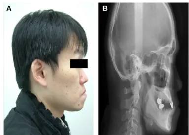
patient also had 4 mm of interocclusal distance. All of this meant that the patient had a loss of occlusal vertical dimension. The patient was diagnosed as skeletodental class III with rampant caries (Fig. 3).
As the remaining teeth were in very poor condition, total extrac- tion was planned for all of the teeth. For improved intermaxillary relation and facial profile, full-arch dental implantation supplemented by orthognathic surgery was needed. However, because there was no midfacial deficiency or facial asymmetry, and given that the patient expressed a desire for less invasive treatment, bilateral sagittal split ramus osteotomy (BSSRO) of the mandible was planned as the
Fig. 1. Intraoral photographs of patient before treatment. (A) Maxillary occlusal view, (B) Right side, (C) Frontal view, (D) Left side, (E) Mandibular occlusal view.
B C
E A
D
sole treatment. For reducing total treatment period and guide for mandibular setback, initiation of treatment was full arch extraction of the maxillary teeth and immediate implantation with socket elevation. Immediate implantation of the mandibular teeth was planed after BSSRO because of considering post operative relapse after BSS- RO.
Prior to the orthognathic surgery, all of the remaining teeth in the maxilla were extracted, and 10 dental implants (s-clean Tapered II;
Dentis, Seoul, Korea)were immediately inserted. At the #16, 17, 26, and 27 extraction sites simultaneously socket elevation were per- formed.
The ideal implant positioning for a maxillary edentulous arch includes at least one central incisor position, bilateral canine positions, bilateral first premolar sites, and bilateral sites in the distal half of the first molars according to guidelines for key implant positions.
One week later, provisional fixed prosthodontic restorations were fabricated. The final surgical treatment objective (STO) for the BSS- RO was planned on the basis of these provisional restorations, and BSSRO was performed under general anesthesia. The class I rela- tionship was achieved by setback positioning of the mandible.
At one month after orthognathic surgery, all of the mandibular teeth were extracted and 7 dental implants (s-clean Tapered II; Dentis, Seoul, Korea) were immediately inserted into the #34, 35, 37, 41, 44, 45 and 46 extraction sockets. At the #33 and 43 extraction sites, because primary stability had not been achieved, socket preserva- tion was performed as a preparation to the planned delayed implant placement. Additionally, implantation at the #41 site was per- formed to accommodate a provisional prosthetic restoration.
One week after implantation in the mandible, both maxillary and mandibular impressions for the provisional restoration were formed
in polyvinyl siloxane material (ImprintTMII GarantTM; 3M ESPE, St.
Paul, MN, USA) using a custom impression tray. The occlusal rim was fabricated on a working cast. The increment of the vertical dimen- sion was determined to be 2 mm using a Leaf gauge. The assembly was mounted on a semi-adjustable articulator (HanauTMModular Articulator; Whip Mix Corp., Louisville, KY, USA) using the face-bow and centric record (CR) at the predetermined vertical dimen- sion. A wax-up was done in the ideal form, and the occlusion was given canine guidance (Fig. 4). The wax-up was scanned for fabrication of a CAD/CAM titanium customized abutment (MyplantTM; Raphabio Co., Seoul, Korea). Using the putty index of the wax-up, provisional restorations also were made. The cus- tomized abutment was installed and tightened to 25 Ncm, the provisional restorations were cemented (TempBond; Kerr, Romulus, MI, USA), and occlusal adjustment was performed (Fig. 5).
Five months after socket preservation, dental implants (s-clean Tapered II; Dentis, Seoul, Korea) were inserted at the #33 and 43 sites.
The patient, with his delivered provisional restorations at the cor- rected vertical dimension, had no temporomandibular disorder (TMD), pronunciation or deglutition problems for 2 months.
Therefore, provisional prostheses were used to facilitate fabrication of the definitive restorations, to which end, mandibular final impressions were formed in polyvinyl siloxane material, and a cus- tomized anterior guidance table was established. The CR record was made between the anterior customized abutment using polyvinyl silox- ane (O-BiteTM; DMG, Hamburg, Germany), while the posterior pro- visional restorations were kept in place to maintain the occlusal ver- tical dimension. Another CR record was made, in a similar manner, between the posterior abutments. This assembly was then re- mounted, and the definitive restorations were fabricated, according Fig. 2. Panoramic radiograph of patient before treatment.
Fig. 3. (A) Initial lateral view of face, (B) Initial lateral cephalometric radiograph.
A B
to the custom incisal guide table, as the cemented type for the ante- rior area and the screw-cemented-retained prostheses (SCRP) type for the posterior area. The definitive prostheses were cemented with resin (PremierTMImplant CementTM; Premier Co., Plymouth Meeting, PA, USA) (Fig. 6, Fig. 7).
After final prosthesis cementation, the patient, who had been pro- vided with thorough instruction in proper oral hygiene, was recalled
for follow-up appointments. As needed, occlusal re-adjustment of the restoration was performed. Subsequently, it was determined that the patient’s facial profile and masticatory ability had been considerably improved (Fig. 8). Six months after the definitive-prostheses cementation, the patient demonstrated a favorable prognosis.
Finally, total treatment period took about 14 months in this case (Fig. 9).
Fig. 4. Diagnostic wax-up. The diagnostic wax-up was done in the ideal form. The occlusion was given canine guidance. (A) Right side, (B) Frontal view, (C) Left side.
Fig. 5. Intraoral photographs at provisional prostheses delivery. (A) Maxillary occlusal view, (B) Right side, (C) Frontal view, (D) Left side, (E) Mandibular occlusal view.
B C
E A
D
Fig. 6. Intraoral photographs at definitive prostheses delivery. (A) Maxillary occlusal view, (B) Right side, (C) Frontal view, (D) Left side, (E) Mandibular occlusal view.
B C
E A
D
Fig. 7. Panoramic radiograph at definitive prostheses insertion.
Fig. 8. (A) Final lateral view of face, (B) Final lateral cephalometric radiograph.
A B
Discussion
For the patient’s esthetics and rapid recovery to full functional- ity, the total treatment period needed to be shortened. To achieve this, the following two steps were designed. First, the dental implant was inserted immediately after tooth extraction with socket elevation in maxillary molar position, without allowing for any healing time.
Second, early loading was applied as temporary prostheses were installed. Most researchers in fact have asserted that immediate and delayed implantations have similar success rates found that imme- diate implantation relative to delayed implantation decreases bone resorption.5-8In terms of early loading, whereas some studies have claimed that it has a lower implant success rate compared with con- ventional loading, however, a more recent investigation found no failure, marginal bone loss or postoperative infection rate differences between immediately loaded non-submerged dental implants and delayed loaded submerged implants.9Robling and Turner10report- ed that for successful osseointegration, primary stability during implan- tation is most important. And the key factor determining primary sta- bility is insertion torque.11In the present case, the insertion torque was over 35 Ncm, and was sufficient to obtain primary stability. Also,
In performing immediate implantation, both the vertical and the horizontal position of the implant must be considered, in that heal- ing of the extraction socket alters the peri-implant’s hard tissue.14 In the present case, the rough portion of each of the fixtures in the patient’s premolar area was exposed. Calvo-Guirado et al.15showed that during an 8-week healing period after tooth extraction, there was remarkable hard-tissue remodeling that altered both the buccal and the lingual bone. Caneva et al.16concluded that in order to reduce exposure above the alveolar crest of the rough portion of the fixture, implants should be positioned approximately 1 mm below the alveolar crest and in a lingual position in relation to the middle of the alveolar ridge. Certainly, determination of implant position preparatory to immediate implantation requires consideration of the phenomenon of bone alteration.
Despite efforts, the overall treatment period was 14 months, from December 2012 to February 2014. There were two reasons for this. First, immediate implantation following tooth extraction was not possible for the mandibular canines. For minimal primary sta- bility of those implants, at least 3 - 5 mm of bone generally is required at the implant apex (or, an implant wider than the extraction sock- et has to be used).17 In the present case, the root length of the mandibular canine was about 15 mm, and the root width was about 6.2 mm at the crest bone. Therefore, the types of dental implants that can obtain sufficient primary stability in the mandibular canine area are limited. Thus, implant placement on the mandibular canine was delayed for 5 months. The second reason was the fact that orthognathic surgery was performed prior to the mandibular implan- tation, not simultaneously in the maxilla and mandible. This sequential procedure allows minimizing the influence of early horizontal relapse after BSSRO which ranges widely from 0.2 to 4.2 mm.18,19 That is, when performing implantation after horizontal relapse has completed, the relapse extent can be compensated for by adjusting the axial position of implant.
Alternatively, if a surgical template for implantation and ball stent for orthognathic surgery were made using the diagnostic wax-up already utilized for the provisional prosthesis, simultaneous implant placement in the maxilla and mandible would be possible which would short- en the total treatment period and secure postoperative stability.
However, simultaneous implant placement remains controversial.
Fig. 9. Overall treatment procedure. The overall treatment period was 15 months, from December 2012 to March 2014. The B, C, E, and F periods represent indispensable time. However, the A period could be reduced by immediate loading, and the D period could be reduced by positioning implantsin the #32 and 42 extraction sockets. (A, D periods: variable time, black line; B, C, E, F periods:
indispensable time, red line; M: month(s))
Full-mouth rehabilitation aims to achieve ideal functionality and esthetics for the patient. Given the extensiveness of such treatment, adaptation of the neuromuscular system can be also required in order to accommodate the sometimes-necessary alteration of the occlusal vertical dimension. A newly established occlusal vertical dimension must be evaluated for 1 - 3 months to determine whether the patient is adapting or exceeding the adaptability of the neuromus- cular system.20In the present case, the patient had no problems (e.g.
TMD, pronunciation or deglutition) for 2 months. Therefore, pro- visional prostheses were used to facilitate the fabrication of the defin- itive restorations.
Upon completion of the current patient’s final treatment, not only the facial profile but also the functional occlusal relation was improved. And although the patient needed periodic maintenance and re-evaluation due to poor oral hygiene, he has maintained a favor- able prognosis for 1 year without encountering any complica- tions. However, we could not reduce the overall treatment period to less than 14 months because a limitation of the procedure at #33, 43 extraction site implants insertion. It would be possible reducing total treatment period to 4 months if changing the key position of implant in the mandible but, it is still questionable (Fig. 9).
ORCID
Hye-Rim Ahn http://orcid.org/0000-0003-0813-5711
References
1. Ravindran DM, Sudhakar U, Ramakrishnan T, Ambalavanan N.
The efficacy of flapless implant surgery on soft-tissue profile comparing immediate loading implants to delayed loading implants: A comparative clinical study. J Indian Soc Periodontol 2010;14:245-51.
2. Williams AC, Shah H, Sandy JR, Travess HC. Patients' motivations for treatment and their experiences of orthodontic preparation for orthognathic surgery. J Orthod 2005;32:191-202.
3. Janson M, Janson G, Sant'Ana E, Tibola D, Martins DR.
Orthognathic treatment for a patient with Class III malocclusion and surgically restricted mandible. Am J Orthod Dentofacial Orthop 2009;136:290-8.
4. Lindeboom JA, Tjiook Y, Kroon FH. Immediate placement of implants in periapical infected sites: a prospective randomized study in 50 patients. Oral Surg Oral Med Oral Pathol Oral Radiol Endod 2006;101:705-10.
5. Degidi M, Piattelli A, Carinci F. Immediate loaded dental implants:
comparison between fixtures inserted in postextractive and healed bone sites. J Craniofac Surg 2007;18:965-71.
6. Pieri F, Aldini NN, Fini M, Corinaldesi G. Immediate occlusal loading of immediately placed implants supporting fixed restora-
tions in completely edentulous arches: a 1-year prospective pi- lot study. J Periodontol 2009;80:411-21.
7. Raes F, Cooper LF, Tarrida LG, Vandromme H, De Bruyn H. A case-control study assessing oral-health-related quality of life af- ter immediately loaded single implants in healed alveolar ridges or extraction sockets. Clin Oral Implants Res 2012;23:602-8.
8. Meizi E, Meir M, Laster Z. New-design dental implants: a 1-year prospective clinical study of 344 consecutively placed implants comparing immediate loading versus delayed loading and flap- less versus full-thickness flap. Int J Oral Maxillofac Implants 2014;29:e14-21.
9. Chrcanovic BR, Albrektsson T, Wennerberg A. Immediately loaded non-submerged versus delayed loaded submerged dental implants:
a meta-analysis. Int J Oral Maxillofac Surg 2015;44:493-506.
10. Robling AG, Turner CH. Mechanical signaling for bone mod- eling and remodeling. Crit Rev Eukaryot Gene Expr 2009;19:319- 38.
11. Wang HL, Boyapati L. "PASS" principles for predictable bone regeneration. Implant Dent 2006;15:8-17.
12. Vogl S, Stopper M, Hof M, Wegscheider WA, Lorenzoni M.
Immediate Occlusal versus Non-Occlusal Loading of Implants:
A Randomized Clinical Pilot Study. Clin Implant Dent Relat Res 2015;17:589-97.
13. Lang NP, Pun L, Lau KY, Li KY, Wong MC. A systematic review on survival and success rates of implants placed immediately in- to fresh extraction sockets after at least 1 year. Clin Oral Implants Res 2012;23:39-66.
14. Tomasi C, Sanz M, Cecchinato D, Pjetursson B, Ferrus J, Lang NP, Lindhe J. Bone dimensional variations at implants placed in fresh extraction sockets: a multilevel multivariate analysis. Clin Oral Implants Res 2010;21:30-6.
15. Calvo-Guirado JL, Boquete-Castro A, Negri B, Delgado Ruiz R, Go′mez-Moreno G, Iezzi G. Crestal bone reactions to immedi- ate implants placed at different levels in relation to crestal bone.
A pilot study in Foxhound dogs. Clin Oral Implants Res 2014;
25:344-51.
16. Caneva M, Salata LA, de Souza SS, Baffone G, Lang NP, Botticelli D. Influence of implant positioning in extraction sockets on osseointegration: histomorphometric analyses in dogs. Clin Oral Implants Res 2010;21:43-9.
17. De Rouck T, Collys K, Cosyn J. Single-tooth replacement in the anterior maxilla by means of immediate implantation and pro- visionalization: a review. Int J Oral Maxillofac Implants 2008;23:
897-904.
18. Kobayashi T, Watanabe I, Ueda K, Nakajima T. Stability of the mandible after sagittal ramus osteotomy for correction of prog- nathism. J Oral Maxillofac Surg 1986;44:693-7.
19. Proffit WR, Phillips C, Dann C 4th, Turvey TA. Stability after sur- gical-orthodontic correction of skeletal Class III malocclusion.
I. Mandibular setback. Int J Adult Orthodon Orthognath Surg 1991;6:7-18.
20. Carlsson GE, Ingervall B, Kocak G. Effect of increasing verti- cal dimension on the masticatory system in subjects with natural teeth. J Prosthet Dent 1979;41:284-9.
악교정 수술과 임플란트 보철을 통한 전악재건술의 임상적 치료는 보철과 의사에게 도전적인 과제이다. 본 증례는 골격적 부조화를 가진 다발성 우식증 환자의 진단과 치료에 있어서 치의학의 여러 분야의 협진을 보여주었다. 그 결과로 악교정 수술을 동반한 발치 후 즉시 임플란트 식립 및 조 기 하중을 가하는 임플란트 보철물에 의한 전악 재건이 예견가능하고 효과적인 치료 방법이었기에 이를 보고하고자 한다. (대한치과보철학회지 2016;54:57-64)
주요단어: 완전 구강 회복술; 즉시 임플란트 식립; 조기 임플란트 하중; 악교정 수술
*교신저자: 김복주
49201 부산 서구 대신공원로 26 동아대학교의료원 구강악안면외과학교실 051-240-5475: e-mail, [email protected]
원고접수일: 2015년 7월 29일 / 원고최종수정일: 2015년 8월 27일 / 원고채택일: 2015년 9월 1일
2016 대한치과보철학회
이 글은 크리에이티브 커먼즈 코리아 저작자표시-비영리 3.0 대한민국 라이선스에 따라 이용하실 수 있습니다.
c cc