Clinical Outcomes of Cryopreserved Arterial Allograft Used as a Vascular Conduit for Hemodialysis
This single center cohort study aimed to test the hypothesis that use of a cryopreserved arterial allograft could avoid the maturation or healing process of a new vascular access and to evaluate the patency of this technique compared with that of vascular access using a prosthetic graft. Between April 2012 and March 2013, 20 patients underwent an upper arm vascular access using a cryopreserved arterial allograft for failed or failing vascular accesses and 53 using a prosthetic graft were included in this study. The mean duration of catheter dependence, calculated as the time interval from upper arm access placement to removal of the tunneled central catheter after successful cannulation of the access, was significantly longer for accesses using a prosthetic graft than a cryopreserved arterial allograft (34.4 ± 11.39 days vs. 4.9 ± 8.5 days, P < 0.001). In the allograft group, use of vascular access started within 7 days in 16 patients (80%), as soon as from the day of surgery in 10 patients. Primary (unassisted; P = 0.314) and cumulative (assisted;
P = 0.673) access survivals were similar in the two groups. There were no postoperative complications related to the use of a cryopreserved iliac arterial allograft except for one patient who experienced wound hematoma. In conclusion, upper arm vascular access using a cryopreserved arterial allograft may permit immediate hemodialysis without the maturation or healing process, resulting in access survival comparable to that of an access using a prosthetic graft.
Keywords: Allografts; Cryopreservation; Renal Insufficiency; Vascular Access Devices Tae-Yong Ha,1 Young Hoon Kim,1
Jai Won Chang,2 Yangsoon Park,3 Youngjin Han,1 Hyunwook Kwon,1 Tae-Won Kwon,1 Duck Jong Han,1 Yong-Pil Cho,1 and Sung-Gyu Lee1
1Department of Surgery, University of Ulsan College of Medicine and Asan Medical Center, Seoul, Korea;
2Department of Internal Medicine University of Ulsan College of Medicine and Asan Medical Center, Seoul, Korea; 3Department of Pathology, University of Ulsan College of Medicine and Asan Medical Center, Seoul, Korea
Received: 17 August 2015 Accepted: 12 April 2016 Address for Correspondence:
Yong-Pil Cho, MD
Department of Surgery, University of Ulsan College of Medicine, Asan Medical Center, 88 Olympic-ro 43-gil, Songpa-gu, Seoul 05505, Korea
E-mail: [email protected]
Funding: This study was supported by a grant (grant number 2012-0557) from the Asan Institute for Life Sciences, Asan Medical Center, Seoul, Korea.
http://dx.doi.org/10.3346/jkms.2016.31.8.1266 • J Korean Med Sci 2016; 31: 1266-1272
INTRODUCTION
Morbidity and mortality in end-stage renal disease (ESRD) pa- tients undergoing hemodialysis have been associated with di- alysis efficiency (1). International guidelines recommend an autogenous arteriovenous fistula (AVF), created in the arm, for safe and long-term vascular access (1-3). However, short- and long-term AVF dysfunction, including failed maturation, vein thrombotic occlusion, aneurysmal changes, and infection, are the major causes of morbidity and hospitalization in hemodial- ysis patients. Indeed, the primary patency rate of AVFs at 2 years was recently estimated to be less than 60% (4). Pooled data for the DOQI analysis suggested that the primary patency rate of arteriovenous prosthetic dialysis grafts (AVGs) was approximate- ly 50% at 1 year, although other studies have reported primary patency rates as low as 23% at 1 year and 4% at 2 years (5). There- fore, salvaging a failed or failing vascular access can also prevent the need for use of a central venous catheter or a new prosthetic vascular access. However, urgent dialysis via a temporary cen- tral venous catheter is required and may be life-saving for chron- ic hemodialysis patients with a non-salvageable, failed or failing vascular access. Although various techniques have been report-
ed to reduce temporary hemodialysis catheter-related compli- cations, mechanical and infectious complications related to the insertion of temporary hemodialysis catheter can be fatal (6).
Cryopreserved cadaveric vascular allografts offer the potential advantage of decreased infection (7) and could be used as an effective alternative to salvage a failed or failing vascular access in an infected graft environment.
This single center cohort study assessed the ability of a cryo- preserved iliac arterial allograft from a deceased donor to act as a vascular conduit in patients with failed or failing vascular ac- cess, non-salvageable by any surgical or endovascular revision procedures. This study also evaluated the ability of this tech- nique to permit immediate hemodialysis without the use of a central venous catheter. In addition, primary (unassisted) and cumulative (assisted) access survivals were compared in pa- tients undergoing vascular access using a cryopreserved arteri- al allograft and a prosthetic graft.
MATERIALS AND METHODS Study design and patient population
Between April 2012 and March 2013, 386 vascular accesses were
created to enable hemodialysis at our institution. Of these 386 operations, 20 (5.2%) established an upper arm vascular access using a cryopreserved iliac arterial allograft from a deceased donor. Patients were included in this group if they 1) had ESRD treated chronically by hemodialysis, 2) provided informed con- sent, 3) had no option for salvaging dysfunctional vascular ac- cess and no adequate potential future access site, and 4) were not candidates for renal transplantation. ABO blood type com- patibility was not considered in the inclusion criteria. No tissue matching was performed and no immunosuppression was giv- en to the patients. To compare the patency of this technique, patients with an upper arm vascular access using a prosthetic graft during the same study period were included as a control group. In addition, the demographic and clinical characteristics of the deceased donors and the duration of cryopreservation were obtained from the Korean Network for Organ Sharing (KO- NOS). Potential risk factors, clinical characteristics, and treat- ment outcomes had been recorded prospectively in an Excel database (Microsoft Corp., Redmond, WA, USA) and were ana- lyzed retrospectively as part of this study.
Procurement of vascular tissues from deceased donors and cryopreservation techniques
This study employed human arterial tissues from deceased multi- organ donors. All procedures for vascular tissue procurement and processing were in compliance with Korean legislation (Law 5,858/1999 and Law 11,976/2013) and conformed to the ethical and safety concerns for therapeutic use.
The aorto-iliac arterial allograft was obtained aseptically from each anonymized donor diagnosed with brain death during the course of multi-organ procurement. The aorto-iliac arterial al- lograft was washed with saline solution and stored at 4°C. For cryopreservation, washed arterial allografts were immersed in cryopreservation solution, consisting of 90% culture medium (RPMI 1640) and 10% dimethylsulfoxide (DMSO) in a cryostor- age bag (Medi-Rution, Gyeonggi-do, Korea) at 20°C (8). The bag was sealed in a laminar flow cabinet. Programmed cryopreser- vation was performed in a Controlled Rate Freezing System (Model 14S-B®, SY-LAB Geräte GmbH, Neupurkersdorf, Aus- tria), as described (9). The protocol consisted of a slow, pro- grammed cooling at a mean rate of 1°C/min, to -70°C. The bag was immediately transferred to the gas phase of a liquid nitro- gen compartment, followed by rapid cooling to -196°C. The de- frost protocol was a two-stage rewarming process (9), consist- ing of slow warming by transferring the bag from the nitrogen gas phase to room temperature (20°C) over 30 minutes, follow- ed by rapid warming by immersing the bag in a water bath at 40°C until the contents were completely defrosted. The cryo- protectant liquid was gradually removed in four 3-minute steps by immersion in tapering concentrations of DMSO (10%, 5%, 0%, and 0%) at 4°C.
Surgical technique
Before surgery, all patients provided written informed consent.
Selected patients underwent preoperative duplex scanning or venography. In the allograft group, all operations were perform- ed at the site of the previously placed vascular access under lo- cal anesthesia. Upper arm hemodialysis vascular access grafts required exposure of the axillary vein and brachial artery in the axilla and antecubital fossa, respectively. In the prosthetic graft group, the tapered 4- to 6-mm expanded polytetrafluoroethyl- ene (PTFE) prosthetic graft, tunneled subcutaneously in a strai- ght configuration, was anastomosed to the vein and artery end- to-side using 6-0 Prolene. In the allograft group, the cryopre- served aorto-iliac arterial allograft was thawed (9). To adequate- ly modify the diameter and length of the cryopreserved arterial allograft, the common and/or external iliac arterial segments were isolated and tributaries were ligated. To lengthen the vas- cular conduit, both arterial segments were anastomosed con- tinuously with each other end-to-end using 6-0 Prolene. The cryopreserved arterial allograft, tunneled subcutaneously in a straight configuration, was anastomosed to the vein and artery end-to-side using 6-0 Prolene. Duplex investigation and surveil- lance were performed only when dialysis was poor.
Definitions and statistical analyses
A successful hemodialysis was defined as a graft that had used successfully for at least three dialysis sessions. These patients were prospectively evaluated from the time of access placement until cumulative access failure to determine: 1) duration of cath- eter dependence from access placement to successful cannula- tion of the vascular access, 2) primary (unassisted) access sur- vival, 3) cumulative (assisted) access survival, and 4) total num- ber of interventions during the life of the access (10). Primary access failure was defined as an access never usable for dialysis.
The duration of catheter dependence was calculated as the time interval from upper arm access placement to removal of the tunneled central catheter after successful cannulation of the ac- cess. Primary (unassisted) access survival was calculated from the access placement to the first intervention required to main- tain its patency for dialysis. Cumulative (assisted) access sur- vival was calculated from the access placement to permanent failure, regardless of the number of interventions for the main- tenance of its patency.
The patients’ demographic and clinical characteristics were analyzed using the Mann-Whitney U-test, Pearson’s χ2 test, or Fisher’s exact test, as appropriate. Primary (unassisted) and cu- mulative (assisted) access survivals, stratified by graft materials (prosthetic graft or cryopreserved iliac arterial allograft), were plotted using the Kaplan-Meier method, with patient follow-up censored for death, renal transplant, or transfer to a nonpartici- pating dialysis unit. Access survival in the two groups was com- pared using the log-rank test. All statistical analyses were per-
formed using SPSS software (version 18.0; SPSS, Chicago, IL, USA), with P values ≤ 0.05 considered statistically significant.
Ethics statement
The study protocol was approved by the institutional review board of Asan Medical Center (IRB No.: 2012-0557). Informed consent was confirmed by the board.
RESULTS
Patient population
Of the 386 hemodialysis patients who underwent vascular ac- cess placement, 20 (5.2%) received an upper arm vascular ac- cess of a cryopreserved iliac arterial allograft (allograft group) and 53 (13.7%) underwent upper arm vascular access using an expanded PTFE prosthetic graft (prosthetic graft group) (Fig. 1).
Demographic and clinical characteristics
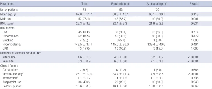
Table 1 summarizes the demographic and clinical characteris- tics of these two patient groups. The percentage of males was significantly higher in the prosthetic graft than in the allograft group (P = 0.001). However, age and other atherosclerosis risk factors were similar in the two groups. Primary access failure did not occur in either group. In the allograft group, the causes of a failed or failing previous vascular access included venous stenosis followed by thrombosis in ten patients (50.0%), diffuse aneurysmal dilation with thrombosis in six (30.0%), and gross vascular access graft infection in four (20.0%). The mean base- line diameters of the vascular conduit on the arterial (6.2 ± 0.7 mm, P < 0.001) and venous (7.1 ± 1.6 mm, P < 0.001) sides in
the allograft group were significantly greater than those in the prosthetic graft group, with all of the latter receiving vascular access using tapered 4- to 6-mm prosthetic grafts.
Table 1. Baseline characteristics of the study population
Parameters Total Prosthetic graft Arterial allograft* P value
No. of patients 73 53 20
Mean age, yr 67.8 ± 11.7 68.9 ± 12.1 65.1 ± 10.7 0.119
Male sex 57 (78.1) 47 (88.7) 10 (50.0) 0.001
BMI, kg/m2 22.3 ± 3.2 22.4 ± 3.3 21.9 ± 2.9 0.634
Risk factors DM Hypertension Smoking Hyperlipidemia† CAD
45 (61.6) 62 (84.9) 4 (5.5) 143.5 ± 37.1
13 (17.8)
32 (60.4) 46 (86.8) 3 (5.7) 145.0 ± 36.0
10 (18.9)
13 (65.0) 16 (80.0) 1 (5.0) 139.4 ± 40.6
3 (15.0)
0.717 0.479 1.000 0.404 1.000 Diameter of vascular conduit, mm
Artery side
Vein side 4.6 ± 1.0
6.3 ± 0.9 4.0 ± 0.0
6.0 ± 0.0 6.2 ± 0.7
7.1 ± 1.6 < 0.001
< 0.001 Clinical factors
CV catheter‡ Time to use, day§ Interventionll Antiplatelet use Follow-up, mon
7 (9.6) 26.1 ± 17.0
1.1 ± 1.2 36 (49.3) 18.6 ± 8.6
6 (11.3) 34.4 ± 11.39
1.1 ± 1.2 26 (49.1) 18.4 ± 8.8
1 (5.0) 4.9 ± 8.5 1.1 ± 1.3 10 (50.0) 18.8 ± 8.3
0.665 < 0.001 0.735 0.943 0.862 Continuous data are expressed as mean ± SD, and categorical data as numbers (%).
BMI, body mass index; DM, diabetes mellitus; CAD, coronary artery disease; CV catheter, ipsilateral central venous catheter.
*Cryopreserved iliac arterial allograft from deceased donor; †Total cholesterol level > 200 mg/Dl; ‡Use of an ipsilateral central venous catheter for hemodialysis at the time of access placement; §Mean duration of catheter dependence until successful cannulation of the upper arm access; llTotal rate of interventions for the life of the access.
AVF creation
n = 270 AVG creation
n = 116
VA revision (n = 55) Thrombectomy (n = 35) Interposition bypass (n = 20)
Other surgery (n = 51) Ligation (n = 25) Branch ligation (n = 14) Prosthetic graft removal (n = 12) Vascular access (VA) surgery
n = 492
VA creation n = 386
Prosthetic graft pre-HD (n = 11) on HD (n = 53)
Cryopreserved allograft (on HD) n = 20
Forearm straight n = 2
Forearm U-loop n = 25
Arm straight n = 84
Thigh U-loop n = 5
Fig. 1. Flow chart of patient inclusion.
AVF, arteriovenous fistula; AVG, arteriovenous graft; HD, hemodialysis; pre-HD, ac- cess placement before initiation of hemodialysis; on HD, access placement after initi- ation of hemodialysis.
At the time of access placement, there was no definite steno- sis or occlusion of the central vein and use of an ipsilateral cen-
tral venous catheter for hemodialysis did not differ in the two groups (P = 0.665) (Table 1). The mean duration of catheter de- pendence was significantly longer in the prosthetic graft than in the allograft group (34.4 ± 11.39 days vs. 4.9 ± 8.5 days, P < 0.001).
In the allograft group, use of vascular access started within 7 days in 16 patients (80.0%), as soon as from the day of surgery in 10 patients, versus none within 7 days in the prosthetic graft group.
The mean duration of patient follow-up in these two groups was similar (18.4 ± 8.8 months vs. 18.8 ± 8.3 months, P = 0.862).
Table 2 summarizes the demographic and clinical characteris- tics, as well as the causes of brain death, of the multi-organ de- ceased donors in this study.
Pathologic findings of the allograft and matured vein Histologic evaluation of the allograft from a malfunctioned up- per arm vascular access using a cryopreserved arterial allograft revealed fibrous intimal thickening with myxoid degeneration, Table 2. Baseline characteristics of the deceased donors
Parameters No. (%) of deceased donors (n = 20)
Mean age, yr 39.6 ± 11.4
Male sex 12 (60.0)
BMI, kg/m2 23.2 ± 3.8
Risk factors DM Hypertension
1 (5.0) 3 (15.0) Causes of brain death
Multiple trauma ICH
Hypoxic damage
13 (65.0) 4 (20.0) 3 (15.0) Duration of cryopreservation, day* 197.8 ± 212.7
Continuous data are expressed as mean ± SD, and categorical data as numbers (%).
BMI, body mass index; DM, diabetes mellitus; ICH, spontaneous intracerebral hemor- rhage.
*Duration from arterial procurement to use as a vascular conduit.
A B C
Fig. 2. Pathologic findings of the allograft and matured vein. (A) Histologic evaluation of the allograft from a malfunctioned upper arm vascular access shows fibrosis and hya- linization of media and fibrous intimal thickening (H&E, ×100). (B) Elastic staining of the allograft reveals fragmentation of internal elastic lamella, widening of interlamellar spac- es and extensive loss of elastic framework in media (Elastic van Gieson, ×100). (C) Histologic evaluation of the matured vein from a malfunctioned autogenous arteriovenous fistula shows extensive fibrous intimal thickening with myxoid degeneration and luminal occlusion (H&E, ×40).
Fig. 3. Kaplan-Meier estimates of primary and cumulative access survivals. (A) Primary (unassisted) and (B) cumulative (assisted) access survival of upper arm vascular ac- cesses using cryopreserved iliac arterial allografts and prosthetic grafts.
Primary (unassisted) access survival
Month
0 6 12 18 24 1.0
0.8
0.6
0.4
0.2
0
Cryopreserved allograft
Prosthetic graft P = 0.314
Cumulative (assisted) access survival
Month
0 6 12 18 24 1.0
0.8
0.6
0.4
0.2
0
Cryopreserved allograft Prosthetic graft
P = 0.673
A B
fragmentation and calcification of internal elastic lamella, and widening of interlamellar spaces. The media showed degenera- tive changes such as fibrosis, hyalinization, extracellular depo- sition of mucoid materials and extensive thinning, fragmenta- tion and loss of elastic fibers. Infiltration of inflammatory cells into adventitia was rarely seen (Fig. 2A and B). These findings were similar to the matured vein from a malfunctioned autoge- nous AVF that revealed extensive fibrous intimal thickening with myxoid degeneration and degenerative changes of the media including fibrosis, hyalinization and calcification (Fig. 2C).
Kaplan-Meier estimates of primary and cumulative access survivals
Kaplan-Meier analysis showed that primary (unassisted, P = 0.314) and cumulative (assisted, P = 0.673) access survivals were similar in the two groups (Fig. 3). The total rate of interventions (from its creation to permanent failure) was similar in the pros- thetic graft and allograft groups (1.1 ± 1.2 times vs. 1.1 ± 1.3 times, P = 0.735). In the allograft group, absolute treatment ef- fect, hazard ratio, and P value for primary (unassisted) access survival and cumulative (assisted) access survival could not be calculated because of the small number of patients, resulting in unreliable estimates. There were no postoperative complica- tions, such as steal phenomenon, related to the use of a cryo- preserved iliac arterial allograft except for one patient who ex- perienced wound hematoma. In the four patients with recent gross vascular access graft infection, none experienced recur- rent infection after placement of vascular access using a cryo- preserved arterial allograft.
DISCUSSION
Successful hemodialysis procedures for ESRD patients receiv- ing chronic renal replacement therapy require functional vas- cular access (5). However, studies have reported rates of prima- ry nonfunction or failure to mature as high as 50%, and the mat- uration process of a new AVF is time-consuming. Although rel- atively easy to place and ready to use, AVGs have a substantial complication rate. Implantation of a prosthetic graft also requires healing process for optimal hemodialysis. Hence, salvaging a failed or failing vascular access to prolong its patency is as im- portant as its initial creation (1). However, use of a central ve- nous catheter is unavoidable in chronic hemodialysis patients with non-salvageable, failed or failing vascular access. Com- pared with vascular access for hemodialysis, central venous catheters have been associated with reduced blood flow, in- creased rates of local and systemic infections, development of central venous stenosis and thrombosis, and increased mor- bidity and mortality rates (11). Therefore, use of a central ve- nous catheter for hemodialysis should be avoided if possible, except as a temporary measure or for patients with short life ex-
pectancy (5).
Arterial allografts were the first widely used vessel grafts (12), but are no longer used clinically because their chronic rejection can result in arterial wall dilation and rupture, making them unsuitable for long-term arterial replacement in vascular sur- gery (13). This rejection process induces intense remodeling of the arterial wall, with medial destruction being the main conse- quence of chronic rejection in arterial allografts. Arterial walls become unable to counter the force exerted by the blood. This can result in thinning of the media, dislocation of the elastic la- mellae, progressive destruction of smooth muscle cells, and in- filtration of inflammatory cells into the adventitia (13). There- fore, medial cell loss, matrix degeneration, and adventitial in- flammation indicate immune injury and response in arterial allografts (13).
Two strategies have been adopted to reduce these arterial changes during rejection: reducing the immunologic response of the host and reducing allograft antigenicity, mainly by tan- ning (13). Although animal experiments suggest that a low-main- tenance dose of cyclosporine prevents aneurysmal changes in arterial allografts, cyclosporine can also induce potentially seri- ous adverse effects in elderly and critically ill patients (14). Cryo- preservation can reduce allograft antigenicity by decellulariza- tion, but the optimal cryopreservation methods have not yet been determined (13,14). Allograft decellularization results in the qualitative and quantitative preservation of the medial elas- tin network, as well as suppressing adventitial inflammatory cell infiltration into allografts (13-17). In this study, although histologic evaluation showed significant degenerative changes in the cryopreserved arterial allograft, these findings were com- parable to those of the matured vein and there were also similar clinical outcomes compared with the prosthetic graft. For the determination of the optimal cryopreservation method and histologic changes of the cryopreserved arterial allograft, fur- ther studies on larger cohorts are warranted.
Several limitations should be noted. First, it was performed at a single center, and there was not adjustment for baseline dif- ferences between the two groups. The choice to use a cryopre- served arterial allograft and the timing to first use a vascular ac- cess were decided without randomization by the vascular sur- geon and the nephrologist, respectively. Furthermore, the mean diameters of the vascular conduit on the arterial and venous sides in the allograft group were significantly greater than those in the prosthetic graft group; the use of a larger vascular conduit may impair optimal performance of the vascular access because of the diverging size discrepancy between native vessels and the conduit. However, we could not obtain a smaller arterial al- lograft, such as femoral or popliteal arterial allograft, from the deceased donors. Second, the number of patients in the allograft group was relatively small, not allowing evaluation of absolute treatment effect, hazard ratio, and P values for primary (unas-
sisted) and cumulative (assisted) access survival. Third, because the immediately usable PTFE graft (a polyetherurethaneurea vascular access prosthetic graft) is not available in Korea (18), we could not compare our results with this type of PTFE graft.
Moreover, we could not compare costs in the two groups be- cause the costs of cryopreserved arterial allograft have not yet been determined in our institution. We did not consider ABO blood type compatibility in the inclusion criteria because of our study design. Although there has been controversy over the im- pact of ABO blood type compatibility, some authors reported that use of the cryopreserved allograft with donor-recipient ABO compatibility for peripheral arterial bypass had significantly better patency rate (19,20). We did not evaluate whether the cryopreserved arterial allograft affected the prognosis of future renal transplantation. It is known that the use of cryopreserved cadaveric vascular allografts for vascular access leads to broad allosensitization as measured by panel reactive antibody assay;
cryopreserved cadaveric vascular allografts should not be used for vascular access in potential renal transplant recipients (7).
Although cryopreserved arterial allografts could be more resis- tant to infection than prosthetic grafts, we could not recommend their routine use for vascular access in all patients undergoing dialysis because they may be considerably more expensive than prosthetic grafts according to other studies (17) and access sur- vival was not significantly superior to that of an access using a prosthetic graft in this study.
In conclusion, use of a cryopreserved arterial allograft may allow immediate hemodialysis without the use of a central ve- nous catheter in patients with a failed or failing vascular access, with access survival not inferior to that of an upper arm vascu- lar access using a prosthetic graft. Cryopreserved arterial allo- grafts may be a safe temporizing measure to help eradicate in- fection in certain clinical circumstances, such as in an infected graft environment, and could permit subsequent use of a pros- thetic graft if necessary.
DISCLOSURE
No potential conflicts of interest relevant to this article were re- ported.
AUTHOR CONTRIBUTION
Conception and design: Ha TY, Kim YH, Chang JW, Cho YP. Ac- quisition of data: Ha TY, Kim YH, Chang JW, Han Y, Kwon H, Kwon TW, Cho YP. Analysis and interpretation of data: Ha TY, Kim YH, Chang JW, Park Y, Han DJ, Cho YP, Lee SG. Writing or revision of the manuscript: Ha TY, Kim YH, Chang JW, Park Y, Cho YP. Final approval: all authors.
ORCID
Tae-Yong Ha http://orcid.org/0000-0001-9932-0212 Young Hoon Kim http://orcid.org/0000-0003-3840-8426 Jai Won Chang http://orcid.org/0000-0003-0296-5992 Yangsoon Park http://orcid.org/0000-0003-2832-3525 Youngjin Han http://orcid.org/0000-0002-5315-0155 Hyunwook Kwon http://orcid.org/0000-0001-5018-5304 Tae-Won Kwon http://orcid.org/0000-0003-3803-0013 Duck Jong Han http://orcid.org/0000-0002-0990-6824 Yong- Pil Cho http://orcid.org/0000-0002-0639-451X Sung-Gyu Lee http://orcid.org/0000-0001-9161-3491 REFERENCES
1. Caroli A, Manini S, Antiga L, Passera K, Ene-Iordache B, Rota S, Remuzzi G, Bode A, Leermakers J, van de Vosse FN, et al. Validation of a patient- specific hemodynamic computational model for surgical planning of vascular access in hemodialysis patients. Kidney Int 2013; 84: 1237-45.
2. Sidawy AN, Spergel LM, Besarab A, Allon M, Jennings WC, Padberg FT Jr, Murad MH, Montori VM, O’Hare AM, Calligaro KD, et al. The Society for Vascular Surgery: clinical practice guidelines for the surgical placement and maintenance of arteriovenous hemodialysis access. J Vasc Surg 2008;
48: 2S-25S.
3. Long B, Brichart N, Lermusiaux P, Turmel-Rodrigues L, Artru B, Boutin JM, Pengloan J, Bertrand P, Bruyère F. Management of perianastomotic stenosis of direct wrist autogenous radial-cephalic arteriovenous access- es for dialysis. J Vasc Surg 2011; 53: 108-14.
4. Rooijens PP, Tordoir JH, Stijnen T, Burgmans JP, Smet de AA, Yo TI. Radi- ocephalic wrist arteriovenous fistula for hemodialysis: meta-analysis in- dicates a high primary failure rate. Eur J Vasc Endovasc Surg 2004; 28:
583-9.
5. Roy-Chaudhury P, Sukhatme VP, Cheung AK. Hemodialysis vascular ac- cess dysfunction: a cellular and molecular viewpoint. J Am Soc Nephrol 2006; 17: 1112-27.
6. Clark EG, Barsuk JH. Temporary hemodialysis catheters: recent advanc- es. Kidney Int 2014; 86: 888-95.
7. Benedetto B, Lipkowitz G, Madden R, Kurbanov A, Hull D, Miller M, Bow L. Use of cryopreserved cadaveric vein allograft for hemodialysis access precludes kidney transplantation because of allosensitization. J Vasc Surg 2001; 34: 139-42.
8. Kim HO, Kim J, Jin HL. Evaluation of domestic cryostorage blood bags.
Korean J Blood Transfus 2006; 17: 48-53.
9. Pegg DE, Wusteman MC, Boylan S. Fractures in cryopreserved elastic ar- teries. Cryobiology 1997; 34: 183-92.
10. Lee T, Barker J, Allon M. Comparison of survival of upper arm arteriove- nous fistulas and grafts after failed forearm fistula. J Am Soc Nephrol 2007;
18: 1936-41.
11. Asif A, Cherla G, Merrill D, Cipleu CD, Briones P, Pennell P. Conversion of tunneled hemodialysis catheter-consigned patients to arteriovenous fis- tula. Kidney Int 2005; 67: 2399-406.
12. Szilagyi DE, McDonald RT, Smith RF, Whitcomb JG, Whitcomb JG. Bio- logic fate of human arterial homografts. AMA Arch Surg 1957; 75: 506-27.
13. Allaire E, Guettier C, Bruneval P, Plissonnier D, Michel JB. Cell-free arteri- al grafts: morphologic characteristics of aortic isografts, allografts, and xe- nografts in rats. J Vasc Surg 1994; 19: 446-56.
14. Schmitz-Rixen T, Megerman J, Colvin RB, Williams AM, Abbott WM. Im- munosuppressive treatment of aortic allografts. J Vasc Surg 1988; 7: 82-92.
15. Bia D, Pessana F, Armentano R, Pérez H, Graf S, Zócalo Y, Saldías M, Perez N, Alvarez O, Silva W, et al. Cryopreservation procedure does not modify human carotid homografts mechanical properties: an isobaric and dy- namic analysis. Cell Tissue Bank 2006; 7: 183-94.
16. Kieffer E, Gomes D, Chiche L, Fléron MH, Koskas F, Bahnini A. Allograft replacement for infrarenal aortic graft infection: early and late results in 179 patients. J Vasc Surg 2004; 39: 1009-17.
17. Madden RL, Lipkowitz GS, Browne BJ, Kurbanov A. A comparison of cry-
opreserved vein allografts and prosthetic grafts for hemodialysis access.
Ann Vasc Surg 2005; 19: 686-91.
18. Kakkos SK, Andrzejewski T, Haddad JA, Haddad GK, Reddy DJ, Nypaver TJ, Scully MM, Schmid DL. Equivalent secondary patency rates of upper extremity Vectra Vascular Access Grafts and transposed brachial-basilic fistulas with aggressive access surveillance and endovascular treatment.
J Vasc Surg 2008; 47: 407-14.
19. Walker PJ, Mitchell RS, McFadden PM, James DR, Mehigan JT. Early ex- perience with cryopreserved saphenous vein allografts as a conduit for complex limb-salvage procedures. J Vasc Surg 1993; 18: 561-9.
20. Zehr BP, Niblick CJ, Downey H, Ladowski JS. Limb salvage with CryoVein cadaver saphenous vein allografts used for peripheral arterial bypass: role of blood compatibility. Ann Vasc Surg 2011; 25: 177-81.