대한관젏경학회지 제 13권 제 2호 2009 Journal of Korean ArthroscopySoc.
Vol니me 13, Number2, June, 2009
증 례
정복 불가능한 슬관절 탈구의 관절경적 치료
가톨릭대학교 성빈 센트병원 정형외과
정진영
Arthroscopic Reduction of Irreducible Knee Dislocation -A Case Report -
Jinyoung Jeong, M.D., Ph.D.
Department of Orthopedic Surgery, St. Vincenfs Hospital, The C隹th이ic University of Korea, Suwon, Korea
Irreducible knee dislocationis a rareinjuryand often need an open procedure with ligaments reconstruction. This report describes acaseof arthroscopic treatment of apatient withtraumatic kneedislocation unable toreduce in a closedmethod. MRI revealed incar
ceration of the medial collateral ligament and capsule inthe medial compartment. Andarthroscopic examinationconfirmed incarcer ated medial capsuloligamentous structures whichprevented the knee from reduction. Arthroscopic procedure withoutligaments reconstruction was complete when the medial condylewaswell visualized and the knee reduced.After 4 weeks of immobilization in extension,rangeofmotion exercise andgradualincreasesin weight bearing was allowed. At the3- year follow-up,mildlaxity was remained but the patient did not have any discomfort of doingADLactivity and showedfull range of motionof the knee.
KEY WORDS:Irreducible kneedislocation,Arthroscopic treatment
도수 정복되지 않는 슬관절탈구는 매우 드믈며, 대개 수술 적 처치를요한다.치료방법으로는 관혈적 정복만을하는 경 우。, 관혈적 정복 및 외고정 장치3, 관혈적 정복과 내측 연부 조직의 봉합顾, 관혈적 정복 및 전,후방십자인대의 봉합® 또 는 모든인대의 봉합을 하는경우3的 등 다양하다. 그러나 관 절경적 변연절제술 만으로 도수 정복되지 않는 슬관절 탈구 를치료한예"는 드믈며 특히 국내에서는보고된바 없다. 따 라서 저자들은도수 정복되지 않는 슬관절 후외측 탈구에 대 하여 관절경적 변연절제술 만으로치료한 예를 보고하는 바 이다.
증 례
62세 여자환자가2미터 높이에서 추락한후우측 슬관절
* Address reprintrequestto Jinyoung Jeong, M.D., Ph.D.
St. Vincent's Hospital, TheCatholic University of Korea, 93 Ji-dong, PaldaLgu, Suwon, Korea
Tel: 82-31-249-8166, Fax: 8흐-31-254-7186 E-mail:osjeong@hotmai1.com
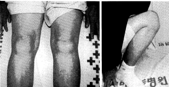
동통 및 관절 운동장애를주소로 내원하였다. 추락한 후 환자 는우측 다리로 설 수 없었으며보행도 불가했다고하였다. 방 사선 검사 상 우측슬관절은후외측으로 탈구되 었으며 전후 방 방사선 검사에서 내측 관절 간격이 넓어져있어 연부 조직 이 삽입되어 있음을짐작할 수있었다(Fig. 1). 타의료기관에 서 수 차례 도수정복을 시도하였으나 실패하여 전원 되었으 며, 내원 시 우측슬관절은 반상출혈을동반한심한 종창과 함께 내측 관절선을 따라서 깊은 주름이 형성되어 있었다 (Fig. 2). 슬관절은 불안정하였으며 심한동통으로 관절운동 은불가한상태였다.자기공명 영상 검사상내측 측부인대 및 주위 관절막이 내측 관절 구획 및 대퇴골 과간에 감돈
(incarceration)되어 정복을 방해하고 있었으며(Fig. 3), 전,후방 십자인대 모두 파열되어 있었다. 도수 정복이 불가 능하다는 판단 하에 관절경적 수술을시행하였으며 , 내측 대 퇴골과가 완전하게 노출되고 슬관절이 정복될 때까지 감돈된 인대, 관절막 및 연부조직을 절제하였다(Fig. 4). 인대 재건술 또는봉합술은 시행하지 않았으며 슬관절이 완전히 정복된 것을확인한후수술을 종료하였다. 슬관절을 신전한상태에 서 장하지 석고로고정하고 약 4주간체중부하를금하였다.
가능한 범위 내에서 조기 관절 운동을허용하였으며 이후 점
161 —
대한관절경학회지 제尚권 제2호 2009년
진적인 체중 부하를 허용하였다. 술 후3년 경과시점에서 경 도의 우측 슬관절 불안정증이 있었으나 일상생활에는 지장 이 없었으며 정상적인 관절 운동범위를 회복하였다(Fig. 5).
고 찰
도수 정복이 불가능한 슬관절의 탈구는 드물며, 대부분이 경골의후외측탈구손상으로 알려져 있다. 슬관절에 상당한 외반력에 작용하면서 손상된 내측 관절막과지대의 틈을 통 하여 대퇴골 내과가빠져 나가며 대부분의 경우에서 내측 측 부인대 및 전,후방십자인대의 파열을동반하나외측 측부인 대와 신경혈관조직 등은 보존된다'或. 이러한 관절 내 감돈된 내측 조직에 의하여 도수정복은 방해되고관혈적 정복을 요 하게 된다. 관절 내 감돈된 내측 조직은 본 예에서와 같이 슬 관절 외부에서 깊은피부주름을 형성하며 이는 도수정복이 성공하기 어려운간접 징후로 볼 수 있다.관혈적 정복의 방법 은 전술한바와같이 다양하며 많은경우에서 관절경을 이용 하여 정복을 방해하는구조물을 관찰하고동시에 파열된 인 대 조직의 재건술 또는 봉합술을 시행하지만 단순히 관절경 적 변연절제술 만으로 치료한 경우를 보고한 예는 드믈다.
Dubberley 등은 도수 정복 불가능한슬관절의 후외측탈구 를 관절경 적 변연절제술로 치료한 2례를 보고하였으며 , 이 중 1례는 추후 불안정 증상의 호소로 전,후방 십자인대 및 내측 측부인대 재건술을 시행하였다. Schaefer 등»은도수정복 불 가능한 슬관절 탈구를 관절경 수술을 통하여 정복 방해 구조 물을관찰하고 정복을 시도하였으나 실패하여 관절을 절개한
후 정복하고 추후단계적인후방십자인대재건술을 시행하였 다고 보고하였다. 그러나 본 증례에서는 정복을 방해하는 구 조물을 절제하여 대퇴골 내과를 완전히 노출시키자 관절이 쉽게 정복이 되었으며 추가적인 관절 절개를필요로하지 않 았다. 탈구와 동반된인대 손상에 대한 봉합 또는 재건에 대해 서는 아직 논란의 여지가많으며空,젊고 활동적인 환자에 있 어서는 초기에 재건하는 것을고려할 수 있겠으나활동력이 감소된 고령에서는 간단히 관절경적 변연절제술만으로도좋 은 결과를기 대할 수 있을 것으로 사료된다•
Fig. 2. Clinical photograph showing a swollen knee joint with ecchymosis and skin furrow over medial joint line.
Fig. 1.(A) Anteroposterior radiographof knee withposterolateral subluxationand medial joint space widening. (B)The lateral radi
ograph revealed that lateral tibialplateauwasposteriorly subluxated.
정복 불가능한 슬관절 탈구의 관절경적 치료 • 정진영
Fig. 3. Magnetic resonance imagingrevealedincarcerationof themedial collateralandcapsule in the medial compartment.
Fig. 4. (A) Arthroscopic photograph of soft tissue interposedin medial compartment. Medial meniscusis visible but medial femoral condyle is not visible. (B) Arthroscopicphotograph after debridement of medial soft tissue, (medialfemoral condyle now visible)
Fig.5. Afterthreeyears, the patientshowed full range of motionof the knee with mild laxity.
— 163 —
대한관절경학회지 제 13권 제2호 2009년
REFERENCES
1) BrennanJJ, KrauseME, MacDona너 WF: Irreducible posterolateraldislocation of the kneegrosslyintactcruci
ate ligaments. Am J Surg, 104:117-120, 1962.
2) Dedmond BT, AlmekindersLC: Operative versus non
operative treatment ofknee dislocations: a meta analysis.
Am J Knee Surg, 14: 33-38, 2001.
3) DubberleyJ, BurnellC,Longstaffe A, MacDonaldPB:
Irreducible knee dislocation treated by arthroscopic debridement. Arthroscopy.il: 316-319, 2001.
4) Kilicoglu O, AKman S, Demirhan M,Berkman M:
Muscularbuttonholing; An unusual cause ofirreducible knee dislocation. Arthroscopy, 17: E22-E25,2001.
5) Kontakis GM, Christoforakis JJ, Katonis PG, Hadjipavlou AG: Irreducible knee dislocation due to interpositionofthe vastus medialis associatedwith neu
rovascular injury. Orthopedics, 26: 645-647, 2003.
6) NystromM,Samimi S, Ha,Eri GB: Two casesof imducible knee dislocation occurring simultaneously in two patients and a review of theliterature. Clin Orthop, 277:197-200,1992.
7) Samimi S, Shahriaree H: Arthroscopic viewofan irre
duciblekneedislocation. Arthroscopy, 9: 322-326, 1993.
8) S사taefer RA, BellafioreVA, Corzatt RD: Irreducible dislocation of the knee.MilMed, 164: 827-829,1999.
9) Sigmeth A, Menth-Chiari WA,Amsuess H:A rarecase of irreducible knee dislocation in aseventy-three-year-old male. J Orthop Trauma,14(1): 70-72, 2000.
10) Wand JS: A physicalsign denotingirreducibility of a dis located knee. JBone Joint surgBr, 71:862-865, 1989.
11) Zafagnini S,lacono F, Presti ML, et al.: A newhinged dynamic distractor, for immediate mobilization after knee dislocations: technicalnote. Arch Orthop TraumaSurg, 128: 1233-1237, 2008.
초 록
외상성 슬관절 탈구 중 도수 정복되지 않는 경우는 매우 드물며 대개는관혈적 정복술을 요한다. 본 증례는 도수 정복되 지않는 슬관절 탈구에 대한관절경적 치료 경험으로 자기공명 영상에서 정복을 방해하는 내측 인대 및 관절낭구조물을 관찰하고 관절경 검사로확인한 후 대퇴골내측와가 완전히 관찰되고관절이 정복 될 때까지 끼어있는 조직을제거하였으 며 인대 봉합이나 재건술은시행하지 않았다. 술 후 약 4주간의 신전상태에서 고정 후 점진적인 관절운동 및 체중부하 보 행을 허용하였다. 3년 추시 관찰에서 경도의 슬관절 불안정성은 있었으나일상생활에 지장없었으며 정상범위의 관절운 동범위를회복하였다.
색인 단어: 정복불가능한 슬관절 탈구, 관절경적 치료
— 164 —