Sung-Do Cho, M.D.,Sang-Hun-Ko, M.D., Soo-Yeon Hwang, M.D., Ju-Yong Lee,M.D.
Department of Orthopaedic Surgery, Ulsan University Hospital, College of Medicine, University of Ulsan, Korea
ABSTRACT: The meniscal cyst of knee joint is a rare disease, and the discoid meniscal cyst has not been reported in Korea. We report 3 cases of the discoid meniscal cyst confirmed by MRI and arthroscopy. In physical examination, all cases have tenderness of the knee joint. One has palpable mass of knee joint and the other one has limitation of knee motion with knee flexion contracture of 10 degrees and positive McMurray test at external rotation of the knee. All cases are complete discoid meniscus, which are, one medial discoid meniscus and two lateral discoid meniscus through MRI. We confirmed horizontal tear of meniscus in all cases and the location of meniscal cysts are anterior horn of meniscus in one and body of meniscus in two.
We could get excellent results in all 3 cases that return to normal knee range of motion.
KEY WORDS: Discoid meniscal cyst
서 론 증례 보고
슬관절의 낭포가 동반된 원판형 반월상 연골은 매우 드문 질환이며 198&4 Avrahar꿔에 의해 최초로 보고된 이후 그 치료 방법에 대해서는 아직까지도 논란의 대상이 되고 있다. 국내에서는 1996션 Kinf에 의헤 1 예 보고된 바 있 으며 최근에는보고된 바가 없다. 저자들은 1997년 3월부 터 2。0囲 2월까지 낭포를 동반한 완전 원판형 반월상 연 골 3예를경험하고 문헌 고찰과 함께 보고하고자 한다
* Address correspondence and reprint requests to Sung-DoCho, M.D.
Department of Orthopaedic Surgery, Ulsan University Hosipital, 290-3 Cheonha-Dong, Dong-Ku, Ulsan 682-714 Tel: 82-52-250-7129, Fax: 82-52-235-2823
E-mail: [email protected]
증례 1
46세의 남자로 내원 2년전부터 간헐적 좌측 슬관절 동통 을 주소로 내원하였다. 외상의 병력은 없었고 이학적 소견 상 대퇴 사두근의 위축이 보였으며 , 외측 관절선 압통을 호 소하였으며 종물 촉지는 되지 않았고 관절 운동 범위는 정 상이었다. 단순 방사선 사진상 특이 소견은 없었으며 자기 공명 영상 검사상 수평 파열이 동반된 외측 원판형 반월상 연골이 관찰되었고 원판형 반월상 연골체부에서 낭포가 관 찰되었다(Fig. 1). 관절경 검사상 외측 완전 원판형 반월 상 연골에 수평 파열이 동반되어 있었다. 저자들은 원판형 반월상 연골 부분 절제술 후 Accessory portal을 통헤 외 측 반월상 연골의 수평파열부 하엽을back bite로 절제하 였으며, 전동 소파기를 이용하여 낭포를 감압하였다. 술 후
Fig. 2.Arthroscopic view of a remaining later저meniscus rim fallowing arthroscopic partial meniscectomy & cyst decompression.
동통은 소실되었으며 종물의 재발은 없었다(Fig 2).
증례 2
24세 남자로 내원 5년전 교통 사고후 발생한 간헐적 좌 측 슬관절 동통을 주소로 내원하였다. 이학적 소견상 내측 관절선 압통을 호소하였고, McMurray검사상 외회전시 양성 소견을 보였고 종물의 촉지는되지 않았다. 단순 방사 선 사진상 특이 소견은 없었으며 자기 공명 영상 검사상 내 측에 원판형 반월상 연골이었고수평 파열이 동반되어 있었 으며 중간 1/3冲서 낭포가 관찰되었다(Fig. 3). 관절겅 김 사상 내측이 완전 원판형 반월상 연골이었으며 수평 판상 파열이 동반되어 있었다(Fig. 4). 저자들은 전동 소파기를 이용하여 원판형 반월상연골부분 절제술후 소식자를이용하 여 낭포를 감압하여 노란색 낭포액을 유출 시켰다(Fig・ 5)
Fig. 5. Yellowish fluid leaked out from the cystic cavity by decompression & partial meniscectomy with shaver.
술 후 증상은 소실되어 만족할 만한 결과를 얻을수 있었다.
증례 3
38세 여자로 내원 5년전부터 간헐적 좌측 슬관절 동통을 주소로 내원하였다. 이학적 검사상 외측 관절선 압통을 호 소하였고굴곡 구축이 1。도였으며 전외측에서 종물이 촉지 되었다.단순 방사선 사진상 특이 소견은 없었으며 자기 공 명 영상 검사상 외측이 완전 원판형 반월상 연골이었으며 전각부에서 낭포가 관찰되었고 수평 파열이 동반되어 있었
다(Fig 6, 7). 관절경 검사상 내측 대퇴과는 Outer- Brideg显류상 grade H의 연골 결손이 관찰되었고 외측 연골은 완전 원판형 반월상 연골이었다. 저자들은 수평 파 열이 동반된 원판형 반월상 연골의 상엽을 부분 절제술 시 행하였으며 전각부의 낭포는 전동 소파기를 이용하여 감압 하였다(Fig. 8). 추시 관찰상 증상은 소실되었으며 종물의 재발은 없었다.
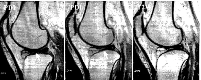
Fig. 6.Previous Saggital view of MRI on left knee showed complete discoid lateral meniscal cyst with horizontal tear.
Fig. 7.Previous Coronal view of MRI on left knee showed complete discoid lateral meniscal cyst with horizontal tear.
Fig.8.Yellowish fluid leaked out from the cystic cavity by decompression & partial meniscectomy with punch forceps & shaver.
1988션 Avraham1]- TorrP, 1996년 KirH과 ChoP는 각각 1예씩 보고하였으며, 저자들의 경우는 자기 공명 영 상 및 관절경을 이용하여 낭포가 동반된 원판형 반월상 연 골 3예를 경험하였다.
성별분포는 일반적으로 원판형 반월상 연골은 3:2로 남 자에게 많다고 하며 반월상 연골 낭종은 Gall。와 Bryant* 2:1로 남자에게 호발하며 저자들의 경우는 원 판형 반월상 연골 낭종의 3예중 2예에서 남자였으며 평균 연령은36세였다.
발생 부위에 따라Larsoi과 Gran痹는 반월상 연골 낭 종의 외측 대 내측 비율이 22:贸도였으며, 원판형 연골은 Watanabfe0에 의하면 외측이 내측에 비해 2배 이상 많은
것으로 보고하고 있으며, 저자들은 내측 1례, 외측 2례를 경험하였다.
발생 원인으로는 여러 학설이 있으며 반월상 연골 낭종의 경우 Zadek과 Jaffe⑶는 선천성 이론을 제시하였고, Smil创는 외상이 주 원인이라고 주장하였으며 , Lichten- steirf은 반월상 연골의 교원성 결체 조직의 국소 퇴행성 변화와 낭종성 변화라고주장하였다.
증상은 여러 저자들&財。이 슬관절 주위 통증을 가장 빈 번한증상으로 보고하였으며 . 이외에도 파행, 불안정감. 운 동제한, 조기 피로 등이 있으며, 저자들의 경우에서도 전례 에서 간헐적 통증이 관찰되었고 운동제한이 있었던 경우도 1 예 있었다.
이학적 소견으로 Gaik針 Bryarf은 종물의 촉지가 가 장 흔한 소견이라 하였고 슬관절 주위의 압통도 비교적 흔 하게 나타났다고 하였으며 , 저자들의 경우 슬관절 주위 압 통이 전례에서 관찰되었고, 종물의 촉지는 1예에서 보였으 며. 나머지 2예에서는 이학적 검사상 낭포를 촉지하지 못 하였으며 자기공명영상으로 확인할 수 있었다. 이 외에도 McMurray 검사는 내측 원판형 반월상 연골 1예에서 외 회전시 양성 소견을 보였으며, 대퇴 사두근 위축이 1 여】, 굴 곡 구축이 1 예였다.
종의 치료와 동일한 방법 으로 치료를 시 행하였다.
술후 추시 관찰상 3예 모두 Glasgov임상 평가 기준에 서 우수였으며, 관절 운동 범위도 정상이었다. 저자들은 수 평파열이 동반된 원판형 연골 낭포를 3예를 경험하였고 만 족할만한 결과를 얻었기에 보고하는 바이며 원판형 반월상 연골과 반월상 연골 낭종과의 관련성에서는 보다많은 연구 가 필요하리 라 사료된다.
REFERENCE
1) 이병일,김대성,신병준, 최창욱: 양측성 내측 원판형 연 골. 대한슬관절학회지, 24:334-342, 1989.
2) Avraham Sand Tom H:Medi시 Didscoid Meniscus with Cyst Formation in a Child. J Pediatric Ortho., 8:471 -473, 1988.
3) Ebner A: Muchen. Med Wehnschi, 51:1737, 1904.
4) Gallo GAand Bryan RS: Cysts of 아)e Semilunar carti
lage of the Knee: A report of 16 cases including arthro- graphic study. Am. J. Surg., 116-65, 1965.
5) Glasgow MMS, Allen PW, BlakewayC:Arthroscopic treatment of cysts of the lateral meniscus, J Bone Joint Surg, 75-B:299-302, 1993.
6) LarsonRL and Grana WA: The knee (Form, Function, Pathology & Treatment), 445~447, 1993.
7) KimSJ and ChoiCH: Bilateral complete discoid medial menisci combined with anomalous insertion and cyst for
mation. Arthroscopy, 9:704-706, 1993.
8) Lichtenstein L: Disease of Bone and Joints, 2nd ed. St.
Louis, CVMosby, 1975.
9) SmillieIS:Injuries of the knee joint. 5th ed. London, Churchill-Livingstone, 94, 1978.
10) Taylor H: Cysts of the fibrocartilages of the knee joint. J Bone Joint Surg, 17-A:558-596, 1935.
11) Watanabe M: Arthroscopy of the knee joint in disorders the knee, 145, 1975.
12) Young RB: The external semilunar cartilage as a com - 13) Zadeck I and Jaffe HL: Cysts of the semilunar cartilage
plete dis. In, 179, 1889. of the knee. Arch Surg, 54:188, 1947.
슬관절의 반월상 연골 낭포는 비교적 드문 질환이며, 특히 원판형 연골낭포는 최근에 국내에서 보고된 바가 없다.
저자들은 자기 공명 영상 및 관절경을이용하여 원판형 연골 낭포로 진단하였던 3예의 환자를보고하고자 한다. 주 증상으로 전례에서 간헐적 슬관절 동통을 호소하였고, 1예에서 외상의 병력이 있었다. 이학적 검사상전례에서 관 절선 압통이 있었고, 1예에서는 촉진시 종물이 의심되었으며 다른 1예에서는 10도 굴곡 구축의 관절 운동 제한을 보였고, McMurray 검사상 내측 원판형 반월상 연골 1예에서 외회전시 양성 소견을 보였다. 원판형 반월상 연골 분류는 전례에서 완전형이었으며, 내측원판형 반월상 연골이 1예, 외측이 2예였고, 자기 공명 영상 소견상 전례에 서 수평 파열의 양상을 보였으며 낭포의 발생 위치는 전각부가 1예, 중간부가2예였다. 치료는3예 모두 반월상 연 골 낭포의 치료와 동일하게 관절경하 부분 절제술 및 낭포 감압술을 시행하였다. Glasgow의 임상 평가기준에 의 한 숡후 결과는 3예 모두우수였으며, 관절 운동 범위도 정상으로 회복되어 만족할만한결과를얻을 수 있었다.
색인 단어: 원판형 연골 낭포