Young1) reported the first case of discoid lateral meniscus in 1889. Cave and Staples2) described the first two cases of partially discoid medial meniscus in 1941. Murdoch3) reported the first case of bilateral medial discoid menisci in 1956. Since then, more than 23 cases of bilateral discoid medial menisci have been docu
mented to date. The treatment of choice for symptomatic torn discoid meniscus is arthroscopic partial meniscectomy.
The present study reports our experience of treating four cases of symptomatic discoid medial meniscus, three of which were bilateral. The treatment outcomes were assessed with the Knee Injury and Osteoarthritis Outcome Score (KOOS) and a visual analogue scale (VAS).
Case Reports
1. Case 1
A 17yearold male patient complained of pain in the right knee with locking, giving way and swelling. He did not remem
ber any specific trauma. The symptom duration was 2 years for the right knee and the left knee was asymptomatic. Physical examination of the right knee showed atrophy of the quadriceps femoris muscle compared to the opposite side, medial joint line tenderness and a positive McMurray sign. Plain Xray showed widening of the medial joint space and cupping of the medial tibial plateau. Magnetic resonance imaging (MRI) of both knees showed complete discoid medial meniscus tears and the right knee demonstrated a horizontal cleavage and flap tear (Fig. 1A and B). We performed partial meniscectomy with a fourportal technique using a knife taking care to preserve the 6mm periph
eral margin after confirmation by MRI (Fig. 2). The anterior horn of the meniscus was attached to the normal footprint (Fig. 3). At 2 years after surgery, clinical results were assessed by the KOOS and VAS. Any postoperative complications were not seen and the preoperative locking as well as giving way disappeared. The pre
operative and postoperative average KOOS were 65.2 and 92.4, respectively. The preoperative and postoperative VAS were 6 and 0, respectively.
Discoid Medial Meniscus Tear, with a Literature Review of Treatments
In Soo Song, MD, Jun Bum Kim, MD, Jong Keun Lee, MD, and ByeongSeop Park, MD
Department of Orthopaedic Surgery, Daejeon Sun Hospital, Daejeon, Korea
The present study reports our experience of treating four cases of symptomatic discoid medial meniscus, three of which were bilateral. We performed partial meniscectomy with a fourportal technique using a knife leaving a 6 mm peripheral margin after confirmation of magnetic resonance imaging findings. Clinical results were assessed at the end of 2year followup using the Knee Injury and Osteoarthritis Outcome Score and a visual analogue scale. We obtained satisfactory clinical results without recurrence of the symptoms in all cases.
Keywords: Knee, Discoid medial meniscus, Meniscectomy pISSN 2234-0726 · eISSN 2234-2451
Knee Surgery & Related Research
Received September 2, 2015; Revised (1st) February 18, 2016;
(2nd) April 10, 2016; (3rd) May 7, 2016; Accepted May 10, 2016 Correspondence to: In Soo Song, MD
Department of Orthopaedic Surgery, Daejeon Sun Hospital, 29 Mokjungro, Junggu, Daejeon 34811, Korea
Tel: +82422208460, Fax: +82422208460 Email: [email protected]
237
This is an Open Access article distributed under the terms of the Creative Commons Attribution NonCommercial License (http://creativecommons.org/licenses/bync/4.0/) which permits unrestricted noncommercial use, distribution, and reproduction in any medium, provided the original work is properly cited.
Copyright © 2017 KOREAN KNEE SOCIETY www.jksrr.org
2. Case 2
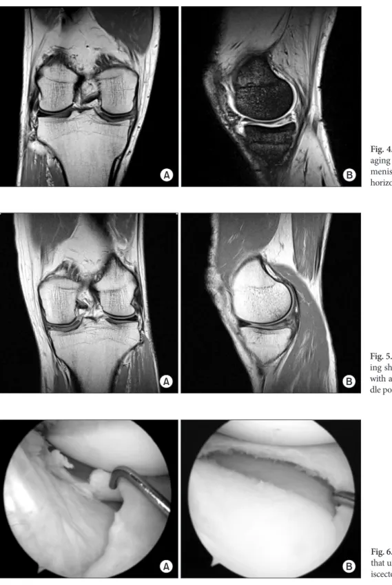
A 41yearold male patient complained of right knee pain. MRI showed a complete discoid medial meniscus with a horizontal cleavage and flap tear in the middle third of the meniscus (Fig. 4).
The opposite knee showed a discoid medial meniscus with a tear in the middle portion, but the patient had no complaints on his left knee and physical examination did not reveal any abnormali
ties (Fig. 5). We tried to preserve the sound meniscus as much as possible to minimal 6 mm (Fig. 6). The preoperative symptoms disappeared and any complications were not seen. The preopera
swelling and effusion on her right knee with intermittent pain.
The range of motion was limited due to the pain during flexion more than 90°. MRI of her right knee showed a complete discoid medial meniscus with a horizontal cleavage and flap tear. We performed a partial meniscectomy. There were no postoperative complications and the preoperative symptoms disappeared. The preoperative and postoperative KOOS were 72.2 and 92.0, re
spectively. The preoperative and postoperative VAS were 5 and 0, respectively.
4. Case 4
A 40yearold male patient complained of pain in his right knee with giving way, locking and swelling. The symptom occurred 2 weeks before his visit to the hospital. Physical examination showed medial joint line tenderness and a positive McMurray sign. Xray showed widening of the medial joint space and cup
ping of the medial tibial plateau. MRI of the right knee showed a complete discoid medial meniscus with a horizontal flap tear. He had been diagnosed with a discoid medial meniscus with a tear in the left knee and treated with arthroscopic partial meniscec
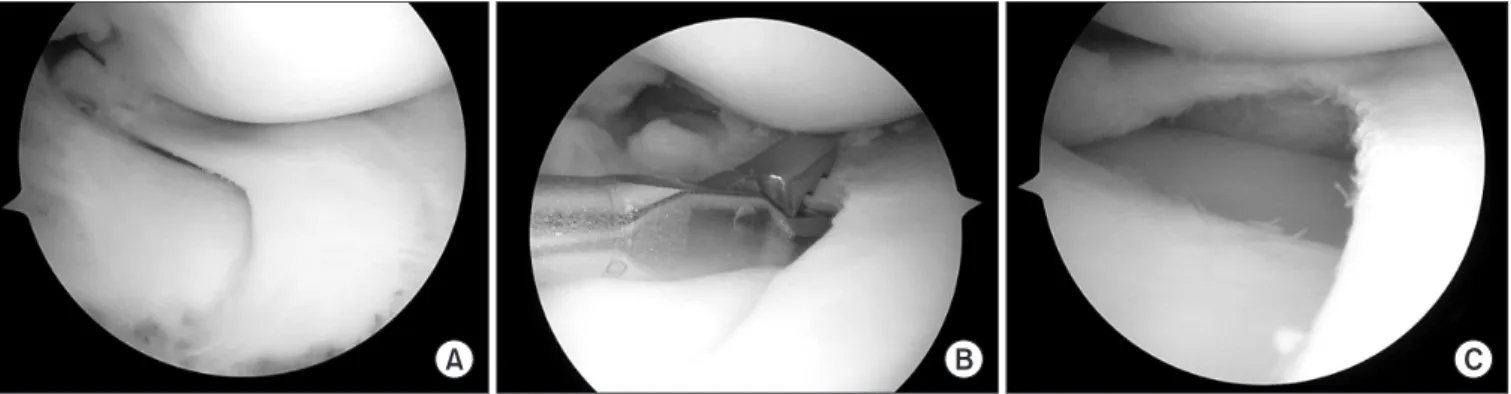
tomy 2 years ago. We performed a partial meniscectomy on the right knee (Fig. 7). There were no postoperative complications and preoperative locking disappeared. The preoperative and postoperative KOOS were 53 and 90.5, respectively, and the pre
operative and postoperative VAS were 6 and 0, respectively.
Discussion
The discoid meniscus of the knee is an abnormally wide and thick meniscus that widely covers the articular surface of the tibial plateau, and is easily damaged as a result. Discoid lateral meniscus is a wellstudied and documented entity of the knee whereas discoid medial meniscus is an extremely rare pathology.
There are less than 70 cases of discoid medial menisci reported in the literature46). The involvement of the bilateral medial me
nisci is also extremely rare. Dickason et al.6) examined 14,731 menisci in a retrospective study and found that of the 8,040 me
dial menisci (excluding the 6,691 lateral menisci), only 10 were discoid menisci (0.12%), and only one had bilateral involvement A
B
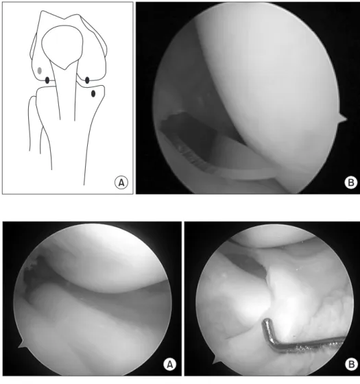
C Fig. 1. (A) The right knee magnetic resonance imaging (MRI) showed a complete discoid medial meniscus with continuity of the meniscus from anterior to posterior horns and a horizontal cleavage and flap tear.
(B) The left knee MRI showed a complete discoid meniscus, which was treated with conservative management. (C) The plane Xray showed widening of the medial joint space and cupping of the medial tibial pla
teau.
(0.012%). Although several studies on bilateral discoid medial menisci have been reported by Asian doctors in recent years, no more than 23 cases of bilateral discoid medial menisci have been reported to date. Tachibana et al.4) recommended systematic MRI of the asymptomatic contralateral knee. They considered the in
cidence of bilateral cases to be underestimated as the abnormality is congenital. Discoid lateral meniscus tends to manifest as hy
permobility (snapping knee) in childhood without tear whereas discoid medial meniscus is asymptomatic in childhood until re
vealed by tear. The ultrastructure of discoid menisci differs from that of normal menisci. These differences in collagen fiber orga
nization and stress redistribution induced by increased meniscal thickness are factors of vulnerability.
Surgical indications are the same for the discoid lateral and medial menisci. Only symptomatic lesions should be operated;
in case of preoperative discovery of asymptomatic discoid me
nisci, abstention is regarded as a rule4). Total meniscectomy had been widely indicated for symptomatic discoid meniscus in the past. Several studies showed excellent results after total menis
cectomy. In 1995, Washington et al.7) reported on the mean 19.8
year followup results with an average duration of 17 years after total meniscectomy for 18 discoid menisci. They concluded that total meniscectomy might offer the best prognosis with no evident degenerative changes on roentgenograms. Conversely, in 1998, Raber et al.8) retrospectively reviewed longterm results of total meniscectomy for discoid lateral meniscus and reported osteoarthritic changes compared with the untreated contralateral knee in 10 of 11 knees. Therefore, although total meniscectomy was recommended as a treatment for symptomatic discoid lat
eral meniscus, there was a risk of progressive osteoarthritis and poor prognosis810). Therefore, partial meniscectomy (so called
“saucerization”) is currently recommend for the treatment of symptomatic discoid meniscus to reduce progressive degenera
tion of the cartilage in the joint. The goal of partial meniscectomy is to remove the central portion of the discoid meniscus and to leave a stable as well as balanced rim. By doing so, the remaining portion performs the function of the meniscus and reduces in
stability resulting from total meniscectomy. The width of the rim
A B
Fig. 2. (A) We used a fourportal technique using the standard anteromedial, anterolat
eral, additional high far lateral, and addi
tional low anteromedial portals. (B) A No.
15 blade facing anteriorly was inserted into the high far lateral portal and was used to cut the anterior horn of the discoid medial meniscus.
A B
Fig. 3. (A) Arthroscopic findings of a thick complete discoid medial meniscus in a patient. (B) The patient underwent partial meniscectomy with basket punch leaving the 6 mm stable rim.
of the remaining meniscus is dependent on the degree of the torn meniscus. The posterior segment and inside the middle segment are known as the most common tear sites of a discoid meniscus.
When these sites were torn, Hayashi et al.9) left 6–8 mm width for complete and incomplete lesions and Vandermeer and Cunning
ham10) left 45 mm width. In our patients, we used a fourportal technique including the standard anteromedial, anterolateral, ad
ditional high far lateral and additional low anteromedial portals.
A No. 15 blade facing anteriorly was inserted into the high far lat
eral portal and was used to cut the anterior horn of the discoidal
A B
Fig. 4. Right knee magnetic resonance im
aging showing a complete discoid medial meniscus of bowtie configuration with a horizontal cleavage and flap tear.
A B
Fig. 6. A complete medial discoid meniscus that underwent reshaping and partial men
iscectomy of the inferior flap.
A B
Fig. 5. Left knee magnetic resonance imag
ing showing a complete discoid meniscus with a horizontal and flap tear in the mid
dle portion.
medial meniscus. Instead of scissors, we used a knife for en bloc resection. As a result, we could determine the remnant margin easily and precisely. Also, we could perform saucerization fast and conveniently without anterior cartilage damage. With this method, we performed partial meniscectomy only on the symp
tomatic knee, not on the asymptomatic opposite knee and tried to leave a 6 mm rim to achieve stability and avoid impingement of the femoral condyle against the rim of the meniscus.
Clinically, the most frequent symptoms of discoid medial me
niscus are medial knee pain, iterative effusion and locking in flexion which are not specific46). Knee snapping is relatively rarer in discoid medial meniscus than in discoid lateral meniscus. Pain and effusion are more associated with the meniscal tear rather than the discoid shape of the meniscus. Locking may occur be
cause of the discoid shape as such, with the thick central region passing forward of the medial condyle.
Plain Xray may show an enlarged joint space in the knee with a discoid medial meniscus5). MRI is the best tool for confirming di
agnosis and exploring the associated meniscal lesions. Diagnosis of discoid medial meniscus requires continuity between the ante
rior and posterior horns on three consecutive 5mm thick sagittal slices and is confirmed by coronal slices showing an abnormally thick meniscus, sometimes extending as far as to the intercon
dylar groove4). Our patients had radiological evidence of discoid medial meniscus described in the literature, such as hypoplasia of the medial tibia and femoral condyles and increased medial joint space. But none of them had a lytic lesion in the distal femoral condyle. Both MRI and arthroscopy did show the presence of a depressed medial tibia plateau and complete discoid medial meniscus with continuity from the anterior horn to the posterior horn.
We think that discoid medial meniscus tears should be man
aged with more active surgical treatment because the medial me
niscus is subject to greater stress due to screw home mechanism
and less mobility. Furthermore, because weight bearing is trans
ferred to the medial side of the knee, patients tend to have more discomfort, such as giving way, locking and pain than patients who have lateral discoid meniscus tears.
In conclusion, we experienced four cases of symptomatic dis
coid medial meniscus and managed the rare deformity using a fourportal arthroscopic partial meniscectomy, which is similar to the technique used in knees with a discoid lateral meniscus.
We think that active surgical treatment for a torn discoid medial meniscus can provide satisfactory results without recurrence of symptoms.
Conflict of Interest
No potential conflict of interest relevant to this article was re
ported.
References
1. Young RB. The external semilunar cartilage as a complete disc. In: Cleland J, MacKay JY, Young RB, eds. Memoirs and memoranda in anatomy, vol. 1. London: Williams and Nor
gate; 1889. p179.
2. Cave EF, Staples OS. Congenital discoid meniscus: a cause of internal impingement of the knee. Am J Surg. 1941;54:3716.
3. Murdoch G. Congenital discoid medial semilunar cartilage.
J Bone Joint Surg Br. 1956;38:5646.
4. Tachibana Y, Yamazaki Y, Ninomiya S. Discoid medial me
niscus. Arthroscopy. 2003;19:E128.
5. Pinar H, Akseki D, Karaoglan O, Ozkan M, Uluç E. Bilateral discoid medial menisci. Arthroscopy. 2000;16:96101.
6. Dickason JM, Del Pizzo W, Blazina ME, Fox JM, Friedman MJ, Snyder SJ. A series of ten discoid medial menisci. Clin Orthop Relat Res. 1982;(168):759.
A B C
Fig. 7. Arthroscopically confirmed discoid medial meniscus (A) that underwent partial meniscectomy (B) preserving a 6 mm remnant margin (C).