Lee-Ryan Eye-Hand Coordination Test 를 이용한 인위적 시력저하가 눈-손 협응능력에 미치는 영향
이기석*
뉴사우스웨일즈대학교, 검안학과, 시드니
투고일(2014년 08월 2일), 수정일(2014년 09월 3일), 게재확정일(2014년 9월 18일)
···
목적: 최근 보고된 Lee-Ryan Eye-Hand Coordination(EHC) Test를 이용하여 정상안의 인위적 시력저하가 눈-손 협응능력 (EHC)에 미치는 영향을 알아보고자 하였다. 방법: 정상시력을 가진 성인 11명(29.46±5.94세)을 대상으로 비우위안에 볼 록렌즈를 가입하여 인위적으로 근거리 약시 상태를 만들어 EHC를 검사하였고, 2주 후 정상 시력상태에서 다시 검사를 하 였다. EHC를 알아보기 위해 Lee-Ryan EHC Test의 각기 다른 난이도의 7가지 모양을 선택하여 총 소요시간과 에러 횟수 를 EHC의 분석 요소로 비교 분석하였다. 결과: 총 소요시간에서는 인위적으로 시력 저하된 눈이 정상 단안 시력보다 더 많은 시간을 소비하였고(p=0.013), 양안시 상태에서는 오히려 정상안 상태보다 저하된 시력을 가지고 더 빨리 검사를 끝 마쳤다(p=0.001). 에러 횟수에서는 소요시간과 같이 저하된 시력이 더 많은 실수를 하였으나(p<0.001), 인위적으로 저하된 시력을 가진 상태에서 단안과 양안시의 차이는 보이지 않았다. 결론: 기존의 EHC 방법들이 갖고 있는 적용하는데 있어 서의 한계들과 달리 간단한 컴퓨터 시스템을 기반으로 한 Lee-Ryan EHC Test로도 EHC의 이상(deficit)을 관찰 할 수 있었 다. 후에 약시와 같은 시력저하에 관련된 분야에 더 많은 연구가 이루어 질 것으로 사료된다.
주제어: Lee-Ryan Eye-Hand Coordination Test, 인위적 시력저하, 눈-손 협응능력
···
서 론
일상생활에서 효율적이고 효과적인 시각(visual)과 운동 (musculoskeletal) 시스템들 간의 정상적인 협력은 매우 중 요하다. 이러한 눈과 손의 협응능력(Eye-Hand Coordination, EHC)의 평가는 어떤 사물에 접근하기 위해 손을 뻗거나 (reaching) 움켜지는(grasping) 목표 지향적인(goal-oriented) 행위와 같이 시기능(visual function)과 관련된 유용한 정 보를 제공한다. 20세기 초 Moore[1]가 작은 구멍에 사물을 넣는 간단한 방법으로 EHC의 능력을 측정하는 방법(Moore EHC Test)을 제안하였고, 그 후 이를 기초로 한 몇 가지 방 법들이 지속적으로 발전과 연구가 되어왔다.[2, 3] 하지만 기 본적으로 이 검사법은 검사자가 직접 검사 시간을 측정하 는 등의 검사자의 편견(experimenter bias)이 발생될 수 있는 문제점을 갖고 있어 높은 객관성을 요구하는 실제 연구의 목적으로는 부족한 면이 있었다. 이 방법과 달리, 보다 높 은 정확성과 측정 만족도를 위해 최근 연구[4, 5]에서는 biomarker(infra-red reflective marker)를 이용하여 약시 (amblyopia)와 같은 복합적인 시기능 이상(visual deficit)이 나타내는 EHC의 특징들을 보고하고 있다. 이 biomarker를
이용한 EHC 특징들을 나타내는 요소들(variables)로 최초 피검사자의 눈을 가린 다음 사물을 보여준 후 그 사물을 집 는 순간의 속도(velocity(mm/s)), 사물을 보고 난 후의 반응 시간(reaction time), 사물을 본 후 집기까지의 총 시간 (movement time)등의 결과를 기초로 분석하고 있다. 하지만 이 방법 또한 고 비용, 제한적 실험 환경 그리고 높은 기술 적 요구와 같은 임상에서 적용하기엔 많은 어려움들이 있 다. 더욱이 이 검사법은 다양한 연령층 특히, 집중과 협조 도(compliance)가 약한 소아를 대상으로 한 검사에서 결과 에 대한 신뢰도를 떨어뜨린다고 할 수 있다. 이에 최근 Lee et al.[6]이 i-Pad®를 이용한 새로운 EHC 검사법(Lee-Ryan EHC Test)을 제안 하였고 이는 기존에 EHC 검사법들이 갖 고 있는 문제점들을 최소화하고, 새 검사법으로서의 반복성 (repeatability)과 다양한 연령층에 대한 적용성(applicability) 을 갖는 것으로 보고하고 있다.
시력저하 및 여러 시각 이상(visual abnormality)의 특징 들을 갖고 있는 약시의 EHC 이상(deficit)은 이미 많은 연 구를 통해 보고가 되었다.[4,5,7] 이런 EHC의 이상은 입체시 감소와 그로 인한 양안시의 감소 또는 손실이 원인으로 알려져 있다. 특히, 정상안의 단안 상태에서 EHC(또는
*Corresponding author: Ki-Seok Lee, TEL: +61-422-435 261 E-mail: [email protected]
<초청논문>
prehension(reaching과 grasping))는 사물을 집는 과정에서 양 안 상태보다 더 많은 시간 소요와 낮은 정점 속도(low peak velocity)의 특징을 보인다.[8] 이는 양안시 상태에서의 안운동 시스템 기능(oculomotor system functions)이 더 우수하며 만 약, 양안시가 불가능하게 되면 이 시스템 기능이 단안의 망 막에 들어오는 움직임 정보(retinal motion information)에 좌 우되기 때문에 단안시 상태에서 물체의 위치와 크기를 판단 하는 능력이 저하된다. 이에 정상안의 단안에 인위적 시력저 하(굴절성 약시 상태)가 시기능 변화에 미치는 연구로서 Odell et al.[9]이 Bangerter filters를 이용하여 인위적으로 저하 된 단안 시력이 입체시 감소 또는 손실과 관련이 있다는 연 구 결과를 발표하였다. 특히, Pardhan et al.[10]이 정상시력의 한 쪽 눈에 볼록렌즈를 가입하여 단안에 인위적 약시상태를 만들어 비정상 양안시 상태(unequal)와 정상 양안시 상태 (equal)에서 사물을 집는 과정에서의 최대 속도, 시간 등을 적외선 카메라(infra-red camera)를 이용하여 비교하였으며, 비정상 양안시가 오히려 정상 단안상태보다 EHC이 더 나빠 진다는 보고를 하였다. 하지만, 보다 간단하고 객관적인 방 법으로 정상안의 인위적 시력저하가 EHC에 미치는 영향에 대한 연구는 아직 보고된 바가 없다.
이에 본 연구는 인위적인 시력저하로 야기된 양안시 결 손이 최근 보고된 Lee-Ryan EHC Test로 기존에 알려진 고 비용 및 고 기술적인 장비 없이 소요시간(time taken) 과 에러 회수(the number of errors)와 같은 간단한 요소들 (variables)로 EHC 이상이 관찰되는지와 그에 따른 EHC 영향을 분석하고자 한다.
대상 및 방법
1. 검사 방법
Lee-Ryan EHC Test를 이용한 EHC 검사는 Lee et al.[6]
의 동일한 실험 환경과 조건에서 실시하였으며, 전체 13
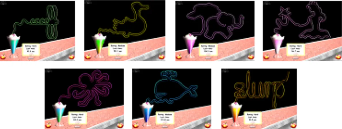
가지 EHC검사 모양들 중 난이도(difficulty)를 고려해 7가 지를 선택하였다. 7가지 모양의 평균 난이도(normalized difficulty value)는 0.6±0.2(0.37~0.83; ‘1’에 가까울수록 검사 난이도가 높음)였다(Fig. 1).
비우위안(non-dominant eye)에 볼록렌즈를 이용하여 근 거리 시력(소수 시력)을 0.3에서 0.5정도의 굴절성 약시 (moderate refractive amblyopia) 상태로 만들었다. EHC 검 사 전에 학습효과(learning effect)[6]를 최소화하기 위해 실 험에 사용 될 7개 모양 외의 적어도 다른 2가지 이상을 가 지고 정상적인 양안시 상태에서 연습을 하게 했다. 인위적 인 시력저하의 EHC를 측정하기 위해 참여 대상자들 모두 2주 간격으로 두 번 반복 실험을 하였다. 첫 번째 검사에 서는 인위적 시력저하상태에서 실시하였고 두 번째 검사 는 정상 시력상태에서 실시하였으며, 두 검사 모두 단안 과 양안 상태에서 각각 EHC 검사를 하였다.
2. 대상
The University of New South Wales Human Research Ethics Committee에서 본 연구에 대한 승인을 하였으며, Declaration of Helsinki에 따라 대상자 모두 연구에 대한 충분한 설명 후에 실험에 대한 동의를 받았다.
호주에 있는 School of Optometry and Vision Science, University of New South Wales Australia에서 광고를 통해 지원한 정상시력을 가진 성인 11명(나이 29.46±5.94세)을 Fig. 1. Seven of 13 different shapes chosen for the study (Dragonfly, Duck, Elephant, Fairy, Octopus, Whale and ‘Slurp’).
Table 1. Details of subjects’ ages and visual functions
Age(years) 29.46±5.94
Gender Male 7
Female 4
Accommodation(D) 9.93±3.46
Stereopsis(second of arc) 28.64±7.10
대상으로 하였다. 대상자들 모두 시력 검사(Snellen chart), 굴절 검사(skiascopy), 조절 검사(push-up test), 눈 정렬상 태(cover test), 색각 검사(Ishihara test), 우위안 검사(the hole-in-the card(Dolman method)), 입체시 검사(Randot stereotest)와 눈과 관련된 건강 상태 등 본 연구에 관련된 시기능 검사를 하였고 참여자들 모두 정상 시기능 상태를 갖고 있었다(Table 1).
3. 분석 및 통계
EHC를 분석하기 위한 측정 요소로서는 7가지 모양을 검사하는 동안에 나타난 소요시간(time taken)과 검사 중 에 발생되는 에러 수(the number of errors)로 EHC의 능력 을 비교 하였다. 데이터 통계 분석은 IBM SPSS Statistics 22프로그램을 이용하였으며, 동일한 대상자들이 단안과 양안시 상태에서 반복된 실험 하에 실시된 검사이기 때문 에 Wilcoxon singed test를 실시하였으며, 다중비교
(multiple comparisons)를 위해 Bonferroni correction을 적 용하였다.
결 과
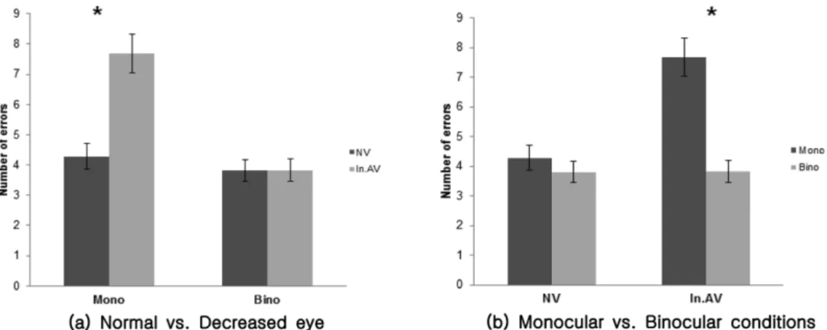
단안시 상태에서 인위적으로 저하된 시력(28.94±11.7 sec)은 정상적인 단안 시력(26.31±10.7 sec) 상태보다 더 많은 시간을 소비 하였지만(p=0.013), 양안시 상태에서는 오히려 시력이 저하된 단안(22.98±10.0 sec)에서 정상시 력(25.95±10.7 sec) 보다 더 빠르게 검사를 끝냈다(p=0.001).
즉, 인위적으로 감소된 시력은 정상안 시력상태와는 달리 단안과 양안시 상태에 따라 각각 다른 검사시간을 소비하 였다(p<0.001)(Fig. 2(a) and 2(b)).
인위적인 시력저하의 단안(7.22±5.6) 상태에서 정상시 력의 단안(4.29±3.7)보다 훨씬 더 많은 실수를 하였으나 (p<0.001), 양안 상태에서는 저하된 시력상태(3.82±3.23)
Fig. 2. Comparison of the time taken between (a) monocular and binocular viewing in normal versus induced-amblyopic eyes and (b) between normal and induced-amblyopic eyes under monocular versus binocular conditions. Error bars represents 95%
confidential interval. (Mono: Monocular, Bino: Binocular, NV: Normal vision, In.AV: Induced artificially decreased vision)
Fig. 3. Comparison of the number of errors between (a) monocular and binocular viewing conditions for the normal eye versus the eye with induced-amblyopia, and (b) between normal and induced-amblyopic eyes under monocular versus binocular conditions. Error bars represents 95% confidential interval. (Mono: Monocular, Bino: Binocular, NV: Normal vision, In.AV:
Induced artificially decreased vision)
에서와 정상 시력상태(3.81±3.1)와 유의한 차이는 없었다.
단안과 양안 비교에서는 인위적 약시의 단안 시력상태가 EHC를 하는데 있어서 더 많은 실수를 보여 단안의 저하 된 시력상태에서 정상적인 EHC를 하는데 더 큰 어려움을 초래하는 것으로 나타났다(p<0.001)(Fig. 3(a) and 3(b)).
고 찰
연구와 임상 분야에서 사용되고 있는 대표적인 두 가지 EHC 검사법들 즉, biomarker와 Moore EHC Test는 각각 의 검사법이 갖고 있는 한계와 문제점들이 있을 뿐만 아 니라 공통적으로 EHC의 능력의 측정과 더불어 검사과정 에서 팔 전체의 운동성을 요구하기 때문에 순수한 EHC의 성격을 관찰한다고 하기엔 문제가 있다고 보인다. 이에 본 연구는 위의 검사법들이 가지고 있는 문제점들을 최소화 한 새로운 검사법인 Lee-Ryan EHC Test를 사용하여 렌즈 를 이용한 방법[9,11]으로 정상안의 인위적 약시상태에서 EHC 이상(deficit)이 관찰되는지 그리고 인위적인 양안시 손실(loss of binocularity)이 실제 EHC에 영향을 미치는지 를 분석하였다.
양안시 조건에 따른 EHC의 능력에 대해 Loftus et al.[12]
의 연구결과에 따르면 단안시 상태에서는 양안시 상태보 다 위치 변화(positional variance)에 대해 증가의 형태를 보였다고 했지만, 본 연구에서는 단안시와 양안시 상태에 서 소요시간과 에러 수에서는 뚜렷한 차이를 보이지 않았 다. 이는 Lee-Ryan EHC Test 검사 환경이 기존 EHC 검 사와는 달리 평면에서 이루어지는 2차원(two dimensions) 의 환경 조건이라 높은 입체시를 요구하지 않거나, 또는 비록 검사 중 대상자들의 몸이나 머리의 움직임을 자제하 도록 하였지만 머리의 움직임이 EHC 검사를 하는데 도움 을 주었을 거라고 사료된다. Jones et al.[13]은 심지어 3차 원(three dimensions)검사 조건에서 머리의 움직임이 자유 로울 때 입체시는 크게 중요하지 않다고 보고가 있어 이 를 더욱 뒷받침 해주고 있다.
정상안의 인위적으로 저하된 시력은 실제 검사를 수행 하는데 정상시력보다 더 많은 시간을 필요로 하였으며, 이 는 볼록렌즈로 인해 감소된 시력상태에서 흐려진 사물의 이미지가 EHC 검사를 어렵게 만들었다고 생각 된다. 이 에 반해 저하된 시력은 양안시 상태에서 EHC 에 영향을 미치지는 않았는데, 이는 아마도 시력이 정상인 눈이 우위 안으로 대신 대체하는 것으로 사료된다.
검사 동안 발생된 에러의 수에서도 저하된 시력이 EHC 에 영향을 주는 것은 감소된 시력상태가 EHC의 정확성을 더욱 떨어트리는 것으로 보인다. 이러한 Lee-Ryan EHC Test를 이용한 시력의 변화와 양안시 상태에 따른 EHC는
정상안의 결과와 확실한 구별을 보였다. 다시 말하면, 실 제 굴절성 약시안이 단지 시력저하뿐만 아니라 다른 복합 적 시기능 이상을 갖고 있지만, 정상시력의 상태에서의 EHC의 능력과는 달리 감소된 시력상태(즉, 굴절성 약시 (moderate refractive amblyopia))가 EHC에 영향을 주어 소 요시간과 에러의 증가로 나타났다고 할 수 있다.
렌즈를 이용한 인위적 시력의 저하(optical blur)로 인한 양안시 변화에 대해 Odell et al.[9]은 Random dot test를 이 용하여 저하된 단안 시력상태에서의 입체시(stereoacuity thresholds)가 감소된다는 보고를 하였고, Pardhan et al.[10]
은 biomarker로 prehension(뻗기(reaching)와 움켜잡기 (grasping)의 행동)의 변화를 측정하여 저하된 단안 상태에 서의 양안시의 prehension 기능이 정상 양안시보다 떨어진 다고 하였다. 본 연구결과 또한 이 결과들과 같은 양상을 보였다. 즉, 인위적으로 변화된 양안시 상태에서 EHC의 능력은 떨어진다. 하지만, 정상안의 인위적 약시안의 양안 시 상태에서는 실제 약시안에서 보이는 억제(suppression) 의 형태가 없으며 두 눈 간의 융합(fusion)을 통한 양안성 (binocularity)을 유지하고 있기 때문에 이 정상안의 변화된 EHC 능력이 실제 약시안의 양안시 상태에서 EHC와 같은 지는 아직 알 수는 없다.
본 연구에서는 Pardhan et al.[10]의 실험과 같이 biomarker 를 이용한 인위적인 굴절이상(refractive errors)으로 인한 EHC 변화를 간단한 방법인 Lee-Ryan EHC Test로도 확인 할 수 가 있었다. 이에 추가 연구에서는 기존의 연구결과
들[4-6]로 통해 알려진 약시와 같은 복합적인 시기능 이상
(deficit)으로 인한 EHC 이상뿐만 아니라 본 연구의 결과 에서 나타난 인위적 시력저하로 인한 EHC와 비교와 분석 을 통해 더 다양한 측면으로의 EHC에 대한 접근이 가능 할 것으로 사료된다. 또한, 비록 본 연구에서 연구의 특성 상 대상자를 성인으로 제한하였지만, 어린이(5~6세)가 EHC를 사용하는데 있어서 성인과 다른 EHC의 특성(피드 포워드(feedforward)를 위해 시력(vision)을 사용)을 보인다 는 연구결과[14,15]를 바탕으로 성인뿐만 아니라 어린이까지 다양한 연령층에서 Lee-Ryan EHC Test에 대한 높은 협조 도(compliance)를 보인[6] 이 검사법으로 나이 따른 EHC의 특성에 대한 연구 영역으로도 확대될 것으로 보인다.
결 론
인위적 단안 약시 상태에서는 총 소요시간과 에러 개수 모두에서 정상 단안보다 EHC를 하는데 있어서 어려움을 보였고, 인위적으로 저하된 시력을 가진 상태에서는 양안 시의 조건과 상관없이 두 요소 모두에서 차이를 보였다.
그러므로 Lee-Ryan EHC Test는 양안시 결손에 의한 EHC에
대해 기존의 biomarker 검사법과 같은 민감도(sensitivity)를 보였으며, 후에 실제 약시안 뿐만 아니라 EHC 결손(deficit) 이 있다고 보고된 뇌졸중(stroke)[16]을 대상으로 정상안과 의 비교를 통해 다양한 EHC의 특성을 연구 할 수 있을 것 으로 사료된다.
감사의 글
본 연구를 위해 참여와 실험에 도움을 준 School of Optometry and Vision Science, UNSW의 동료들과 Lee- Ryan EHC Test의 프로그램을 개발하는데 도움주신 Dr.
Malcolm Ryan에게 감사함을 전합니다.
REFERENCES
[1] Moore JE. A test of eye-hand coordination. J Appl Psy- chol. 1937;21(6):668-672.
[2] Gardner RA, Broman M. The Purdue pegboard: Norma- tive data on 1334 school children. J Clin Child Psychol.
1979;8(3):156-162.
[3] Ruff RM, Parker SB. Gender- and age-specific changes in motor speed and eye-hand coordination in adults: norma- tive values for the Finger Tapping and Grooved Pegboard Tests. Percept Mot Skills. 1993;76(3 Pt 2):1219-1230.
[4] Suttle CM, Melmoth DR, Finlay AL, Sloper JJ, Grant S.
Eye-hand coordination skills in children with and without amblyopia. Invest Ophthalmol Vis Sci. 2011;52(3):1851- 1864.
[5] Grant S, Melmoth DR, Morgan MJ, Finlay AL. Prehen- sion deficits in amblyopia. Invest Ophthalmol Vis Sci.
2007;48(3):1139-1148.
[6] Lee KS, Junghans BM, Ryan M, Khuu S, Suttle CM.
Development of a novel approach to the assessment of eye-hand coordination. J Neurosci Methods. 2014;228:50- 56.
[7] Webber AL, Wood JM, Gole GA, Brown B. The effect of amblyopia on fine motor skills in children. Invest Oph- thalmol Vis Sci. 2008;49(2):594-603.
[8] Servos P, Goodale MA, Jakobson LS. The role of binocu- lar vision of prehension: a kinematic analysis. Vision Res.
1992;32(8):1513-1521.
[9] Odell NV, Hatt SR, Leske DA, Adams WE, Holmes JM.
The effect of induced monocular blur on measures of ste- reoacuity. Journal of AAPOS. 2009;13(2):136-141.
[10] Pardhan S, Gonzalez-Alvarez C. How does unilateral visual loss affect motor responses of reaching and grasp- ing? International Congress Series. 2005;1282:689-693.
[11] Schmidt PP. Sensitivity of random dot stereoacuity and Snellen acuity to optical blur. Optometry Vision Sci.
1994;71(7):466-471.
[12] Loftus A, Servos P, Goodale MA, Mendarozqueta N, Mon-Williams M. When two eyes are better than one in prehension: monocular viewing and end-point variance.
Exp Brain Res. 2004;158(3):317-327.
[13] Jones RK, Lee DN. Why two eyes are better than one: the two views of binocular vision. J Exp Psychol Hum Per- cept Perform. 1981;7(1):30-40.
[14] Smyth MM, Peacock KA, Katamba J. The role of sight of the hand in the development of prehension in childhood.
Q J Exp Psychol. 2004;57(2):269-296.
[15] Watt SJ. Bradshaw MF, Clarke TJ, Elliott KM. Binocular vision and prehension in middle childhood. Neuropsycho- logia. 2003;41(4):415-420.
[16] Gao KL, Ng SS, Kwok JW, Chow RT, Tsang WW. Eye- hand coordination and its relationship with sensori-motor impairments in stroke survivors. J Rehabil Med. 2010;
42(4):368-373.
Effect of Artificially Decreased Visual Acuity upon Eye-Hand Coordination using Lee-Ryan Eye-Hand Coordination Test
Ki-Seok Lee
*
School of Optometry and Vision Science, University of New South Wales, Sydney 2052, Australia (Received August 2, 2014: Revised September 3, 2014: Accepted September 18, 2014)
Purpose: The aim of this study was to explore the effect of artificially decreased eye in normal vision on eye- hand coordination (EHC) when using the Lee-Ryan Eye-Hand Coordination Test recently reported. Methods:
Eleven adults with normal vision aged 29.46±5.94 years participated for this study where a non-dominant eye artificially induced moderate refractive amblyopic vision at near by adding a plus lens conducted EHC tasks and then did the test again under normal vision following 2 weeks. To investigate the ability of EHC, 7 tasks including individually different level of difficulty in the Lee-Ryan EHC Test were selected to compare and analyze EHC in terms of two independent variables such as time taken and the number of errors. Results: In time taken, subjects with artificially decreased vision took more time than normal vision under monocular conditions (p=0.013), while those with the decreased vision completed their tasks faster than normal vision under binocular conditions (p=0.001). In the number of errors, subjects with the decreased vision made more mistakes (p<0.001) as shown in time taken, whereas there was no difference between monocular and binocular viewing conditions in the decreased vision. Conclusions: Unlike previous EHC tests including limitations for application, deficit in EHC can be screened by the Lee-Ryan EHC Test developed based on simple computer-based system. Therefore, it is considered that further studies relevant to deficits in visual function such as amblyopia will be carried out in clinics as well as research.
Key words: Lee-Ryan Eye-Hand Coordination Test, Artificially decreased vision, Eye-hand coordination