Dykellic Acid Inhibits Phorbol Myristate Acetate-induced Matrix Metalloproteinase- 9 Expression by Inhibiting Nuclear Factor B Transcriptional Activity
1Ju-Hyung Woo, Jong-Wook Park, Sung-Hee Lee, Young-Ho Kim, In Kyu Lee, Edward Gabrielson, Sang-Han Lee, Ho-Jae Lee, Yung-Hee Kho, and Taeg Kyu Kwon2
Department of Immunology, School of Medicine, Keimyung University, Taegu 700-712, Korea [J-H. W., J-W. P., S-H. L., Y-H. K., I. K. L., T. K. K.]; Department of Pathology and Oncology, Johns Hopkins University School of Medicine, Baltimore, Maryland 21231 [E. G.]; and Korea Research Institute of Bioscience and Biotechnology, Yusong, Taejon 305-333, Korea [S-H. L., H-J. L., Y-H. K.]
ABSTRACT
Proteolytic degradation of the extracellular matrix and tumor metas- tasis correlate with expression of endopeptidases known as matrix metal- loproteinases (MMPs). Expression of MMPs is regulated by cytokines and signal transduction pathways, including those activated by phorbol my- ristate acetate. We found that dykellic acid, a fungal metabolite, signifi- cantly inhibits the phorbol myristate acetate-induced increase in MMP-9 expression and activity. These effects of dykellic acid are time- and dose-dependent, and correlate with decreased MMP-9 promoter activity and mRNA expression. Whereas this compound does not affect DNA binding activity of nuclear factorB (NFB), dykellic acid does inhibit transactivation of NFB. These data demonstrate a role for NFB in the regulation of MMP-9 expression and the ability of dykellic acid to sup- press this action of NFB.
INTRODUCTION
The MMPs3are a family of ⬎20 zinc-dependent endoproteinases that are capable of degrading almost all of the components of the extracellular matrix (1, 2). MMPs can be divided into four categories based on substrate preference: collagenases, gelatinases, stromelysins, and membrane-associated MMPs. Among human MMPs reported previously, gelatinase-A (MMP-2) and gelatinase-B (MMP-9) are key enzymes for degrading type IV collagen, which is a major component of the basement membrane (3, 4). Expression levels of MMP-2 and MMP-9 are associated with tumor metastasis for various human cancers (5, 6). Recent studies show a positive correlation between expression of MMP-9 and tumor metastasis for colorectal cancer and for several types of epithelial cancer (7, 8), thus suggesting an im- portant functional role for these proteinases in the metastasis process.
The mechanisms of MMP-9 gene activation in human cancer cells are not well defined. It is known that the human MMP-9 promoter contains cis-acting regulatory elements and transcription factors in- cluding AP-1 (⫺533 bp, ⫺79 bp), NFB (⫺600 bp), and Sp1 (⫺588 bp), which participate in the regulation of the MMP-9 gene (6). MMPs are synthesized as inactive precursors and are activated by proteolytic cleavage (9 –11). Therefore, the regulation of MMPs occurs at three levels: gene expression, proenzyme processing, and inhibition of enzymatic activity.
The present study is based on a finding that dykellic acid suppresses MMP-9 gene transcription. Dykellic acid, a novel fungal metabolite, was first isolated from the culture filtrate of Westerdykella multispora
F50733 and has not been described previously to have this effect (12, 13). Extending these initial findings, we investigated the molecular mechanism by which dykellic acid inhibits MMP-9 expression.
MATERIALS AND METHODS
Cells and Materials. Human Caski cells were obtained from the American Type Culture Collection (Rockville, MD). The culture medium used through- out these experiments was DMEM, containing 10% FCS, 20 mM HEPES buffer, and 100g/ml gentamicin. Dykellic acid was identified and isolated from W. multispora F50733 (12, 13). Anti-inhibitor of NFB and anti-NFB antibodies were purchased from Santa Cruz Biotechnology Inc. (Santa Cruz, CA). Lipofectamine reagent was from Life Technologies, Inc. (Rockville, MA). Luciferase assay and-galactosidase assay systems were from Promega (Madison, WI).
Western Blot Analysis. Cellular lysates were prepared by suspending 1⫻ 106cells in 100l of lysis buffer [137 mMNaCl, 15 mMEGTA, 0.1 mM
sodium orthovanadate, 15 mMMgCl2, 0.1% Triton X-100, 25 mM4-morpho- linepropanesulfonic acid, 100Mphenylmethylsulfonyl fluoride, and 20M leupeptin, adjusted to (pH 7.2)]. The cells were disrupted by sonication and extracted at 4°C for 30 min. The proteins were electrotransferred to Immo- bilon-P membranes (Millipore Corporation, Bedford, MA). Detection of spe- cific proteins was carried out with an enhanced chemiluminescence Western blotting kit following the manufacturer’s instructions. Densitometric measure- ments of the bands in Western blot analysis were performed using digitalized scientific software program UN-SCAN-IT purchased from Silk Scientific Corporation (Orem, UT).
Gelatin Substrate Gel Zymography. To determine the effect of dykellic acid on PMA-induced MMP-9 activity, cells were treated with various con- centrations of dykellic acid in the presence of 75 nM PMA and MMP-9 expression was evaluated by zymography. Zymography was performed by the procedure described by Overall et al. (14) with minor modification. The human cell lines were suspended in their respective medium containing 10% fetal bovine serum and plated at 8⫻ 105cells/35 mm2dish. Dishes were incubated until⬃80% confluent, the medium was aspirated, and then fresh serum-free medium was added to each dish, with and without dykellic acid. Supernatants were collected after incubation for 24 h. Proteins were subjected to SDS-PAGE in 10% polyacrylamide gels that were copolymerized with 1 mg/ml of gelatin.
After electrophoresis, the gels were washed several times in 2.5% Triton X-100 for 1 h at room temperature to remove the SDS, then incubated for 24 – 48 h at 37°C in buffer containing 5 mMCaCl2and 1MZnCl2. The gels were stained with Coomassie blue (0.25%) for 30 min, and then destained for 1 h in a solution of acetic acid and methanol. The proteolytic activity was evidenced as clear bands (zones of gelatin degradation) against the blue background of stained gelatin.
Cloning of Human MMP-9 Promoters. A 0.7 kb segment at the 5⬘- flanking region of the human MMP-9 gene was amplified by PCR using specific primers from the human MMP-9 gene (GenBank accession no.
D10051): 5⬘-ACATTTGCCCGAGCTCCTGAAG (forward/SacI) and 5⬘- AGGGGCTGCCAGAAGCTTATGGT (reverse/HindIII). The pGL2-Basic vector containing a polyadenylation signal upstream from the luciferase gene was used to construct expression vectors by subcloning PCR-amplified DNA of MMP-9 promoter into the SacI/HindIII site of the pGL2-Basic vector. Point mutations of the AP-1 and NFB binding sites to the MMP-9 promoter were generated by a two-step PCR method using the following primers: AP-1-1 (5⬘-CTGACCCCTGAGTCAGCACTTG to 5⬘-CTGACCCCTGAGTTGGCA- CTTG), AP-1-2 (GAAGCTGAGTCAAAGAAGGCT to 5⬘-GAAGCTGAGT- Received 12/17/02; accepted 4/17/03.
The costs of publication of this article were defrayed in part by the payment of page charges. This article must therefore be hereby marked advertisement in accordance with 18 U.S.C. Section 1734 solely to indicate this fact.
1Supported by Grant No. R13-2002-028-01002-0 from the Medical Research Center Program of the Korea Science and Engineering Foundation and partially by a grant from Korea Research Foundation (KRF-2001-041-F00009).
2To whom requests for reprints should be addressed, at Department of Immunology, School of Medicine, Keimyung University, 194 DongSan-Dong Jung-Gu, Taegu, 700- 712, Korea. Phone: 82-53-250-7846; Fax: 82-53-255-1398; E-mail: [email protected].
3The abbreviations used are: MMP, matrix metalloproteinase; NFB, nuclear factor
B; PMA, phorbol myristate acetate; AP, activator protein; EMSA, electrophoretic mobility shift assay; RT-PCR, reverse transcription-PCR; PDTC, pyrrolidine dithiocar- bamate; WT, wild-type.
3430
TGAAGAAGGCT), and NFB (CCCAGTGGAATTCCCCAGCCT to CC- CAGTGGAATTGGCCAGCCT). KpnI and HindIII sites were included in PCR products so that after KpnI and HindIII digestion of the PCR products they could be subcloned into the pGL2-Basic KpnI/HindIII site. Clones rep- resenting each point mutation were sequenced to ensure the accuracy of the PCR amplification procedure.
Plasmids, Transfections, and Luciferase Gene Assays. AP-1 and NFB reporter constructs were purchased from Clontech (Palo Alto, CA). Expression plasmids GAL4p65TA1 and GAL4p65TA1⫹TA2, which use the Rous sar- coma virus promoter to drive expression of a chimeric protein with the GAL4 DNA binding domain (amino acids 1–147) fused to the p65 transactivating domains, were generated by Schmitz and Baeuerle (15) and obtained from Albert Baldwin (University of North Carolina, Chapel Hill, NC). In brief, cells were plated onto six-well plates at a density of 5⫻ 105cells/well and grown overnight. Cells were cotransfected with 2g of various plasmid constructs and 1g of the pCMV--galactosidase plasmid for 5 h by the Lipofectamine
method. After transfection, cells were cultured in 10% FCS medium with vehicle (DMSO) or drugs for 24 h. Luciferase and-galactosidase activities were assayed according to the manufacturer’s protocol (Promega). Luciferase activity was normalized for -galactosidase activity in cell lysate and ex- pressed as an average of three independent experiments.
Nuclear Extract Preparation and EMSA. Preparation of nuclear extracts from control or drug-treated cells was carried out as described previously (16).
The following oligonucleotide 5⬘-AGTTGAGGGGACTTTCCCAGGC corre- sponding to the NFB site was used as probe. The reaction mixture for EMSA contained 20 mMTris-HCl (pH 7.6), 1 mMDTT, 2 mMMgCl2, 1 mMEDTA, 10% glycerol, 1% NP40, 1g of poly(deoxyinosinic-deoxycytidylic acid) and 5g of nuclear proteins. Nonlabeled WT oligonucleotide was added into the reaction mixture and incubated for 10 min at room temperature.32P-labeled probe DNA (300,000 cpm) was added, and the binding reaction was allowed to proceed for another 20 min. Mixtures were resolved on 8% polyacrylamide gels at 150 V for 4 h. Gels were dried and subjected to autoradiography.
RNA Isolation and RT-PCR. To determine whether the reduced amounts of MMP-9 activity were a result of decreased levels of mRNA encoding this collagenase, we compared the levels of MMP-9 in Caski cells, which were treated with or without various concentrations of dykellic acid in the presence of 75 nMPMA. MMP-9 mRNA expression was determined by RT-PCR. Total
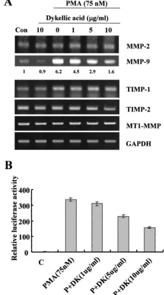
Fig. 1. Effect of dykellic acid on MMP-9 activity. A, Caski cells were treated with various concentrations of PMA. Conditional medium was collected after 24 h, and gelatin zymography was performed. B, Caski cells were treated with various concentrations of dykellic acid in the presence of PMA (75 nM). Conditional medium was collected after 24 h followed by gelatin zymography. C, conditional medium was collected and concen- trated. MMP-9 was identified by Western blot analysis using anti-MMP-9 antibody. The MMP-9 levels shown are representative of three independent experiments. The values below the figure represent change in protein expression of bands.
Fig. 2. Repression of PMA-induced MMP-9 transcription and promoter activity by dykellic acid. A, Caski cells were treated with or without various concentrations of dykellic acid in the presence of PMA (75 nM). Total RNA was isolated, and RT-PCR analysis was performed. The MMP-9 mRNA levels shown are representative of three independent experiments. The values below the figure represent change in mRNA ex- pression of the bands normalized to GAPDH. B, WT-MMP-9 promoter-containing re- porter vector was transfected and treated with various concentrations of dykellic acid in the absence or presence of PMA (75 nM). The cells were lysed and luciferase activity measured. Data represent the mean of at least three independent experiments; bars,⫾SD.
Table 1 Primer sequences used for RT-PCR of MMPs, TIMPs,aand MT1-MMP expressions
Genes Sequences PCR product (bp)
MMP-2 447
Sense 5⬘-ACAAAGAGTGGCAGCAA-3⬘
Antisense 5⬘-CACGAGCAAAGGCATCATCC-3⬘
MMP-9 590
Sense 5⬘-CACTGTCCACCCCTCAGAGC-3⬘
Antisense 5⬘-GCCACTTGTCGGCGATAAGG-3⬘
TIMP-1 400
Sense 5⬘-GGGGACACCAGAAGTCAACCAGA-3⬘
Antisense 3⬘-CTTTTCAGAGCCTTGGAGGAGCT-3⬘
TIMP-2 590
Sense 5⬘-TGCAGCTGCTCCCCGGTGCAC-3⬘
Antisense 5⬘-TTATGGGTCCTCGATGTCGAG-3⬘
MT1-MMP 550
Sense 5⬘-CCCTATGCCTACATCCGTGA-3⬘
Antisense 5⬘-TCCATCCATCACTTGGTTAT-3⬘
GAPDH 300
Sense 5⬘-CGGACTCAACGGATTTGGTCGTAT-3⬘
Antisense 5⬘-AGCCTTCTCCATGGTTGGTGAAGA-3⬘
aTIMP, tissue inhibitor of metalloproteinase; GAPDH, glyceraldehyde-3-phosphate dehydrogenase.
3431
cellular RNA was extracted from Caski cells using the TRIzol reagent (Life Technologies, Inc.). A cDNA was synthesized from 2g of total RNA using Moloney murine leukemia virus reverse transcriptase (Life Technologies, Inc., Gaithersburg, MD). PCR primers are described in Table 1. PCR products were analyzed by agarose gel electrophoresis and visualized by ethidium bromide.
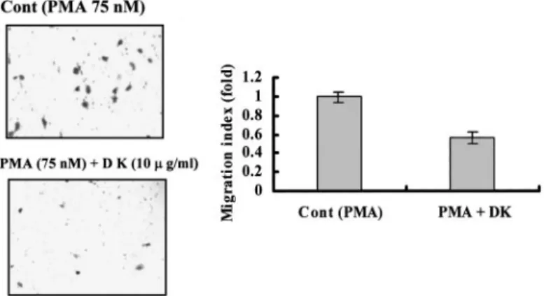
Invasion Assay. Five⫻ 104cells/chamber were used for each invasion assay. Invasion assays were performed using modified Boyden chambers with polycarbonate nucleopore membrane (Corning, Corning, NY). Precoated fil- ters (6.5 mm in diameter, 8m pore-size, Matrigel 100 g/cm2) were rehy- drated with 250l of medium, and 5 ⫻ 104cells in 200l medium with or without dykellic acid in the presence of PMA were seeded into the upper part of each chamber. After incubation for 24 h at 37°C, nonmigratory cells on the upper surface of the filter were wiped with a cotton swab, and migrated cells on the lower surface of the filter were fixed and stained with 0.125% Coo- massie Blue in a methanol:acetic acid:water mixture (45:10:45 v/v/v). Random fields were counted under a light microscope.
RESULTS
Dykellic Acid Inhibits MMP-9 Activity. Caski cells, a human cervical cancer cell line that releases basal levels of MMP-9 when cultured in serum-free medium, were treated with various concentra- tions of PMA for 20 h. Whereas the level of MMP-2 expression is not significantly altered by PMA, PMA induces the expression and se- cretion of large amounts of latent MMP-9 as determined by gelatin zymography (Fig. 1A). The expression and secretion of MMP-9 is induced by PMA in a dose-dependent manner, with a dramatic in- crease in MMP-9 activity after treatment with 75 nMPMA. As shown in Fig. 1B, dykellic acid decreases PMA-induced MMP-9 activity in a dose-dependent manner, whereas the activity of a control metallo- proteinase, of which the size was identical to the Mr72,000 type IV
collagenase MMP-2 (17), is not reduced in dykellic acid-treated cells.
We assayed the conditioned medium for MMP-9 by Western blotting to exclude the presence of a physiological inhibitor such as a tissue inhibitor of metalloproteinase family members (Ref. 5; Fig. 1C).
Similar to zymography data, expression levels of MMP-9 protein gradually decrease in a dose-dependent manner indicating that the reduced MMP-9 enzyme activity is because of smaller amounts of the protein.
Dykellic Acid Inhibits Transcription of MMP-9. Treatment of Caski cells with dykellic acid also induces a decrease in the levels of PMA-stimulated MMP-9 mRNA (Fig. 2A). RT-PCR shows that steady-state MMP-9 mRNA levels are lower in dykellic acid-treated cells compared with cells not treated with dykellic acid. The effect of dykellic acid on MMP-9 expression was additionally investigated using HEK293 cells transiently transfected with a luciferase reporter gene linked to the 0.7-kb fragment of MMP-9 promoter sequence. As shown in Fig. 2B, luciferase gene expression is activated up to 23-fold in these cells treated with PMA compared with untreated cells. Treat- ment of cells with dykellic acid (10g/ml) decreases PMA-mediated luciferase activity up to 2-fold, indicating that the 0.7-kb fragment of the MMP-9 promoter contains dykellic acid response elements.
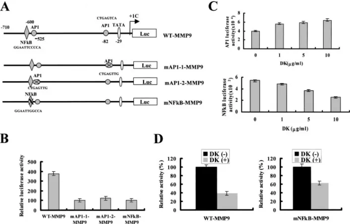
Effects of Dykellic Acid on NFB and AP-1 Activities. AP-1 and NFB point-mutated MMP-9 promoters show a diminished response to treatment with PMA, compared with the constructs made using a WT MMP-9 promoter (Fig. 3, A and B). In subsequent experiments, cells were transiently transfected with reporter vectors that included either the AP-1 or NFB binding sites. Luciferase activity in the cells with the NFB construct is significantly reduced by treatment with dykellic acid, whereas luciferase activity in cells with the AP-1
Fig. 3. Effects of dykellic acid on the activities of AP-1 and NFB. A, schematic structure of MMP-9 promoter constructs used for testing luciferase activity. Mutations were introduced into the NFB or AP-1 binding sites of WT-MMP-9 by 2-bp changes. B, WT or mutant MMP-9 promoters were transfected and treated with 75 nMPMA for 24 h, and luciferase activity measured. C, to elucidate the effects of dykellic acid on AP-1 and NFB activities, a reporter vector that has AP-1 (top panel) or NFB (bottom panel)-binding sites was transfected. The cells were treated with or without various concentrations of dykellic acid in presence of PMA (75 nM). Luciferase activity was measured. D, WT-MMP9 or mNFB-MMP9 plasmid was transfected, and treated with or without dykellic acid in presence of PMA (75 nM). Luciferase activity was measured. Data represent the mean of at least three independent experiments; bars,⫾SD.
3432
construct is slightly increased by treatment with dykellic acid (Fig.
3C). As shown in Fig. 3D, dykellic acid-mediated MMP-9 promoter inhibition measured by dykellic acid-driven luciferase activity is significantly greater in the WT MMP-9 construct than in the construct with the NFB-mutated MMP-9 promoter.
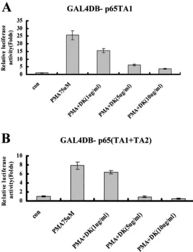
Dykellic Acid Does Not Inhibit DNA Binding of NFB, But Inhibits Activation through NFB Transactivation Domains TA1 and TA2. To determine whether dykellic acid inhibits activation of NFB through the inhibition of DNA binding of NFB, we examined the effect of dykellic acid on PMA-induced binding of NFB by EMSA. Dykellic acid did not affect the intensity of the NFB-DNA complex induced by PMA or its migration in Caski cells (data not shown). We investigated whether dykellic acid inhibits transcriptional activation through a chimeric transcription factor in which the trans- activating domain of p65 is fused to the yeast GAL4 DNA binding domain. Schmitz and Baeuerle (15) identified that the COOH-terminal portion of NFB p65 contains two transactivation domains, TA1 and TA2, and showed that chimeric fusion proteins with the TA1 (p65 amino acids 522–551) or TA1⫹TA2 (p65 amino acids 286–521) domains fused to the DNA binding domain of GAL4 (amino acids 1–147) confer PMA-inducible transcriptional activation onto a GAL4 reporter gene (15). We transiently transfected HEK293 cells with a GAL4 response element-luciferase reporter construct together with expression constructs encoding either the GAL4 DNA binding do- main alone (GAL4DB), GAL4DB-p65 TA1, or GAL4DB-p65
(TA1⫹TA2) followed by subsequent stimulation with increasing doses of dykellic acid. The expression construct for GAL4DB alone shows no transactivation of the GAL4 response element (data not shown). Constitutive transcription by the chimeric transcription fac- tor, GAL4DB-p65 TA1, is enhanced⬃25-fold after stimulation with PMA, and the enhancement of transcription is inhibited by dykellic acid (Fig. 4A). We observed a similar pattern of regulation using the chimeric transcription factor GAL4BD-p65 (TA1⫹TA2). Constitu- tive transcription is enhanced⬃8-fold after stimulation with PMA, and the PMA enhancement of transcription is inhibited by dykellic acid (Fig. 4B).
Inhibition of PMA-induced MMP-9 Activation by Other NFB Inhibitors. To examine effects of other well-known NFB inhibitors on PMA-induced MMP-9 activation, Caski cells were treated with
␣-lipoic acid, and PDTC in the presence of 75 nMPMA and MMP-9 expression was evaluated by zymography. As shown in Fig. 5, inter- estingly, ␣-lipoic acid and PDTC decrease MMP-2 and MMP-9 activity in a dose-dependent manner. However, PDTC and ␣-lipoic acid have no specific inactivation of MMP-9 activity (Fig. 5). Taken together, these data suggest that dykellic acid is unique in its ability to inhibit NFB activation and suppress MMP-9 gene expression.
Effect of Dykellic Acid on in Vitro Invasion of Caski Cells.
Treatment of cells with 10g/ml dykellic acid neither induced mor- phological changes nor inhibited the growth of Caski cells (data not shown). In the in vitro invasion assay, as shown in Fig. 6, dykellic acid inhibited migration by⬎40%. Therefore, the effect of dykellic acid on in vitro invasion inhibition was correlated well with its effect on MMP-9 inhibition.
DISCUSSION
The expression of proteases such as MMP-9 is regulated by diverse growth factors, cytokines, and xenobiotics such as PMA. Studies have shown that the mechanism responsible for PMA-mediated responses may involve direct alteration of transcription factors, but these mech- anisms are not completely understood. Small molecular weight inhib- itors that target the pathways that regulate MMP-9 expression could improve our understanding of these pathways and potentially be of clinical utility for the treatment of cancer. Using a cervical cancer cell line, our study demonstrates the ability of dykellic acid to reduce the expression of MMP-9.
We also investigated the molecular mechanism by which dykellic acid inhibits PMA-mediated expression of MMP-9 using AP-1 and
Fig. 4. Dykellic acid inhibits transcriptional activity of p65. HEK293 cells were transiently transfected with 1g of GAL4RE luciferase reporter plasmid and 1 g of expression plasmids for either GAL4DB-p65 TA1 (A) or GAL4DB-p65 (TA1⫹TA2; B).
The cells were treated with or without the indicated concentrations of dykellic acid in the absence or presence of PMA (75 nM). The mean normalized luciferase activity of GAL4DB-p65 (TA1⫹TA2) in untreated PMA cells was 500-fold higher than that of GAL4DB-p65 TA1. Data represent the mean of at least three independent experiments;
bars,⫾SD.
Fig. 5. Effect of␣-lipoic acid and PDTC on MMP-9 activity. Caski cells were treated with various concentrations of␣-lipoic acid in the presence or absence of PMA (75 nM).
Conditional medium was collected 24 h after treatment followed by gelatin zymography.
Caski cells were treated with vehicle, PMA (75 nM), PDTC (100M), and PMA plus PDTC. Conditional medium was collected after 24 h followed by gelatin zymography.
3433
NFB reporter constructs and found that NFB activity, but not AP-1 activity, is significantly reduced by treatment with dykellic acid. Thus, dykellic acid suppresses expression of MMP-9 via inhibition of NFB transactivation. To our knowledge, this is the first report of a small molecule that inhibits transcriptional activation by NFB. Whereas our study is unique in the demonstration of a small molecule inhibitor of NFB having an effect on the MMP-9 promoter, a previous study demonstrated the stimulation of the MMP-9 promoter by the activa- tion of the AP-1 motif at⫺79 bp, and either tumor necrosis factor␣ or PMA activation of NFB and Sp1 binding sites at ⫺600 bp and
⫺558 bp, respectively (9). We show that NFB is a crucial transac- tivator for MMP-9 gene expression, as PMA-driven luciferase activity is reduced significantly in both NFB and AP-1 mutant promoters.
Our experiments have also clarified the mechanism by which dykellic acid controls NFB transcriptional activation. NFB signal- ing involves stimulation-induced degradation of cytoplasmic inhibitor of NFB (18), releasing p65 for translocation from the cytoplasm into the nucleus, where p65 interacts with p50 and binds specifically to the NFB target DNA sequence. After specific binding to DNA, tran- scriptional activation of NFB is regulated through phosphorylation of p65 at several distinct sites (19, 20). We found that dykellic acid does not affect the DNA binding of NFB, but it does block trans- activation of NFB. We extended our analysis of the inhibition of NFB transcriptional activation by demonstrating that dykellic acid inhibits the PMA-induced activation of chimeric transcription factors containing the NFB p65 TA1 and TA2 transactivation domains.
Thus, our data indicate that dykellic acid-mediated MMP-9 transcrip- tional down-regulation critically depends on intact NFB binding sites within the MMP-9 promoter region.
Dykellic acid is a low molecular weight compound, which selec- tively targets NFB transactivation without affecting AP-1 during
suppression of PMA-induced MMP-9 expression. This compound will undoubtedly be a useful tool for laboratory investigations of NFB activity. Furthermore, considering the strong evidence support- ing a role for NFB in PMA-induced MMP-9 up-regulation, addi- tional studies determining the potential efficacy of dykellic acid in inhibiting invasion are warranted.
REFERENCES
1. Stetler-Stevenson, W. G., Hewitt, R., and Corcoran, M. Matrix metalloproteinases and tumor invasion: from correlation and causality to the clinic. Semin. Cancer Biol., 7: 147–154, 1996.
2. Chambers, A. F., and Matrisian, L. M. Changing views of the role of matrix metalloproteinases in metastasis. J. Natl. Cancer Inst., 89: 1260 –1270, 1997.
3. Zucker, S., Lysik, R. M., Zarrabi, M. H., and Moll, U. M(r) 92, 000 type IV collagenase is increased in plasma of patients with colon cancer and breast cancer.
Cancer Res., 53: 140 –146, 1993.
4. Bernhard, E. J., Gruber, S. B., and Muschel, R. J. Direct evidence linking expression of matrix metalloproteinase 9 (92-kDa gelatinase/collagenase) to the metastatic phe- notype in transformed rat embryo cells. Proc. Natl. Acad. Sci. USA, 91: 4293– 4297, 1994.
5. Sato, H., Takino, T., Okada, Y., Cao, J., Shinagawa, A., Yamamoto, E., and Seiki, M.
A matrix metalloproteinase expressed on the surface of invasive tumour cells. Nature (Lond.), 370: 61– 65, 1994.
6. Sato, H., and Seiki, M. Regulatory mechanism of 92 kDa type IV collagenase gene expression which is associated with invasiveness of tumor cells. Oncogene, 8:
395– 405, 1993.
7. Waas, E. T., Lomme, R. M., DeGroot, J., Wobbes, T., and Hendriks, T. Tissue levels of active matrix metalloproteinase-2 and -9 in colorectal cancer. Br. J. Cancer, 86:
1876 –1883, 2002.
8. Lee, P. P., Hwang, J. J., Murphy, G., and Ip, M. M. Functional significance of MMP-9 in tumor necrosis factor-induced proliferation and branching morphogenesis of mam- mary epithelial cells. Endocrinology, 141: 3764 –3773, 2000.
9. Nagase, H., and Woessner, J. F., Jr. Matrix metalloproteinases. J. Biol. Chem., 274:
21491–21494, 1999.
10. Kleiner, D. E., and Stetler-Stevenson, W. G. Matrix metalloproteinases and metas- tasis. Cancer Chemother. Pharmacol., 43: Suppl., S42–51, 1999.
11. Brew, K., Dinakarpandian, D., and Nagase, H. Tissue inhibitors of metalloprotein- ases: evolution, structure and function. Biochim. Biophys. Acta, 1477: 267–283, 2000.
12. Lee, H. J., Chun, H. K., Chung, M. C., Lee, C. H., Rhee, J. S., and Kho, Y. H.
Biosynthesis of dykellic acid: origin of the carbon skeleton. J. Antibiot., 53: 78 – 80, 2000.
13. Han, S. B., Lee, H. J., Kho, Y. H., Jeon, Y. J., Lee, S. H., Kim, H. C., and Kim, H. M.
New immunosuppressive activity of dykellic acid. J. Antibiot., 54: 840 – 843, 2001.
14. Overall, C. M., Wrana, J. L., and Sodek, J. Independent regulation of collagenase, 72-kDa progelatinase, and metalloendoproteinase inhibitor expression in human fibroblasts by transforming growth factor-. J. Biol. Chem., 264: 1860–1869, 1989.
15. Schmitz, M. L., and Baeuerle, P. A. The p65 subunit is responsible for the strong transcription activating potential of NF- B. EMBO J., 10: 3805–3817, 1991.
16. Baek, W. K., Park, J. W., Lim, J. H., Suh, S. I., Suh, M. H., and Kwon, T. K.
Molecular cloning and characterization of human BUB3 promoter. Gene, 295: 117–
123, 2002.
17. Strongin, A. Y., Collier, I., Bannikov, G., Marmer, B. L., Grant, G. A., and Goldberg, G. I. Mechanism of cell surface activation of 72-kDa type IV collagenase. Isolation of the activated form of the membrane metalloprotease. J. Biol. Chem., 270: 5331–
5338, 1995.
18. Baldwin, A. S., Jr. The NF-B and I B proteins: new discoveries and insights.
Annu. Rev. Immunol., 14: 649 – 683, 1996.
19. Vanden Berghe, W., Plaisance, S., Boone, E., De Bosscher, K., Schmitz, M. L., Fiers, W., and Haegeman, G. p38 and extracellular signal-regulated kinase mitogen-acti- vated protein kinase pathways are required for nuclear factor-B p65 transactivation mediated by tumor necrosis factor. J. Biol. Chem., 273: 3285–3290, 1998.
20. Wang, D., and Baldwin, A. S., Jr. Activation of nuclear factor-B-dependent tran- scription by tumor necrosis factor-␣ is mediated through phosphorylation of RelA/
p65 on serine 529. J. Biol. Chem., 273: 29411–29416, 1998.
Fig. 6. Effect of dykellic acid on Matrigel invasion by Caski cells. For invasion assay, the lower and upper parts of Transwells were coated with Matrigel. Caski cells cultured in the presence or absence of either PMA or dykellic acid at indicated concentrations were placed in the upper well. Invasiveness of the cells was determined by measuring ability to pass through a layer of Matrigel-coated filter. After 24 h, cells on the bottom side of the filter were fixed, stained, and counted as described under “Materials and Methods.” Data represent the mean of at least three independent experiments; bars,⫾SD.
3434
2003;63:3430-3434.
Cancer Res
Ju-Hyung Woo, Jong-Wook Park, Sung-Hee Lee, et al.
κ B Transcriptional Activity Factor
Matrix Metalloproteinase-9 Expression by Inhibiting Nuclear Dykellic Acid Inhibits Phorbol Myristate Acetate-induced
Updated version
http://cancerres.aacrjournals.org/content/63/12/3430
Access the most recent version of this article at:
Cited articles
http://cancerres.aacrjournals.org/content/63/12/3430.full.html#ref-list-1
This article cites 19 articles, 8 of which you can access for free at:
Citing articles
http://cancerres.aacrjournals.org/content/63/12/3430.full.html#related-urls
This article has been cited by 4 HighWire-hosted articles. Access the articles at: