211 http://dx.doi.org/10.4196/kjpp.2011.15.4.211
ABBREVIATIONS: MMP-9, matrix metalloproteinase-9; PI3K, phosphatidylinositol 3-kinase; MAPK, mitogen-activated protein kinase.
Received June 7, 2011, Revised August 16, 2011, Accepted August 28, 2011
Corresponding to: Sung-Soo Kim, Department of Pharmacology, College of Medicine, Kangwon National University, Hyoja-2-dong, Chuncheon 200-701, Korea. (Tel) 82-33-250-8851, (Fax) 82-33- 255-8809, (E-mail) [email protected]
Thrombin-induced Migration and Matrix Metalloproteinase-9 Expression Are Regulated by MAPK and PI3K Pathways in C6 Glioma Cells
Jiyoung Kim1, Jae-Won Lee1, Song-In Kim1, Yong-Joon Choi1, Won-Ki Lee1, Myung-Ja Jeong1, Sang-Hoon Cha2, Hee Jae Lee1, Wanjoo Chun1, and Sung-Soo Kim1
1Department of Pharmacology, College of Medicine, 2Department of System Immunology, College of Biomedical Science, Kangwon National University, Kangwon 200-701, Korea
Glioblastoma multiforme is one of the most common and aggressive tumors in central nervous system. It often possesses characteristic necrotic lesions with hemorrhages, which increase the chances of exposure to thrombin. Thrombin has been known as a regulator of MMP-9 expression and cancer cell migration. However, the effects of thrombin on glioma cells have not been clearly understood. In the present study, influences of thrombin on glioma cell migration were examined using Boyden chamber migration assay and thrombin-induced changes in MMP-9 expression were measured using zymography, semi-quantitative RT-PCR, and W estern blotting. Furthermore, underlying signaling pathways by which thrombin induces MMP-9 expression were examined. Thrombin-induced migration and MMP-9 expression were significantly potentiated in the presence of wortmannin, a PI3K inhibitor, whereas MAPK inhibitors suppressed thrombin-induced migration and MMP-9 expression in C6 glioma cells. The present data strongly demonstrate that MAPK and PI3K pathways evidently regulate thrombin-induced migration and MMP-9 expression of C6 glioma cells. Therefore, the control of these pathways might be a beneficial therapeutic strategy for treatment of invasive glioblastoma multiforme.
Key Words: Thrombin, MMP-9, C6 glioma cells, MAPK, PI3K
INTRODUCTION
Gliomas constitute nearly 60% of primary brain tumors and usually infiltrate normal brain parenchyma. Gliomas are categorized as their histologic features, and the major type of gliomas are astrocytomas [1]. Glioblastoma multi- forme (GBM) is the most common type of astrocytomas aris- ing in adults and clinically very aggressive [2]. These tu- mors typically show necrosis and hemorrhage. Patients di- agnosed as GBM survive less than 1 year due to diffusely infiltrating nature of GBM. Glioma cells migrate away from the primary tumor site so that surgical resection of a tumor mass is not effective for remission [3].
Matrix metalloproteinases (MMPs) are critically involved in many physiological and pathological processes such as tissue remodeling and cancer progression [4]. Especially, MMP-2 and MMP-9 (known as gelatinases) have been re- ported to play crucial roles in tumor angiogenesis and metastasis. It has been also reported that enhanced gelati- nase expression in various types of cancers corresponds to increased invasion and metastasis [5].
Thrombin, a trypsin-like serine protease, is the most copi- ous enzyme participating in the coagulation cascade. In ad- dition to its hemostatic roles, thrombin modulates a diver- sity of pathophysiological functions such as inflammation, cell migration and proliferation, apoptosis and so on. There are many evidences that thrombin regulates MMP-9 ex- pression [6-8] and cancer cell migration [9-11]. Neverthe- less, the effect of thrombin on glioma cell migration is not fully understood even though thrombin released by hemor- rhagic site of GBM may have a important role in invasion.
In the present study, we focused on demonstrating the ef- fects of thrombin on the migration of C6 rat glioma cells, which are morphologically similar to GBM [12], and possi- ble underlying signaling pathways that thrombin induces MMP-9-mediated migration.
METHODS Reagents
Thrombin and wortmannin were obtained from Sigma- Aldrich (St. Louis, MO, USA) and Calbiochem (Darmstadt, Germany), respectively. SB203580, SP600125, U0126 and MMP-9 inhibitor, batimastat, were purchased from Tocris
Bioscience (Ellisville, MO, USA).
Cell culture and thrombin treatment
The C6 rat glioma cell line was cultured in Dulbecco's modified Eagle's medium (DMEM; GIBCO BRL, Grand Island, NY, USA) in the presence of 10% fetal bovine serum (FBS; Hyclone Laboratories, Inc.), 100 U/ml of penicillin, 100 mg/ml of streptomycin and 2 mM of glutamine (GIBCO BRL, Grand Island, NY, USA). Cells were maintained at 37oC in a humidified atmosphere with 5% CO2. All in- hibitors were pretreated in serum-deprived culture medium for 1 hr followed by thrombin treatment as indicated.
Cell migration assay
Cell migration assay was performed by BD FalconTM Cell Culture Inserts (BD Bioscience, Bedford, MA; pore size, 8-μ m) in 24-well culture plates. Cell culture inserts were coat- ed with fibronectin (Sigma, St. Louis, MO, USA; 3μg/ml) both upper and lower surfaces. Approximately 0.7×10⁵ cells in 400μl of serum-free medium were seeded in the upper chamber and 400μl of the same medium was placed in the lower chamber. When seeded, cells were respectively pre- treated with appropriate concentration of inhibitors;
SB203580, SP600125, U0126, and wortmannin. After 1 hr of pretreatment, thrombin was added to the lower chamber and the plates were incubated for 6 hr at 37oC in 5% CO2
incubator. Subsequently, cells were fixed in 3.7% form- aldehyde solution for 10 min and permeabilized by 0.2%
Triton X-100 for 10 min. Then, cells on the lower surface were stained with 0.2% crystal violet and rinsed with PBS several times. Non-migrating cells in the upper chamber were attentively removed by cotton swabs. The migrating cells were counted by light microscopy.
Western blot analysis
After removed the culture media, the C6 rat glioma cells were harvested and lysed with PRO-PREPTM Protein Extraction Solution (iNtRON Biotechnology, Inc.). The ob- tained protein samples were quantified by the BCA protein assay and the same amounts of total cellular proteins were resolved on 10% SDS-polyacrylamide gels. After SDS- PAGE, proteins were transferred to polyvinylidene di- fluoride (PVDF) membranes (Amersham Biosciences, Pis- cataway, NJ, USA). The membranes were incubated in blocking solution (5% skim milk in TBST) for 1 hr at room temperature and then probed overnight with suitable anti- bodies at 4oC; anti-MMP-9 (Affinity BioReagents, Golden, CO, USA; 1 : 1,000), p-Akt, Akt (Cell Signaling Technology, Inc.; 1 : 1,000), or β-actin (Sigma-Aldrich, St. Louis, MO, USA; 1 : 5,000). After three washes with TBST, the mem- branes were incubated with either horseradish peroxidase (HRP)-conjugated goat anti-rabbit or donkey anti-mouse secondary antibody (Jackson ImmunoResearch) for 2 hr at room temperature. The blots were detected by enhanced chemiluminescence using KodakⓇ BioMaxTM XAR Film.
RNA isolation and semi-quantitative RT-PCR analysis Isolation of total cellular RNA from C6 glioma cells was performed using the Total RNA Extraction Kit (iNtRON Biotechnology, Inc.) according to the manufacturer's ins- tructions. Total RNA was quantified by NanoDrop spec-
trophotometer (Thermo Fischer Scientific Inc., Wilmington, DE, USA) and 2μg of total RNA was immediately re- verse-transcribed by OmniscriptⓇ RT Kit (QIAGEN GmbH, Hilden, Germany) with oligo-(dT) 15 primers (Promega, Madison, WI, USA). The cDNA was amplified by polymerase chain reaction (PCR) with appropriate primers (MMP-9 for- ward, TCCAGTAGACAATCCTTGCA and reverse, CTC- CGTGATTCGAGAACTTC; GAPDH forward, GACAACT- TTGGCATCGTGGA and reverse, ATGCAGGGATGATG- TTCTGG). 30 cycles of PCR was performed by nTaq polymer- ase under the following conditions: denaturing at 94oC for 30 sec, annealing at 62oC for 45 sec, and elongation at 72oC for 30 sec. The PCR products (MMP-9, 110 bp; GAPDH, 133 bp) were electrophoresed on a 2% agarose gel containing ethi- dium bromide and visualized by UV transillumination.
Gelatin zymography
The gelatin zymography was carried out in 8% poly- acrylamide gels containing 1 mg/ml of gelatin. C6 glioma cells were plated in 60 mm culture dishes and incubated overnight in serum- free medium. Cell culture supernatants were collected and concentrated by AmiconⓇ Ultra Centri- fugal Filter (Millipore, Bedford, MA) subsequent to 24 hr-thrombin treatment. These samples were mixed with non-reducing 5x loading buffer without heating and electro- phoresed on the gel as mentioned above. After PAGE, gels were thoroughly washed with enzyme renaturing buffer (2.5% Triton X-100 solution) at room temperature for 4 times every 15 min. The gels were then transferred into incubation buffer (50 mM Tris-HCl, 0.15 M NaCl, 10 mM CaCl2, and 0.02% NaN3) and placed in a 37oC shaking in- cubator for 24 hr followed by Coomassie brilliant blue R-250 staining and destaining procedures.
Statistical analysis
All data are expressed as the mean±standard deviation (SD). Statistical significance was analyzed by two-tailed Student’s t-test. Data with values of p<0.05 were consid- ered as statistically significant. Single (* and #) and double (** and ##) marks represent statistical significance in p
<0.05 and p<0.01, respectively.
RESULTS
Thrombin induced the migration of C6 glioma cells In the previous studies, it has been demonstrated that thrombin induces cell motility in many cell lines [9,13,14].
To investigate the effect of thrombin on the migration of C6 glioma cells, we used the Boyden chamber migration assay. Approximately 0.7×10⁵ cells were plated in the upper chamber of cell culture inserts and incubated with or with- out 25 U/ml of thrombin. After 6 hr of incubation, the cells migrating through the membrane toward thrombin contain- ing serum-free media were stained with crystal violet and the number of migrated cells was counted (Fig. 1). The number of migrated cells was significantly increased in the presence of thrombin compared to control, suggesting that thrombin has a chemotactic effect on C6 glioma cells.
Fig. 1. Thrombin stimulates C6 glioma cell migration. (A) Representative images of C6 cell migration assay. Thrombin (25 U/ml) treatment for 6 hr exhibited approximately 2 fold increase in number of migrating cells. Migrating cells and unmigrating cells were separated by polyethylene tere- phthalate (PET) membranes. Microscopy images were detected the migrating cells on the lower surface of the membrane. (B) Quantitative analysis of migration assay.
The cell migration was quantified by counting the cells that migrating through the membranes. Cell counting data were expressed as a percentage in comparison with control group.
The experiments were conducted in triplicate and data are shown as the means±SD. **p<0.01 indicates statistically significant difference with control.
Fig. 2. MAPK inhibitors (MAPKIs) inhibit thrombin-induced C6 glioma cell migration. (A) Representative images of the MAPKI effects on thrombin-induced migration. SB203580, SP600125, and U0126 were pretreated for 1 hr at the concentrations of 20, 20, and 10μM, respectively, followed by thrombin treatment for 6 hr. (B) Quantitative analysis of migration assay. The cell migration was quantified by counting the cells that migrating through the membranes. All data were obtained from three independent experiments and presented as the means±SD. *p<0.05 indicates statistically significant difference with control. #p<0.05 and ##p<0.01 indicate statistically signiticunt difference with thrombin.
MAPK inhibitors had an inhibitory effect on thrombin-induced migration of C6 glioma cells Involvement of MAPK pathway in the regulation of can- cer cell migration has been investigated by many studies [15-17]. In the present study, to determine the underlying mechanism for chemotactic action of thrombin, C6 cells were treated with some MAPK pathway inhibitors:
SB203580 (20μM), a specific inhibitor of p38-MAPK;
SP600125 (20μM), a selective inhibitor of c-Jun N-terminal kinase (JNK); U0126 (10μM), a selective inhibitor of
MEK/ERK. All inhibitors were pretreated for 1 hr in serum free media with indicated concentrations and thrombin was subsequently treated for 6 hr. As shown in Fig. 2, throm- bin-induced migrating cells were reduced with pretreat- ment of the MAPK inhibitors. Inhibition of p38 with SB203580 showed significant inhibition of C6 migration (Fig. 2). Especially, inhibition of JNK and ERK with SP600125 and U0126, respectively, exerted such a strong inhibition that the number of migrated cells was reduced almost to control level (Fig. 2). These results suggest that MAPK pathway may regulate thrombin-induced migration of C6 glioma cells.
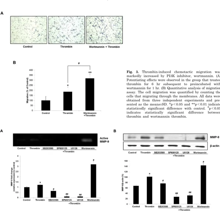
Fig. 3. Thrombin-induced chemotactic migration was markedly increased by PI3K inhibitor, wortmannin. (A) Potentiating effects were observed in the group that treated thrombin for 6 hr subsequent to preincubated with wortmannin for 1 hr. (B) Quantitative analysis of migration assay. The cell migration was quantified by counting the cells that migrating through the membranes. All data were obtained from three independent experiments and pre- sented as the means±SD. *p<0.05 and **p<0.01 indicate statisticodly significant difference with control. #p<0.05 indicates statistically significant difference between thrombin and wortmannin thrombin.
Fig. 4. Effects of thrombin and MAPK or PI3K inhibitors treatment on MMP-9 expression in protein level. All inhibitors were pretreated for 1 hr at the indicated concentrations followed by 24 hr of thrombin (5 U/ml) treatment: SB203580 and SP600125, 20μM; U0126 and wortmannin, 10μM. C6 cells were incubated with inhibitors in the serum free media. (A) MMP-9 activities were analyzed by zymography.
Thrombin-induced MMP-9 expression was blocked by MAPK inhibitors and increased by PI3K inhibitor. (B) The cellular level of MMP-9 was examined by western blot analysis. β-actin was used as an internal control. *p<0.05 indicates statistically signiticant difference with control. #p<0.05 and ##p<0.01 indicate statistically signiticant difference with thrombin.
Thrombin-induced C6 cell migration was significantly increased with wortmannin, a PI3K inhibitor The previous studies have been shown that PI3K path- way is also related to the cancer cell motility [18,19]. On this account, we investigated the effect of the PI3K path- way on thrombin-induced C6 cell migration with a PI3K inhibitor wortmannin, an antifungal antibiotic. 100 nM of wortmannin was used for migration assay and other ex- perimental procedures were the same as mentioned above.
Wortmannin significantly potentiated the migratory activ- ity induced by thrombin (Fig. 3). This result may suggest that the PI3K pathway negatively regulates thrombin-in- duced C6 cell migration.
MMP-9 expression was correlated with C6 cell migration propensity
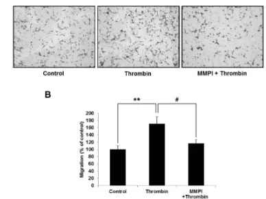
To elucidate whether the thrombin-induced migration and MMP-9 expression are directly related, we used an MMP-9 inhibitor (MMPI), batimastat, for migration assay.
Fig. 6. Thrombin-induced migration was directly blocked by batimastat, an MMP-9 inhibitor (MMPI). (A) Repre- sentative images of the effect of MMPI on thrombin-induced C6 cell migration. 0.5μM of batimastat was pretreated for 1 hr and thrombin was subsequently treated for 6 hr. (B) Quantitative analysis of migration assay. The cell migra- tion was quantified by counting the cells that migrating through the membranes. All data were obtained from three independent experiments and presented as the means±SD.
**p<0.01 and #p<0.05 indicate statistically signficant difference between control and thrombin, and thrombin and MMPI thrombin, respectively.
Fig. 5. Effects of thrombin and MAPK or PI3K inhibitors treatment on MMP-9 expression in mRNA level. C6 cells were pretreated with inhibitors for 1 hr, followed by thrombin (5 U/ml) treatment for 4 hr. Subsequently, RNA samples were isolated and subjected to semi-quantitative RT-PCR for MMP-9 with a housekeeping gene, GAPDH, as an internal control. MMP-9 mRNA expression patterns were similar to western blot and zymographic data.
The suppressive effect of MMPI is represented in Fig. 6.
It clearly showed that MMP-9 is directly involved in cell migration stimulated by thrombin.
To determine whether MMP-9 is regulated by MAPK and PI3K pathways in the thrombin-induced migration of C6 glioma cells, we examined the changes of MMP-9 ex- pression in multiple stages such as mRNA, protein, and ex- tracellular levels. C6 cells were pre-incubated with the MAPK or PI3K inhibitors for 1 hr and thrombin was sub- sequently treated for 24 hr in serum-free media. After in- cubation, the conditioned media were collected and con- centrated for zymography and the cells were harvested and lysed for western blot analysis. Gelatinolytic activities of MMP-9 were visualized by zymography. As shown in Fig.
4A, thrombin induces MMP-9 secretion and it is suppressed by MAPK inhibitors whereas thrombin-induced MMP-9 se- cretion was potently increased by wortmannin (Fig. 4A).
These results closely correspond to migration assay data.
Western blot analysis also presented the same tendency to zymographic data. To further confirm, we measured MMP-9 expression in mRNA level using semi-quantitative RT-PCR assay. After pretreated the inhibitors for 1 hr, C6 cells were incubated with thrombin for 4 hr. MMP-9 mRNA was ex- pressed in the same manner as MMP-9 protein expression (Fig. 5). These data strongly support that thrombin-medi-
ated MMP-9 expression is regulated by MAPK and PI3K pathways.
DISCUSSION
Glioblastoma multiforme (GBM) is one of the most ag- gressive CNS tumors with very poor prognosis because of its highly-infiltrative nature [20]. GBM has a histologically char- acteristic feature which forms a serpentine pattern of ne- crosis and vascular or endothelial cell proliferation. These ne- crotic lesions often contain hemorrhagic components [2].
These conditions lead glioma cells to have more chances of exposure to thrombin. Previous studies have been re- vealed that thrombin is involved in cancer cell migration and invasion [9,10,21]. In the present study, we found that thrombin enhances migration of C6 rat glioma cells by Boyden chamber migration assay.
To determine the mechanism of the thrombin effect, we considered MAPK and PI3K pathways as pivotal factors based on the former studies [22,23]. Thrombin-induced mi- gratory ability was significantly blocked by MAPK inhibitors.
Wortmannin, a specific inhibitor of the PI3K pathway, on the other hand, surprisingly reinforced the thrombin-medi- ated cell migration. We used 100 nM and 1μM of wortman- nin for migration assay, but 1μM was less potent than 100 nM of wortmannin in inducing migratory ability (data not shown). These results might be caused by excessive PI3K pathway inhibition in 1μM of wortmannin treatment.
Because PI3K pathway is critical in cell survival, its in- ordinate suppression subjects to cellular damage. The C6 cells treated with 1μM of wortmannin presented slight morphological changes and with 10μM showed more severe changes in migration assay.
As MMPs are key components of cell migration and in- vasion [5,24], we investigated the relationship between C6 cell migration and MMP-9. Thrombin-induced migration was effectively suppressed by an MMP-9 inhibitor, bati- mastat. It supports that MMP-9 is directly involved in cell migration stimulated by thrombin. However, batimastat alone could not totally block the thrombin-induced migra- tion. This implies that there are more factors responsible
for thrombin-induced cell migration other than MMP-9.
Thrombin induced MMP-9 expression in protein and mRNA level, parallel to increase in C6 cell migration. Induced MMP-9 expression was significantly attenuated by MAPK inhibitors and augmented by the PI3K inhibitor. Our re- sults indicate that these pathways regulate thrombin-medi- ated MMP-9 expression and it affects migratory activity of C6 cells. That is, the activator of PI3K pathway, such as the platelet-derived growth factor (PDGF), or MAPK in- hibitors might inhibit the thrombin-induced MMP-9 ex- pression and migration. A previous study actually demon- strated that PDGF inhibits MMP-9 activity in C6 cells [25].
Wortmannin, for itself, did not increase both MMP-9 ex- pression and cell migration (Data not shown).
In conclusion, the present study suggests that thrombin mediates C6 glioma cell migration by increasing expression of MMP-9. In addition, MAPK and PI3K pathways might be involved in thrombin-induced cell migration and MMP-9 expression. The migratory ability of cells is an important factor for cancer metastasis. Together with the previous studies, this study suggests that inhibition of MAPK path- way or potentiation of PI3K pathway could be a valuable therapeutic target for the treatment of metastatic glio- blastoma.
ACKNOWLEDGEMENTS
This study was supported by Kangwon National Univer- sity (Sung-Soo Kim).
REFERENCES
1. Louis DN, Ohgaki H, Wiestler OD, Cavenee WK, Burger PC, Jouvet A, Scheithauer BW, Kleihues P. The 2007 WHO classification of tumours of the central nervous system. Acta Neuropathol. 2007;114:97-109.
2. Lefranc F, Brotchi J, Kiss R. Possible future issues in the treatment of glioblastomas: special emphasis on cell migration and the resistance of migrating glioblastoma cells to apoptosis.
J Clin Oncol. 2005;23:2411-2422.
3. Holland EC. Glioblastoma multiforme: the terminator. Proc Natl Acad Sci USA. 2000;97:6242-6244.
4. Kessenbrock K, Plaks V, Werb Z. Matrix metalloproteinases:
regulators of the tumor microenvironment. Cell. 2010;141:
52-67.
5. Björklund M, Koivunen E. Gelatinase-mediated migration and invasion of cancer cells. Biochim Biophys Acta. 2005;1755:
37-69.
6. Kawakita K, Kawai N, Kuroda Y, Yasashita S, Nagao S.
Expression of matrix metalloproteinase-9 in thrombin-induced brain edema formation in rats. J Stroke Cerebrovasc Dis.
2006;15:88-95.
7. Choi MS, Kim YE, Lee WJ, Choi JW, Park GH, Kim SD, Jeon SJ, Go HS, Shin SM, Kim WK, Shin CY, Ko KH. Activation of protease-activated receptor1 mediates induction of matrix metalloproteinase-9 by thrombin in rat primary astrocytes.
Brain Res Bull. 2008;76:368-375.
8. Radjabi AR, Sawada K, Jagadeeswaran S, Eichbichler A, Kenny
HA, Montag A, Bruno K, Lengyel E. Thrombin induces tumor invasion through the induction and association of matrix metalloproteinase-9 and beta1-integrin on the cell surface. J Biol Chem. 2008;283:2822-2834.
9. Chiang HS, Yang RS, Huang TF. Thrombin enhances the adhesion and migration of human colon adenocarcinoma cells via increased beta 3-integrin expression on the tumour cell surface and their inhibition by the snake venom peptide, rhodostomin. Br J Cancer. 1996;73:902-908.
10. Kaufmann R, Junker U, Junker K, Nuske K, Ranke C, Zieger M, Scheele J. The serine proteinase thrombin promotes migration of human renal carcinoma cells by a PKA-dependent mechanism. Cancer Lett. 2002;180:183-190.
11. Heider I, Schulze B, Oswald E, Henklein P, Scheele J, Kaufmann R. PAR1-type thrombin receptor stimulates mig- ration and matrix adhesion of human colon carcinoma cells by a PKCepsilon-dependent mechanism. Oncol Res. 2004;14:
475-482.
12. Grobben B, De Deyn PP, Slegers H. Rat C6 glioma as experimental model system for the study of glioblastoma growth and invasion. Cell Tissue Res. 2002;310:257-270.
13. Bizios R, Lai L, Fenton JW 2nd, Malik AB. Thrombin-induced chemotaxis and aggregation of neutrophils. J Cell Physiol.
1986;128:485-490.
14. Pankonin G, Teuscher E. Stimulation of endothelial cell migration by thrombin. Biomed Biochim Acta. 1991;50:1073-1078.
15. Reddy KB, Nabha SM, Atanaskova N. Role of MAP kinase in tumor progression and invasion. Cancer Metastasis Rev.
2003;22:395-403.
16. Jadeski LC, Chakraborty C, Lala PK. Nitric oxide-mediated promotion of mammary tumour cell migration requires sequential activation of nitric oxide synthase, guanylate cyclase and mitogen-activated protein kinase. Int J Cancer. 2003;106:
496-504.
17. Frankenberry KA, Somasundar P, McFadden DW, Vona-Davis LC. Leptin induces cell migration and the expression of growth factors in human prostate cancer cells. Am J Surg. 2004;
188:560-5.
18. Barber MA, Welch HC. PI3K and RAC signalling in leukocyte and cancer cell migration. Bull Cancer. 2006;93:E44-52.
19. Vasko VV, Saji M. Molecular mechanisms involved in differen- tiated thyroid cancer invasion and metastasis. Curr Opin Oncol. 2007;19:11-7.
20. Kanu OO, Hughes B, Di C, Lin N, Fu J, Bigner DD, Yan H, Adamson C. Glioblastoma Multiforme Oncogenomics and Signaling Pathways. Clin Med Oncol. 2009;3:39-52.
21. Chen HT, Tsou HK, Tsai CH, Kuo CC, Chiang YK, Chang CH, Fong YC, Tang CH. Thrombin enhanced migration and MMPs expression of human chondrosarcoma cells involves PAR receptor signaling pathway. J Cell Physiol. 2010;223:737-745.
22. Wang Z, Castresana MR, Newman WH. Reactive oxygen species-sensitive p38 MAPK controls thrombin-induced migra- tion of vascular smooth muscle cells. J Mol Cell Cardiol. 2004;
36:49-56.
23. Wang L, Luo J, He S. Induction of MMP-9 release from human dermal fibroblasts by thrombin: involvement of JAK/STAT3 signaling pathway in MMP-9 release. BMC Cell Biol. 2007;8:14.
24. Watanabe H. Extracellular matrix--regulation of cancer invasion and metastasis. Gan To Kagaku Ryoho. 2010;37:2058-2061.
25. Estève PO, Robledo O, Potworowski EF, St-Pierre Y. Induced expression of MMP-9 in C6 glioma cells is inhibited by PDGF via a PI 3-kinase-dependent pathway. Biochem Biophys Res Commun. 2002;296:864-869.