Korean J Anesthesiol Vol. 54, No. 3, March, 2008
Received:January 17, 2008
Corresponding to:Young Ho Jang, Department of Anesthesiology and Pain Medicine, School of Medicine, Keimyung University, 194, Dongsan-dong, Jung-gu, daegu 700-712, Korea. Tel: 82-53-250-7287, Fax: 82-53-250-7240, E-mail: [email protected]
Effect of A Kappa-opioid Receptor Agonist U50488H Given at Early Reperfusion Phase in Isolated Rat Hearts
Departments of Anesthesiology and Pain Medicine, and *Internal Medicine, School of Medicine, Keimyung University, Daegu, Korea
Yong Cheol Lee, M.D., Young Ho Jang, M.D., Jin Mo Kim, M.D., Ae Ra Kim, M.D., Chan Jin Kim, M.D., and Yoon Nyun Kim, M.D.*
Background: The experiment was performed to determine the role of κ-opioid receptor (OR) agonist U50488H given at early reperfusion.
Methods: Isolated hearts were subjected to 30 minutes of regional ischemia and 120 minutes of reperfusion. Hearts were assigned randomly to one of the three groups: 1) Control (n = 9), 2) U50-1 (n = 8); 1μM of U50488H, and 3) U50-10 (n
= 8); 10μM of U50488H. U50488 was perfused for a period of 5 min before and 30 min after reperfusion.
Results: U50488H significantly reduced infarct size as a percentage of ischemic area (12.2 ± 1.9% in U50-1 and 7.2 ± 1.7%
in U50-10, P < 0.001) compared to the control hearts (27.2 ± 1.2%). After 2 hrs of reperfusion, left ventricular developed pressure was significantly recovered by U50488H (62.6 ± 5.7% in U50-1 and 68.6 ± 4.7% in U50-10, P = 0.018 and 0.002, respectively) compared to the control (46.3 ± 4.4%). Rate-pressure product was improved by 10μM U50488H (62.3 ± 5.5%, P = 0.007) but not by 1μM U50488H (50.0 ± 4.1%) compared to the control (44.7 ± 4.5%). U50488H significantly increased the +dP/dtmax
(77.9 ± 5.5% in U50-1 and 78.0 ± 4.3 in U50-10, P = 0.005 and 0.001 vs. control, respectively). The −dP/dtmin also improved by 10μM U50488H (64.7 ± 4.8%, P = 0.003) compared to control (47.0 ± 2.7%).
Conclusions: U50488H given at early reperfusion phase reduces both infarct size and myocardial stunning in isolated rat hearts.
(Korean J Anesthesiol 2008; 54: S 29∼34)
Key Words: ischemia, myocardium, opioid receptor, reperfusion.
INTRODUCTION
Opioids have been widely used in the anesthesia field as anesthetic adjuncts or for pain control. Because pretreatment is seldom possible in an acute myocardial infarction, pharmacological therapies targeting reperfusion has generated considerable recent interest. In this regard, opioids may play a key strategic role because these drugs are widely used in clinical field and can protect against post-ischemic myocardial injury at the time of reperfusion.
1)Based on binding studies, there is evidence that both δ- and κ-opioid receptors (OR) are located in the myocardium.
2,3)It has previously been demonstrated that the activation of OR by
ischemic preconditioning (IPC) or by opioid-induced pharmaco- logical pretreatment provides cardioprotection against ischemia- reperfusion (I/R) injury via activation of the δ-OR subtypes, especially the δ
1-OR.
4-6)However, the cardioprotective role of κ-OR remains unclear and there continues to be controversy regarding its role in myocardial I/R injury.
6-9)Recently, Wang et al.
10)demonstrated that ischemic postconditioning (Post-C) improved cardiodynamics in isolated rat hearts. They suggested that the functional recovery by Post-C was induced via activation of κ-OR and opening of mitochondrial K
ATP(mK
ATP) chan- nels. Therefore, it is highly suggested that the direct treatment of U50488H, a selective κ-OR agonist, given at early reper- fusion might also improve cardiodynamic parameters in I/R induced hearts.
The objective of this study was to investigate the protective
effect of U50488H on infarct limitation and post-reperfusion
contractile recovery during the reperfusion phase in isolated
perfused rat hearts.
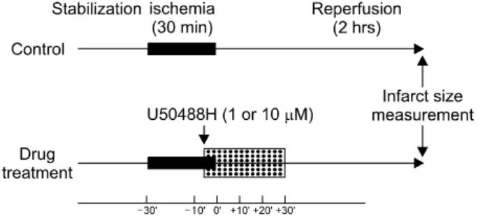
Fig. 1. Experimental protocol. For measurement of hemodynamic data and infarct size by κ-opioid receptor activation, isolated rat hearts are exposed to 30 min ischemia followed by 2 hrs reper- fusion. Two concentrations of κ-opioid receptor agonist U50488H (1 and 10μM) are perfused for a period of 5 min before and 30 min after reperfusion (hatched rectangle).
MATERIALS AND METHODS
The experimental procedures and protocols used in this study were reviewed and approved by our Institutional Animal Care and Use Committee, 2007.
Langendorff isolated heart perfusion preparation
Male Wistar rats weighing 280−320 g were obtained from Korea Taconic Co., Republic of Korea. They received 100 mg/kg of pentobarbital sodium and 300 IU of heparin intra- peritoneally. Hearts were isolated and perfused as described in our previous report.
11)In brief, a midline thoracotomy was performed and the heart was quickly mounted on a Langen- dorff apparatus by the aortic root. The heart was perfused with modified Krebs-Henseleit (KH) solution containing (in mM) 118.5 NaCl, 4.7 KCl, 1.2 MgSO
4, 1.8 CaCl
2, 24.8 NaHCO
3, 1.2 KH
2PO
4, and 10 glucose. All these chemicals were obtained from Sigma-Aldrich Chemical, USA. The solution was filtered through a Nargene 2.0μm microfilter (Nalge Nunc Interna- tional Corp., USA) and was equilibrated with 95% O
2/5% CO
2for 30 min before the experiment and pH maintained at appro- ximately 7.4. The whole perfusion system was heated to 38
oC by means of water jacketing. Perfusion was performed under a hydrostatic pressure of 100 cmH
2O by adjusting the height of the reservoir.
Making a myocardial regional ischemia-repefusion model
In order to induce myocardial regional ischemia, the proximal portion of left coronary artery (LCA) was first localized bet- ween the left atrial appendage and the right ventricular outflow tract. This was then followed by the passage of a 4-0 polypro- pylene suture around the major trunk of the LCA or its pro- minent branches. The ends of the thread were passed through a small PE50 tube to form a snare. All hearts were then allowed to stabilize for at least 30 minutes. Ischemia was induced by pulling the snare and then fixing it by clamping the tubing with a small hemostat. Myocardial ischemia was confirmed by regional cyanosis, a substantial decrease in left ventricular developed pressure (LVDP), and a fall in coronary flow (CF). Reperfusion was initiated by releasing the ends of the suture. All hearts were subjected to 30 minutes of regional ischemia and 120 minutes of reperfusion.
Assessment of cardiac function
In isolated hearts, an air-bubble free, KH buffer-filled latex balloon connected to a pressure transducer with tubing was inserted into the left ventricle through the left atrial appendage.
Balloon volume was adjusted to give a left ventricular end- diastolic pressure (LVEDP) of 5−10 mmHg at the beginning of the experiment and was kept constant throughout the experiment. LVDP was calculated as the difference between left ventricular systolic pressure (LVSP) and LVEDP. CF was measured by a timed collection of the perfusate drippings from the right heart into a graduated cylinder.
Hemodynamic data were continuously recorded with the MP150 system (BIOPAC Systems Inc., USA). The rate-pressure product (RPP) was calculated as the LVDP × heart rate (HR).
The maximum and minimum of first derivative of left ventricular pressure (+dP/dt
maxand −dP/dt
min) were analyzed using ana- lysis software (Acqknowledge, version 3.9.0., BIOPAC Systems Inc., CA).
Experimental protocol
Hearts were randomly divided into three groups: (1) Control;
no intervention either before or after LCA occlusion, (2) U50-1;
1μM of U50488H, and (3) U50-10; 10μM of U50488H (Fig.
1). A standard selective κ-OR agonist U50488H (Tocris Bios-
cience, USA) was perfused for a period of 5 min before and
30 min after reperfusion. U50488H was dissolved in distilled
water and prepared in stock solution. The final concentrations,
which did not exceed 2.5 mM, were stored at 4
oC according
to the manufacturer’s guidance. On the day of each experiment,
Fig. 2. Area at necrosis (AN) as percentage of area at risk (AAR) as evaluated by triphenyltetrazolium chloride staining following 30 min regional ischemia and 2 hrs reperfusion in isolated rat heart model. Rat hearts were subjected to control and treated with 1 (U50-1) or 10μM (U50-10) of U50488H. Both concentrations of U50488H given at early reperfusion phase significantly decreases AN/AAR. Values are expressed as mean ± SEM. *: P < 0.001 vs. control.
Table 1. Morphometrics for Isolated Rat Hearts
Group Body weight Heart weight LV volume AAR volume AAR/LV AN volume
(gm) (gm) (cm3) (cm3) (%) (cm3)
Control (n = 9) 305.0 ± 5.8 1.42 ± 0.02 0.519 ± 0.020 0.303 ± 0.014 60.8 ± 2.9 0.082 ± 0.004 U50-1 (n = 8) 307.8 ± 9.8 1.42 ± 0.03 0.498 ± 0.019 0.291 ± 0.020 59.2 ± 5.1 0.036 ± 0.005*
U50-10 (n = 8) 301.9 ± 4.8 1.40 ± 0.01 0.486 ± 0.029 0.279 ± 0.019 59.2 ± 5.7 0.022 ± 0.006*
Values are mean ± SEM. n: number of hearts. LV: left ventricle, AAR: area at risk, AN: area at necrosis, U50-1: 1μM U50488H, U50-10: 10μM U50488H. There were no differences in body weight, heart weight, volumes of LV and AAR, and AAR/LV among groups.
*: P < 0.001 vs. Control.
the compound was diluted with KH solution to the required final concentrations.
Determination of area at risk and infarct size
At the end of each experiment (2 hrs after reperfusion), the area at risk (AAR) and area at necrosis (AN) were measured as described in our previous study.
11)In brief, the LCA perfusion circuit was reoccluded, and diluted fluorescent polymer microspheres with 2−9μm diameter (Duke Scientific Corp., USA) were infused to demarcate the AAR as the tissue without fluorescence. The hearts were weighed, frozen at −20° C for 1−
3 hrs, and cut into 2 mm thick transverse slices using a rat heart slice matrix (Zivic Instruments, USA). The slices were incubated in 1% 2,3,5-triphenyltetrazolium chloride (TTC, Sigma-Aldrich Chemical, USA) in sodium phosphate buffer (pH = 7.4) at 37
oC for 20 min. The slices were immersed in 10%
formalin to enhance the contrast between viable (red) and ne- crotic (pale) tissue and then compressed to a uniform 2 mm thickness by placing them (basal side) between two glass plates separated by a 2 mm space. The myocardial AAR was identified by illuminating the slices with U.V. light. The infarcted (unstained) and risk (no fluorescent area) zone regions were traced on a clear acetate transparent sheet and quantified with the UTHSCSA Image Tool 3.0 version. Volumes of the left ventricle, infarct zone, and risk zone were calculated by multiplying each area with slice thickness and summing the products. Infarct volume was expressed as a percentage of the AAR volume. All measurements were performed in a blinded fashion.
Statistical analysis
All values were expressed as means ± SEM. Data analysis was performed with SPSS 13.0 version. Data were analyzed using one-way analysis of variance with the Least Significant Difference test. Differences were considered to be statistically
significant when P values were less than 0.05.
RESULTS
A total of 28 rat hearts were used for this experiment. All hearts were perfused within 30−40 seconds after excision.
Three hearts were excluded from data analysis for the following reasons: a CF > 18 ml/min (1), LVDP < 80 mmHg (1), and HR < 250 beats/min (1) during the stabilization period.
Therefore, we report the data for 25 successfully completed experiments (9 in Control, 8 in U50-1, and 8 in U50-10, respectively).
There were no significant group differences with respect to
body weight, heart weight, and volumes of left ventricle and
AAR (Table 1). As shown in Fig. 2, infarct size in the
control hearts was 27.2 ± 1.2% of the AAR. Both concen-
Table 2. Baseline Coronary Flow and Hemodynamic Data
Group CF HR LVDP RPP +dP/dtmax −dP/dtmin
Control (n = 9) 14.0 ± 0.62 83.1 ± 8.1 103.5 ± 8.9 29.1 ± 2.3 2.69 ± 0.15 −2.67 ± 0.20 U50-1 (n = 8) 13.6 ± 0.92 92.1 ± 7.1 103.8 ± 3.8 30.2 ± 1.0 2.60 ± 0.10 −2.66 ± 0.10 U50-10 (n =8) 14.0 ± 1.02 92.1 ± 7.1 103.8 ± 3.8 30.2 ± 1.0 2.60 ± 0.10 −2.66 ± 0.10 Values are mean ± SEM. n: number of hearts. CF: coronary flow (ml/min), HR: heart rate (beats/min), LVDP: left ventricular developed pressure (mmHg), RPP: rate-pressure product (mmHg/min/103), +dP/dtmax: maximum positive left ventricular pressure derivative (mmHg/s/103),
−dP/dtmin: minimum negative left ventricular pressure derivative (mmHg/s/103), U50-1: 1μM U50488H, U50-10: 10μM 50488H. There were no differences in baseline CF and hemodynamics among groups.
Fig. 3. Recovery of the left ventricular developed pressure (LVDP) and rate-pressure product (RPP) after 2 hrs reperfusion in isolated rat hearts. Rat hearts were subjected to control and treated with 1 (U50-1) or 10μM (U50-10) of U50488H. U50488H treatment at reperfusion phase significantly increases the LVDP and RPP. Value are expressed as means ± SEM. *: P < 0.05 vs. control.
trations of a κ-OR agonist U50488H given at early reperfusion phase significantly reduced myocardial infarction (12.2 ± 1.9%
in U50-1 and 7.2 ± 1.7% in U50-10, P < 0.001 vs. control).
The baseline hemodynamic data are summarized in Table 2.
CF and hemodynamic indexes concerning HR, LVDP, RPP, + dP/dt
max, and −dP/dt
minwere comparable in all groups under baseline conditions. The changes in LVDP and RPP after 2 hrs of reperfusion are presented in Fig. 3. LVDP was decreased to 46.3 ± 4.4% after 2 hrs of reperfusion compared to baseline value in control hearts. U50488H treatment at reperfusion phase significantly recovered the LVDP compared to the control hearts (62.6 ± 5.7% in U50-1, P = 0.018, 68.6
± 4.7% in U50-10, P = 0.002). RPP in the control hearts was decreased to 44.7 ± 4.5% after 2 hrs of reperfusion. The atte- nuation of RPP was improved by 10μM U50488H (62.3 ± 5.5%, P = 0.007) but not by 1μM U50488H (50.0 ± 4.1%).
The changes in the recovery of the first derivative of left ventricular pressure after 2 hrs reperfusion are shown in Fig.
4. After 2 hrs of reperfusion, +dP/dt
maxand −dP/dt
minin the control hearts were decreased to 49.2 ± 3.6% and 47.0 ±
2.7% compared to baseline levels, respectively. Both concen- trations of U50488H significantly increased +dP/dt
max(77.9 ± 5.5% in U50-1 and 78.0 ± 4.3% in U50-10, P = 0.005 and 0.001 vs. control, respectively). The treatment of 10μM U50488H effectively improved −dP/dt
min(64.7 ± 4.8%, P = 0.003) com- pared to the control hearts (47.0 ± 2.7%).
DISCUSSION
There remains controversy regarding the role of κ-OR ago-
nists in myocardial I/R injury. Previous studies have demon-
strated that pharmacological preconditioning with a κ-OR ago-
nist bremazocin increased infarct size
9)and another κ-OR agonist
U50488H exacerbated ischemic-reperfusion arrhythmias following
coronary occlusion in the isolated rat hearts.
12)Conversely,
Peart et al.
7)demonstrated that exogenously application of three
different κ-OR agonists (U50488H, ICI204448, and BRL52537)
10 min before the onset of ischemia reduced infarct size in
intact myocardial infarction rat models. Taken together, these
results do not completely rule out roles for κ-OR agonists in
Fig. 4. Recovery of the maximum (+dP/dtmax) and minimum (−dP/dtmin) of first derivative of left ventricular pressure after 2 hrs reper- fusion in isolated rat hearts. Rat hearts were subjected to control and treated with 1 (U50-1) or 10μM (U50-10) of U50488H. U50488H treatment at reperfusion significantly enhanced the recovery of the +dP/dtmax and −dP/dtmin. Value are expressed as means ± SEM. *: P
< 0.05 vs. control.
cardioprotection. However, most of the studies to investigate the role of OR agonists in myocardial I/R injury thus far were mainly focused on the nonspecific OR agonist or δ-OR agonist.
1,13)Furthermore, there is scanty literature that have investigated the effects of a κ-OR agonist U50488H admini- stered solely at reperfusion.
In our present study, the AN/AAR in control hearts was 27.2 ± 1.2%. Although the AN/AAR in our control hearts was smaller than those reported by others,
1,14)this is in agreement with our recently reported study.
15)Although the exact reason for this discrepancy is unknown, differences in the deter- mination of risk and infarct area may account for it. U50488H given at early reperfusion phase was shown to significantly reduce myocardial infarction in our isolated rat hearts. These results are consistent with a recent report by Gross group,
16)which showed that either δ- or κ-OR agonist administered as a single bolus 5 min before reperfusion could provide infarct size sparing effects in intact rat heart. Combined with our data, it is strongly suggested that κ-OR agonist U50488H targeting the reperfusion phase may play a role to prevent lethal reperfusion injury.
While treatment of κ-OR agonists during the reperfusion phase provides anti-infarct effects, little is known about its cardiodynamic effect following reperfusion. Recently, Wang et al.
10)reported that Post-C improved the cardiodynamic parameters via activating κ-OR and mK
ATPby indirect antagonist study.
Peart and Gross
17)reported that U50488H confers cardio- protection with respect to LVDP, +dP/dt
max, and −dP/dt
minin isolated mice hearts undergoing 20 min global ischemia
followed by 45 min reperfusion. However, it is not clear whether the contractile recovery after reperfusion is caused by the treatment prior to the ischemic period or due to its effects during the reperfusion period in their study (they used U50488H for 10 min prior to global ischemia and resumed it at the onset of reperfusion). Therefore, we investigated the functional recovery effects by κ-OR agonist U50488H targe- ting only the reperfusion period. Both concentrations of U50488H (1 and 10μM) given at early reperfusion phase significantly improved the functional recovery of LVDP, RPP,
+dP/dt
max, and −dP/dt
minafter 2 hrs of reperfusion in our study. The LVDP recovered to more than 80% of its baseline levels by U50488H. The maximum and minimum of the first derivative of left ventricular pressure were significantly impro- ved up to 46% compared to control hearts by U50488H after 2 hrs of reperfusion. The functional recovery was greater in the 10μM U50488H. However, one should take into consi- deration that the higher concentration may lead to more unde- sirable effects.
In summary, a κ-OR agonist U50488H given at early reper- fusion could significantly reduce both infarct size and myo- cardial stunning in isolated rat hearts. These results may benefit future clinical strategies when treating patients with ischemic heart disease.
REFERENCES
1. Gross ER, Hsu AK, Gross GJ: Opioid-induced cardioprotection occurs via glycogen synthase kinase β inhibition during reper-
fusion in intact rat hearts. Circ Res 2004; 94: 960-6.
2. Krumins SA, Faden AI, Feuerstein G: Opiate binding in rat hearts:
modulation of binding after hemorrhagic shock. Biochem Biophys Res Commun 1985; 127: 120-8.
3. Ventura C, Bastagli L, Bernardi P, Caldarera CM, Guarnieri C:
Opioid receptors in rat cardiac sarcolemma: effect of phenyle- phrine and isoproterenol. Biochim Biophys Acta 1989; 987: 69-74.
4. Okubo S, Tanabe Y, Takeda K, Kitayama M, Kanemitsu S, Kukreja RC, et al: Ischemic preconditioning and morphine attenuate myocardial apoptosis and infarction after ischemia- reperfusion in rabbit: role of δ-opioid receptor. Am J Physiol Heart Cric Physiol 2004; 287: H 1786-91.
5. Peart JN, Patel HH, Gross GJ: Delta-opioid receptor activation mimics ischemic preconditioning in the canine heart. J Cardiovasc Pharmacol 2003; 42: 78-81.
6. Schultz JE, Hsu AK, Gross GJ: Ischemic preconditioning in the intact rat heart is mediated by δ1 but not µ or κ opioid receptors.
Circulation 1998; 97: 1282-9.
7. Peart JN, Gross ER, Gross GJ: Effect of exogenous kappa-opioid receptor activation in rat model of myocardial infarction. J Cardiovasc Pharmacol 2004; 43: 410-5.
8. Wang GY, Wu S, Pei JM, Yu XC, Wong TM: Kappa- but not delta-opioid receptors mediate effects of ischemic preconditioning on both infarct and arrhythmia in rats. Am J Physiol Heart Circ Physiol 2001; 280: H 384-91.
9. Aitchison KA, Baxter GF, Awan MM, Smith RM, Yellon DM, Opie LH: Opposing effects on infarction of delta and kappa opioid receptor activation in the isolated rat heart: implications for
ischemic preconditioning. Basic Res Cardiol 2000; 95: 1-10.
10. Wang J, Gao Q, Shen J, Ye TM, Xia Q: Kappa-opioid receptor mediates the cardioprotective effect of ischemic postconditioning.
Zhejiang Da Xue Xue Bao Yi Xue Ban 2007; 36: 41-7.
11. Park SS, Zhao H, Jang Y, Mueller RA, Xu Z: N6-(3-Iodobenzyl)- adenosine-5'-N-methylcarboxamide confers cardioprotection at reperfusion by inhibiting mitochondrial permeability transition pore opening via glycogen synthase kinase 3β. J Pharmacol Exp Ther 2006; 318: 124-31.
12. Wong TM, Lee AY, Tai KK: Effects of drugs interacting with opioid receptors during normal perfusion or ischemia and reperfusion in the isolated rat heart-an attempt to identify cardiac opioid receptor subtype(s) involved in arrhythmogenesis. J Mol Cell Cardiol 1990; 22: 1167-75.
13. Chang WL, Lee SS, Su MJ: Attenuation of post-ischemia reperfusion injury by thaliporphine and morphine in rat hearts. J Biomed Sci 2005; 12: 611-9.
14. Gross ER, Hsu AK, Gross GJ: GSK3 inhibition and KATP channel opening mediate acute opioid-induced cardioprotection at reper- fusion. Basic Res Cardiol 2007; 102: 341-9.
15. Jang Y, Xi J, Wang H, Mueller RA, Norfleet EA, Xu Z: Postcon- ditioning prevents reperfusion injury by activating opioid receptors.
Anethesiology 2008; 108: 243-50.
16. Gross ER, Gross GJ: Ligand triggers of classical preconditioning and postconditioning. Cardiovasc Res 2006; 70: 212-21.
17. Peart JN, Gross GJ: Exogenous activation of delta- and kappa-opioid receptors affords cardioprotection in isolated murine heart. Basic Res Cardiol 2004; 99: 29-37.