INTRODUCTION
Remifentanil is a potent ultra-short-acting synthetic opi- oid that is widely used during general anesthesia including cardiac anesthesia for fast-tracking. Remifentanil precondi- tioning (R-Pre) could effectively provide cardioprotection against ischemia-reperfusion injury (I/R) in rat hearts [1,2].
Additionally, adenosine, an endogenous nucleotide, has been shown to increase by several-fold during ischemia and protect the myocardium from reperfusion injury [3,4].
The cardioprotective signaling pathways by R-Pre and adenosine appear to be similar. The opioid receptor (OPR) and adenosine receptor (ADR) are guanine nucleotide bind- ing protein (G protein)-coupled receptors (GPCRs). The activation of GPCRs converges on a key event in the cardio- protective process hypothesized to be stimulation of protein kinase C (PKC) [5]. The OPR has been reported to stimulate
phospholipase C [6]. The cardioprotection of R-Pre is medi- ated via PKC and mitochondrial adenosine triphosphate-de- pendent potassium (mKATP) channels [7]. Additionally, ADRs are linked to signal transduction pathways including phospho- lipase C, PKC, and the mKATP channel in adenosine mediated protection [8-10]. Functional coupling between the OPR fam- ily and ADRs has been previously demonstrated. In isolated rat hearts, improvement of post-ischemic cardiac function by a synthetic opioid analgesic, fentanyl, was reduced by the non- selective OPR antagonist naloxone and the selective A1ADR antagonist 1,3-dipropyl-8-cyclopentylxanthine (DPCPX) [11].
In addition, the cardioprotective effect of the A1ADR ago- nist 2-chloro-N6-cyclopentyladenosine and morphine were attenuated by the δ-OPR antagonist 7-benzylidenenaltrexone maleate and DPCPX, respectively [8]. These examples sug- gest the possibility that there is cross-talk between the OPRs and ADRs in the cardioprotection mediated by remifentanil.
However, the specific type of ADR responsible for cross-talk with OPR in R-Pre remains unclear among the four subtypes (A1, A2A, A2B, and A3).
The purpose of this study was to determine whether there is cross-talk between OPRs and ADRs in the cardiac
*Corresponding author: Sang-Jin Park. Department of Anesthesiology and Pain Medicine, College of Medicine, Yeungnam University, 170, Hyeonchung-ro, Nam-Gu, Daegu 705-703, Republic of Korea.
Tel: +82-53-620-3366, Fax: 82-53-626-5275, E-mail: [email protected] Submitted: 10 September 2015 / Accepted: 23 October 2015
Remifentanil-induced preconditioning has cross-talk with A 1 and A 2B adenosine receptors in ischemic-reperfused
rat heart
Yong-Cheol Lee1, Jiyoon Jung2, Sang-Jin Park2*
1Department of Anesthesiology and Pain Medicine, School of Medicine, Keimyung University, Daegu, Republic of Korea, 2Department of Anesthesiology and Pain Medicine, College of Medicine, Yeungnam University, Daegu, Republic of Korea
ABSTRACT
The purpose of this study was to determine whether there is a cross-talk between opioid receptors (OPRs) and adenosine receptors (ADRs) in remifentanil preconditioning (R-Pre) and, if so, to investigate the types of ADRs involved in the cross-talk. Isolated rat hearts received 30 min of regional ischemia followed by 2 hr of reperfusion. OPR and ADR antagonists were perfused from 10 min before R-Pre until the end of R-Pre.
The heart rate, left ventricular developed pressure (LVDP),velocity of contraction (+dP/dtmax), and coronary flow (CF) were recorded. The area at risk and area of necrosis were measured. After reperfusion, the LVDP, +dP/dtmax, and CF showed a significant increase in the R-Pre group compared with the control group (no intervention before or after regional ischemia). These increases in the R-Pre group were blocked by naloxone, a nonspecific ADR antagonist, an A1 ADR antagonist, and an A2B ADR antagonist. The infarct size was reduced significantly in the R-Pre group compared with the control group. The infarct-reducing effect in the R-Pre group was blocked by naloxone, the nonspecific ADR antagonist, the A1 ADR antagonist, and the A2B ADR antagonist. The results of this study demonstrate that there is cross-talk between ADRs and OPRs in R-Pre and that A1 ADR and A2B ADR appear to be involved in the cross-talk.
KEY WORDS: Adenosine; cross-talk; remifentanil; reperfusion; preconditioning
DOI: http://dx.doi.org/10.17305/bjbms.2016.738 Bosn J Basic Med Sci. 2016;16(1):64-70. © 2016 ABMSFBIH
protection mediated by R-Pre. In addition, we attempted to investigate the specific subtypes of ADR involved in the cross- talk with OPR in R-Pre.
MATERIALS AND METHODS
The experimental procedures and protocols used in this study were reviewed and approved by the Institutional Animal Care and Use Committee.
Drugs and chemicals
Remifentanil (Ultiva®) was purchased from GlaxoSmithKline Manufacturing (Parma, Italy). The nonspe- cific OPR antagonist naloxone was purchased from Reyon Pharmaceutical Corporation (Seoul, Republic of Korea).
The nonspecific ADR antagonist 8-(p-sulfophenyl) the- ophylline hydrate (8-SPT) was purchased from Santa Cruz Biotechnology (Santa Cruz, CA, USA). The A1ADR antago- nist DPCPX, A2AADR antagonist 4-(2-[7-amino-2-[2-furyl]
[1,2,4]triazolo[2,3-a][1,3,5]triazin-5-yl-amino]ethyl)phenol (ZM241385), A2BADR antagonist N-(4-acetylphenyl)-2-[4- (2,3,6,7-tetrahydro-2,6-dioxo-1,3-dipropyl-1H-purin-8-yl) phenoxy]acetamide (MRS1706), and A3ADR antagonist 1,4-dihydro-2-methyl-6-phenyl-4-(phenylethynyl)-3,5-pyr- idinedicarboxylic acid 3-ethyl-5-[(3-nitrophenyl)methyl]
ester (MRS1334) were purchased from Tocris Bioscience (Minneapolis, MN, USA). The fluorescent polymer micro- spheres were purchased from Duke Scientific (Palo Alto, CA, USA). The other chemicals and 2,3,5-triphenyltetrazolium chloride (TTC) were obtained from Sigma-Aldrich Chemical (St. Louis, MO, USA).
Naloxone and 8-SPT were dissolved in distilled water. The ADR antagonists were dissolved in dimethyl sulfoxide. The stock chemicals were stored at -20°C and diluted with Krebs- Henseleit (KH) solution to the required final concentrations on the day of each experiment.
Experimental procedure
Two researchers participated in the study. The first researcher was aware of the group assignment of each rat heart, whereas the second researcher was not. Male Sprague- Dawley rats weighing 300-350 gm obtained from Koatech Corporation (Cheongwon-gun, Republic of Korea) were used. The rats received intraperitoneal administration of 50 mg/kg of pentobarbital sodium and 300 IU of heparin. The rats’ hearts were then isolated and mounted to a Langendorff apparatus and perfused with a modified KH solution contain- ing (in mM) 118.5 NaCl, 4.7 KCl, 1.2 MgSO4, 1.8 CaCl2, 24.8 NaHCO3, 1.2 KH2PO4, and 10 glucose. A snare was made at the level of the proximal length of the left coronary artery
(LCA) and its major branches. Regional ischemia was induced by pulling the snare and was confirmed by regional cyanosis and a substantial decrease in left ventricular developed pres- sure (LVDP). Reperfusion was initiated by releasing the snare.
The rat hearts received 30 min of regional ischemia fol- lowed by 2 hr of reperfusion. The hearts were randomly assigned to one of the following groups according to a com- puter-generated random table: 1) CON: control, no interven- tion before or after LCA occlusion, 2) R-Pre: remifentanil pre- conditioning with 100 ng/mL of remifentanil hydrochloride in three cycles of administration for 5 min interspersed with 5-min drug-free periods, 3) R-Pre+NAL: 100 μM of pretreat- ment with naloxone in the R-Pre group, 4) R-Pre+SPT: 10 μM of 8-SPT pretreatment in the R-Pre group, 5) R-Pre+DPCPX:
200 nM of DPCPX pretreatment in the R-Pre group, 6) R-Pre+ZM: 100 nM of ZM241385 pretreatment in the R-Pre group, 7) R-Pre+M1706: 15 nM of MRS1706 pretreatment in the R-Pre group, 8) R-Pre+M1334: 100 nM of MRS1334 pre- treatment in the R-Pre group. The research object number in each group was eight.
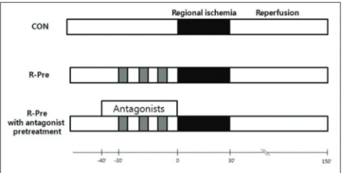
The OPR and ADR antagonists were perfused from 10 min before R-Pre until the end of R-Pre (40 min) (Figure 1). The concentrations of all of the antagonists were based on previ- ous studies performed on isolated working rat hearts that had no effect on infarct size in hearts subjected to I/R [4,12-16].
Measurements
The second researcher, who was blinded to the group assignment, measured the cardiac function and infarction size of the heart. In the isolated hearts, an air-bubble free, KH buffer-filled latex balloon was inserted into the left ven- tricle (LV) through the left atrial appendage. The volume of the balloon was adjusted using the BIOPAC system (BIOPAC Systems Inc., Goleta, CA, USA) to provide and sustain a left
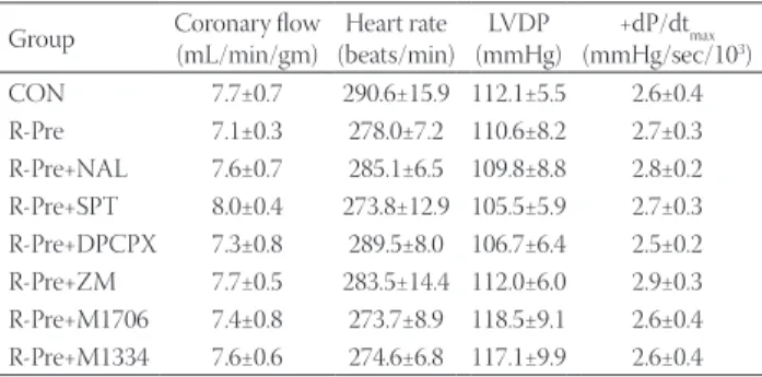
TABLE 1. Baseline coronary flow and cardiodynamic data Group Coronary flow
(mL/min/gm) Heart rate (beats/min) LVDP
(mmHg) +dP/dtmax (mmHg/sec/103) CON 7.7±0.7 290.6±15.9 112.1±5.5 2.6±0.4 R-Pre 7.1±0.3 278.0±7.2 110.6±8.2 2.7±0.3 R-Pre+NAL 7.6±0.7 285.1±6.5 109.8±8.8 2.8±0.2 R-Pre+SPT 8.0±0.4 273.8±12.9 105.5±5.9 2.7±0.3 R-Pre+DPCPX 7.3±0.8 289.5±8.0 106.7±6.4 2.5±0.2 R-Pre+ZM 7.7±0.5 283.5±14.4 112.0±6.0 2.9±0.3 R-Pre+M1706 7.4±0.8 273.7±8.9 118.5±9.1 2.6±0.4 R-Pre+M1334 7.6±0.6 274.6±6.8 117.1±9.9 2.6±0.4 Values are expressed as mean±SEM. The research object number in each group was eight. There were no significant differences among the groups. CON: Untreated control heart; R-Pre: Remifentanil pre- conditioning; NAL: Nonspecific opioid receptor antagonist naloxone;
SPT: Nonspecific adenosine receptor (ADR) antagonist 8-(p-sulfophenyl) theophylline hydrate; DPCPX: A1ADR antagonist; ZM: A2AADR antago- nist ZM241385; M1706: A2BADR antagonist MRS1706; M1334: A3ADR antagonist MRS1334; LVDP: Left ventricular developed pressure;
+dP/dt : Velocity of contraction
ventricular end-diastolic pressure (LVEDP) of 5 to 10 mmHg from the beginning of the experiment. The heart rate (HR), left ventricular systolic pressure (LVSP), LVEDP, and velocity of contraction (+dP/dtmax) were continuously recorded using the BIOPAC system. LVDP was calculated as the difference between the LVSP and the LVEDP. Coronary flow (CF) was measured by the timed collection of the perfusate dripping from the heart into a graduated cylinder.
After 2 hr of reperfusion, the snare was retightened and a fluorescent polymer microsphere was injected to distinguish the normal, non-ischemic region and the area at risk (AR). The hearts were removed from the Langendorff system, drained and weighed. They were then frozen for 3 hr at -20°C. The hearts were cut into 2 mm thick transverse slices using a rat heart slicer matrix (Zivic Instruments, Pittsburgh, PA, USA).
The slices of the LV were incubated in TTC in sodium phos- phate buffer (pH = 7.4) at 37°C for 20 min and subsequently immersed in 10% formalin to enhance the contrast. The LV was removed from the remaining tissue. The area at risk in the LV was identified by illumination with ultraviolet light.
The area of necrosis (AN, unstained with TTC) in the LV was traced on a clear acetate transparent sheet and quanti- fied using UTHSCSA ImageTool, Version 3.0 (Department of Dental Diagnostic Science at The University of Texas Health Science Center, San Antonio, TX, USA). The areas were con- verted into volumes by multiplying them by slice thickness.
The AN volumes were expressed as a percentage of the AR volume. All of the morphometric measurements were blindly performed by an independent technician. The primary end point was the AN in the LV. Secondarily, the CF, HR, LVDP, and +dP/dtmax were measured.
Exclusion criteria
Any heart with a HR < 250 beats/min, CF > 18 mL/min or < 8 mL/min, or LVDP < 80 mmHg when the LVEDP was maintained at 5-10 mmHg at the end of stabilization was
excluded from the study. Any heart exhibiting arrhythmia during the stabilization period was excluded as well.
Statistical analysis
The data are presented as the mean ± SEM. The data were analyzed using one-way analysis of variance (ANOVA) with Dunnett’s post-hoc testing. Null hypotheses of no difference were rejected if the p values were less than 0.05. The data anal- ysis was performed using a personal computer statistical soft- ware package (SPSS for Windows, version 21.0; IBM, Armonk, NY, USA).
RESULTS
A total of 67 rat hearts were used in the experiments. Three hearts were excluded for the following reasons: HR < 250 beats/min (n = 2) and LVDP < 80 mmHg (n = 1) after the sta- bilization period. The number of hearts that successfully com- pleted the infarct experimental study was 64, and the research object number in each group was eight. Thirty-seven hearts (6 in CON, 4 in R-Pre, 4 in R-Pre+NAL, 6 in R-Pre+SPT, 4 in R-Pre+DPCPX, 4 in R-Pre+ZM, 4 in R-pre+M1706, and 5 in R-Pre+M1334) experienced episodes of ventricular fibrillation (VF) during early reperfusion and typically reverted sponta- neously to a sinus rhythm. A statistical analysis was not per- formed for the occurrence of VF because of the small sample size in each group.
Coronary flow
No significant differences in the baseline CF were observed among the groups, with an average of 7.1 to 8.0 mL/min/gm (Table 1). After reperfusion for 2 hr, the CF was compared to the baseline level (Figure 2). In the control group, the CF decreased to 50.7 ± 5.5% from the baseline level. In the R-Pre group, the CF increased significantly compared with the con- trol group after reperfusion (80.0 ± 5.1%, p < 0.01). The nonspe- cific OPR antagonist naloxone (51.1 ± 3.9%) and the nonspecific ADR antagonist 8-SPT (47.7 ± 5.8%) significantly attenu- ated the increase in the CF of the R-Pre group (p < 0.05 and p < 0.001 vs. R-Pre, respectively). Additionally, the increase in the CF of the R-Pre group was blocked by the A1ADR antag- onist DPCPX (56.8 ± 5.7%, p < 0.05) and the A2BADR antago- nist MRS1706 (54.4 ± 4.5%, p < 0.01); it was not blocked by the A2AADR antagonist ZM241385 (71.1 ± 5.8%) and the A3ADR antagonist MRS1334 (72.5 ± 6.7%).
Cardiac functional recovery data
The baseline values of HR, LVDP, and +dP/dtmax after sta- bilization are shown in Table 1. No differences in the baseline cardiodynamic parameters were observed among the groups.
FIGURE 1. Experimental protocols. Hearts were subjected to 30 min of regional ischemia and 2 hr of reperfusion. R-Pre was induced by 100 ng/mL of remifentanil hydrochloride in three cycles of administration for 5 min interspersed with 5-min drug-free periods (gray rectangles). Adenosine or opioid recep- tor antagonists were perfused from 10 min before R-Pre. CON:
untreated control hearts; R-Pre: remifentanil preconditioning.
Figure 3 shows the recovery of HR, LVDP, and +dP/dtmax com- pared to baseline levels. After reperfusion for 2 hr, the HR, LVDP, and +dP/dtmax in the control hearts were 81.8 ± 3.8%, 40.7 ± 3.6%, and 38.4 ± 3.9% of the baseline levels, respectively.
No significant differences were observed among the groups in HR. LVDP was significantly increased in the R-Pre group compared with the control group (53.2 ± 2.4%, p < 0.05).
Naloxone (41.0 ± 3.2%) and 8-SPT (35.7 ± 2.7%) completely abrogated the increase of LVDP in the R-Pre group (p < 0.05 and p < 0.001, respectively). Additionally, DPCPX (40.2 ± 3.0%) and MRS1706 (39.4 ± 3.4%) abrogated the increase of LVDP in the R-Pre group (p < 0.01). However, ZM241385 (53.4 ± 2.7%) and MRS1334 (52.1 ± 1.3%) did not block the increase of LVDP in the R-Pre group.
Compared with the control group, +dP/dtmax showed a significant increase in the R-Pre group (49.2 ± 2.8%, p < 0.05).
Naloxone (37.8 ± 4.0%, p < 0.01), 8-SPT (34.3 ± 3.7%, p < 0.001), DPCPX (39.9 ± 2.4, p < 0.05), and MRS1706 (40.4 ± 1.7, p < 0.05) completely blocked the increase of +dP/dtmax in the R-Pre group. However, ZM241385 (50.3 ± 2.1%) and MRS1334 (51.2 ± 2.0%) did not block the increase of +dP/dtmax in the R-Pre group.
Morphometric analysis
No significant differences in body weight and heart weight were observed among the groups (Table 2). The risk volume averaged 0.387 cm3 to 0.446 cm3 with no statistically signifi- cant differences among the groups. The AR/LV ranged from 58.7% to 64.7% with no significant differences among all of the groups, implying that the changes in infarct size were not
related to the degree of AR in our experiments. As shown in Figure 4, the AN in the control hearts was 34.9 ± 2.6% of the AR, and the AN/AR in the R-Pre group was signifi- cantly reduced compared with the untreated control hearts (20.7 ± 2.5%, p < 0.01). This infarct-reducing effect of the R-Pre group was significantly reversed by naloxone (37.0 ± 3.1%, p < 0.01) and 8-SPT (35.6 ± 2.9%, p < 0.01). Figure 5 shows the effect of four subtypes of ADR antagonists on the anti-infarct effect of R-Pre. The addition of DPCPX (38.0 ± 2.8%, p < 0.01) or MRS1706 (39.6 ± 3.1%, p < 0.001) before R-Pre prevented the infarct-sparing effect in the R-Pre. However, the administra- tion of ZM241385 (22.6 ± 1.6%) or MRS1334 (22.7 ± 3.0%) had no significant effect on the AN/AR compared with the R-Pre group (20.7 ± 2.5%).
DISCUSSION
This study showed that activation of the OPR by R-Pre produced cardiac protection against I/R injury, and this effect FIGURE 2. Percent changes in coronary flow after 2 hr of reperfu-
sion compared to baseline levels in isolated rat hearts. R-Pre signifi- cantly increases the recovery of coronary flow compared to CON after reperfusion. Increase in coronary flow by R-Pre is blocked by NAL and SPT. DPCPX and M1706 also block the increase in cor- onary flow by R-Pre. The research object number in each group was eight. CON: Untreated control hearts; R-Pre: Remifentanil preconditioning; NAL: Nonspecific opioid receptor antagonist naloxone; SPT: Nonspecific adenosine receptor (ADR) antagonist 8-(p-sulfophenyl) theophylline hydrate; DPCPX: A1ADR antagonist;
ZM: A2AADR antagonist ZM241385; M1706: A2BADR antagonist MRS1706; M1334: A3ADR antagonist MRS1334; *: p < 0.05 vs. CON.
FIGURE 3. Percent changes in heart rate, LVDP, and +dP/dtmax after 2 hr of reperfusion compared to baseline levels in isolated rat hearts. R-Pre significantly increases the recovery of LVDP and +dP/dtmax compared to CON after reperfusion. Increases in LVDP and +dP/dtmax by R-Pre are blocked by NAL and SPT.
DPCPX and M1706 also block increases in LVDP and +dP/dtmax by R-Pre. The research object number in each group was eight.
LVDP: Left ventricular developed pressure; +dP/dtmax: Velocity of contraction; CON: untreated control hearts; R-Pre: remifentanil preconditioning; NAL: Nonspecific opioid receptor antagonist naloxone; SPT: Nonspecific adenosine receptor (ADR) antagonist 8-(p-sulfophenyl)theophylline hydrate; DPCPX: A1ADR antagonist;
ZM: A2AADR antagonist ZM241385; M1706: A2BADR antagonist MRS1706; M1334: A3ADR antagonist MRS1334; *: p < 0.05 vs. CON.
was blocked by the nonspecific ADR antagonist 8-SPT as well as by the nonspecific OPR antagonist naloxone. In addi- tion, selective A1ADRand A2BADR antagonists (DPCPX and MRS1706) blocked the cardioprotective effect of R-Pre. The results of this study suggest that there are functional inter- actions between OPRs and ADRs in the cardiac protection
mediated by R-Pre in isolated rat hearts and that A1ADR and A2BADR, in particular, are involved.
Adenosine, an endogenous nucleotide, is released from the myocardium during I/R and relieves ischemic damage.
The ADRs consist of four subtypes (A1, A2A, A2B, and A3ADR) and all of the subtypes play roles in the cardioprotective TABLE 2. Morphometric data
Group Body weight (gm) Heart weight (gm) LV volume (cm3) AR volume (cm3) AR/LV (%)
CON 328.8±8.1 1.68±0.06 0.708±0.064 0.405±0.024 58.9±3.8
R-Pre 327.5±9.8 1.71±0.06 0.690±0.052 0.446±0.034 64.7±2.2
R-Pre+NAL 321.3±6.7 1.63±0.07 0.650±0.020 0.387±0.016 59.7±2.2
R-Pre+SPT 320.9±5.2 1.62±0.05 0.700±0.017 0.434±0.020 61.8±2.1
R-Pre+DPCPX 318.1±3.3 1.63±0.05 0.678±0.016 0.405±0.017 59.7±2.0
R-Pre+ZM 318.8±7.8 1.62±0.05 0.662±0.037 0.388±0.028 58.7±3.5
R-Pre+M1706 322.5±6.2 1.69±0.05 0.702±0.024 0.422±0.027 60.0±2.8
R-Pre+M1334 319.4±7.7 1.65±0.06 0.681±0.037 0.413±0.033 60.4±3.4
Values are expressed as mean±SEM. The research object number in each group was eight. There were no significant differences among the groups.
CON: Untreated control heart; R-Pre: Remifentanil preconditioning; NAL: Nonspecific opioid receptor antagonist naloxone; SPT: Nonspecific adenosine receptor (ADR) antagonist 8-(p-sulfophenyl) theophylline hydrate; DPCPX: A1ADR antagonist; ZM: A2AADR antagonist ZM241385; M1706: A2BADR antago- nist MRS1706; M1334: A3ADR antagonist MRS1334; LV: Left ventricle; AR: Area at risk
FIGURE 4. AN and AR evaluated by 2,3,5-triphenyltetrazolium chloride staining following 30 min of occlusion and 2 hr of reper- fusion in isolated rat hearts. The research object number in each group was eight. (A) Sequential left ventricle slices of a represen- tative object in each group. Pale area represents an area of necro- sis with 2,3,5-triphenyltetrazolium chloride staining. (B) Percent of AN over AR. Each circle represents one heart. Horizontal bars depict mean of the group. Values are expressed as mean ± SEM.
AN/AR is significantly reduced by R-Pre compared to CON. This infarct-reducing effect of R-Pre is significantly reversed by NAL and SPT. AN: Area of necrosis; AR: area at risk; CON: untreated con- trol hearts; R-Pre: remifentanil preconditioning; NAL: nonspecific opioid receptor antagonist naloxone; SPT: nonspecific adenos- ine receptor antagonist 8-(p-sulfophenyl)theophylline hydrate;
*: p < 0.05 vs. CON.
FIGURE 5. AN and AR by pretreatment of four different sub- types of adenosine receptor antagonist in isolated rat hearts. The research object number in each group was eight. (A) Sequential left ventricle slices of a representative object in each group.
Pale area represents AN with 2,3,5-triphenyltetrazolium chlo- ride staining. (B) Percent of AN over AR. Each circle represents one heart. Horizontal bars depict mean of the group. Values are expressed as mean ± SEM. AN/AR is significantly reduced by R-Pre compared to CON. This infarct-reducing effect of R-Pre is significantly reversed by DPCPX and M1706. AN: area of necrosis;
AR: area at risk; CON: untreated control hearts; R-Pre: remifentanil preconditioning; DPCPX: A1 adenosine receptor (ADR) antago- nist; ZM: A2A ADR antagonist ZM241385; M1706: A2B ADR antago- nist MRS1706; M1334: A3 ADR antagonist MRS1334; *: p < 0.05 vs.
CON.
B A
B A
effects mediated by adenosine [3,4,8,15,16]. According to a previous report, the cardiac protection produced by admin- istration before an ischemic insult of an ADR agonist or the nonselective OPR agonist morphine was blocked by an ADR or OPR antagonist [8]. Additionally, it has been reported that the protective effect of fentanyl, a preferential μ-OPR agonist, in preconditioning against myocardial ischemic injury was abolished by an ADR antagonist [11]. These examples and the results of this study suggest the existence of a functional cross- talking effect between ADRs and OPRs in the cardiac protec- tion mediated by R-Pre.
The cellular mechanisms whereby the ADR antagonists block R-Pre mediated cardiac protection are unclear. A pos- sible hypothesis is that the interaction of remifentanil with OPRs could cause release of adenosine, which in turn acts on ADRs to produce a cardioprotective effect [11]. Previous studies have shown that concentrations of cortical A1ADR were increased following treatment with morphine in mice [17], and morphine induced a concentration-dependent release of adenosine in the central nervous system [18]. Such release of adenosine by remifentanil might occur in the heart.
Therefore, it is possible that ADRs and OPRs are coupled functionally.
In this study, we investigated the specific subtypes of ADR involved in the cross-talk with OPR in R-Pre using selective ADR antagonists. We found that the cardioprotective effect of remifentanil was abolished by the selective A1ADR antagonist DPCPX and the A2BADR antagonist MRS1706; however, the A2AADR antagonist and the A3ADR antagonist failed to atten- uate the cardioprotective effect of R-Pre. A previous study proposed, as well, that the A1ADR was involved in morphine’s δ-OPR mediated cardiac protection [8]. In addition, fentanyl, a μ-OPR agonist like remifentanil, has been reported to improve post-ischemic cardiac mechanical function and this effect was blocked by the selective A1ADR antagonist DPCPX [11].
These results correspond to the findings of this study in that an A1ADR antagonist abolished the anti-infarct effect of R-Pre and there were functional interactions between the A1ADR and OPRs in the cardiac protection mediated by R-Pre.
Additionally, this study showed that A2BADR appeared to have cross-talk with OPRs in the cardiac protection mediated by R-Pre. A2BADR is generally found in vascular and blood cells and to mediate vasodilatory and anti-inflammatory actions [19]. Recent studies demonstrated that activation of the A2BADR against myocardial I/R provided an anti-infarct effect and that activation of PKC in the heart was involved in the process of protection [4,20,21]. Contrary to our results, a selective A3ADR antagonist was reported to block the car- dioprotective effect of morphine, indicating that A3ADR is involved in δ-OPR mediated cardiac protection [8]. These conflicting results might be because of differences in the
opioids used in the studies. Peart and Gross [8] used the nonselective OPR agonist morphine, whereas remifentanil, a selective μ-OPR agonist, was utilized in this study. In addition, the previous studies on cross-talk with ADRs in morphine or fentanyl aimed to determine whether ADRs were involved in the mechanisms of their cardioprotection so they only examined A1ADR or A3ADR [8,11]. Furthermore, the role of A2BADR has remained considerably unexplored compared with the active investigation of the roles of other subtypes in cardiac protection at I/R. The specific mechanisms by which ADRs interact with OPRs in the cardioprotective effect remain unknown. Therefore, the additional study on the rest of the ADR subtypes in morphine or fentanyl mediated car- dioprotection might be helpful in understanding a functional coupling of OPR and ADR in the heart. Additionally, further studies are necessary to investigate the mechanisms involved in cross-talk between OPRs and ADRs, including A2BADR, in the cardioprotective effect of R-Pre.
A limitation of this study is its lack of immunoblot anal- ysis for detection of the expression of specific receptors.
Immunoblot analysis, using techniques such as Western blot, could directly demonstrate whether there is cross-talk between the two receptors. We concluded that cross-talk between OPRs and ADRs in R-Pre exists from the changes of cardiac functional data and infarct size, applying the antag- onists that target OPRs and ADRs. The changes of hemody- namic data after reperfusion might be occasionally various or conflict with the results of immunoblot analysis and infarct size comparison because of the negative chronotropic effect of opioids. However, in this study, the results of hemody- namic changes after applying the specific antagonists that target OPRs and ADRs corresponded well with the changes of the myocardial infarct size. These coincident results could be helpful in supporting our conclusion. Previous reports regarding the cross-talk between ADR and OPR also obtained the conclusion using indirect evidence such as the improve- ment of cardiac function and a reduction in infarct size [8,11].
Cardiac functional data and morphometric analysis of infarct size could serve as reasonable evidence of cross-talk between OPRs and ADRs in R-Pre.
In conclusion, this study provides evidence that there is cross-talk between ADRs and OPRs in the cardiac protec- tion mediated by R-Pre in isolated rat hearts. Among the four subtypes of ADRs, the A1 ADR and the A2B ADR appear to be involved in cross-talk between ADRs and OPRs in R-Pre. In addition, the results suggest that OPR and ADR might work together to afford cardioprotection in R-Pre.
DECLARATION OF INTERESTS
The authors declare no conflict of interests.
REFERENCES
[1] Zhang Y, Irwin MG, Wong TM. Remifentanil pre- conditioning protects against ischemic injury in the intact rat heart. Anesthesiology 2004;101(4):918-923.
http://dx.doi.org/10.1097/00000542-200410000-00017.
[2] Kim HS, Kim SY, Kwak YL, Hwang KC, Shim YH. Hyperglycemia attenuates myocardial preconditioning of remifentanil. J Surg Res 2012;174(2):231-237. http://dx.doi.org/10.1016/j.jss.2011.01.018.
[3] Rork TH, Wallace KL, Kennedy DP, Marshall MA, Lankford AR, Linden J. Adenosine A2A receptor activa- tion reduces infarct size in the isolated, perfused mouse heart by inhibiting resident cardiac mast cell degranula- tion. Am J Physiol Heart Circ Physiol 2008;295:H1825–1833.
http://dx.doi.org/10.1152/ajpheart.495.2008.
[4] Xi J, McIntosh R, Shen X, Lee S, Chanoit G, Criswell H, et al. Adenosine A2A and A2B receptors work in con- cert to induce a strong protection against reperfusion injury in rat hearts. J Mol Cell Cardiol 2009;47(5):684-690.
http://dx.doi.org/10.1016/j.yjmcc.2009.08.009.
[5] Downey JM, Davis AM, Cohen MV. Signaling pathways in ischemic preconditioning. Heart Fail Rev 2007;12(3-4):181-188.
http://dx.doi.org/10.1007/s10741-007-9025-2.
[6] Lee JW, Joshi S, Chan JS, Wong YH. Differential coupling of μ-, δ-, and κ-opioid receptors to Gα16-mediated stimu- lation of phospholipase C. J Neurochem 1998;70:2203-2211.
http://dx.doi.org/10.1046/j.1471-4159.1998.70052203.x.
[7] Zhang Y, Irwin MG, Wong TM, Chen M, Cao CM. Remifentanil preconditioning confers cardioprotection via cardiac κ- and δ-opioid receptors. Anesthesiology 2005;102:371-378.
http://dx.doi.org/10.1097/00000542-200502000-00020.
[8] Peart JN, Gross GJ. Adenosine and opioid receptor-mediated cardioprotection in the rat: evidence for cross-talk between receptors. Am J Physiol Heart Circ Physiol 2003;285(1):H81-89.
http://dx.doi.org/10.1152/ajpheart.00985.2002.
[9] Parsons M, Young L, Lee JE, Jacobson KA, Liang BT. Distinct car- dioprotective effects of adenosine mediated by differential cou- pling of receptor subtypes to phospholipases C and D. FASEB J 2000;14:1423-1431. http://dx.doi.org/10.1096/fj.14.10.1423.
[10] Peart J, Willems L, Headrick JP. Receptor and non-recep- tor-dependent mechanisms of cardioprotection with ade- nosine. Am J Physiol Heart Circ Physiol 2003;284:H519-527.
http://dx.doi.org/10.1152/ajpheart.00717.2002.
[11] Kato R, Ross S, Foëx P. Fentanyl protects the heart against ischaemic
injury via opioid receptors, adenosine A1 receptors and KATP channel linked mechanisms in rats. Br J Anaesth 2000;84:204-214.
http://dx.doi.org/10.1093/oxfordjournals.bja.a013404.
[12] Chen Z, Li T, Zhang B. Morphine postconditioning protects against reperfusion injury in the isolated rat hearts. J Surg Res 2008;145(2):287-294. http://dx.doi.org/10.1016/j.jss.2007.07.020.
[13] Strande JL, Hsu A, Su J, Fu X, Gross GJ, Baker JE. Inhibiting protease-activated receptor 4 limits myocardial ischemia/
reperfusion injury in rat hearts by unmasking adenos- ine signaling. J Pharmacol Exp Ther 2008;324(3):1045-1054.
http://dx.doi.org/10.1124/jpet.107.133595.
[14] Ebrahimi S, Faghihi M, Keshavarz M, Kadkhodaee M, Mirershadi F, Asadi B. Anti-infarct effect of magnesium is not mediated by adenosine A1 receptors in rat globally ischaemic isolated hearts. Clin Exp Pharmacol Physiol 2004;31:868-872.
http://dx.doi.org/10.1111/j.1440-1681.2004.04128.x.
[15] Monahan TS, Sawmiller DR, Fenton RA, Dobson JGJ. Adenosine A2a-receptor activation increases contractility in isolated perfused hearts. Am J Physiol Heart Circ Physiol 2000;279:H1472–1481.
[16] Park SS, Zhao H, Jang Y, Mueller RA, Xu Z. N6-(3-iodobenzyl)- adenosine-5'-N-methylcarboxamide confers cardioprotection at reperfusion by inhibiting mitochondrial permeability transition pore opening via glycogen synthase kinase 3 beta. J Pharmacol Exp Ther 2006;318(1):124-131. http://dx.doi.org/10.1124/jpet.106.101477 [17] Kaplan GB L-MK, Sears MT. Alterations of adenosine A1 recep-
tors in morphine dependence. Brain Res 1994;657:347-350.
http://dx.doi.org/10.1016/0006-8993(94)90990-3.
[18] Sandner-Kiesling A, Li X, Eisenach JC. Morphine- induced spinal release of adenosine is reduced in neu- ropathic rats. Anesthesiology 2001;95:1455-1459.
http://dx.doi.org/10.1097/00000542-200112000-00026.
[19] Headrick JP, Hack B, Ashton KJ. Acute adenosiner- gic cardioprotection in ischemic-reperfused hearts.
Am J Physiol Heart Circ Physiol 2003;285:H1797–1818.
http://dx.doi.org/10.1152/ajpheart.00407.2003.
[20] Kuno A, Critz SD, Cui L, Solodushko V, Yang XM, Krahn T, et al. Protein kinase C protects preconditioned rabbit hearts by increasing sensitivity of adenosine A2b-dependent signaling during early reperfusion. J Mol Cell Cardiol 2007;43(3):262-271.
http://dx.doi.org/10.1016/j.yjmcc.2007.05.016.
[21] Philipp S, Yang XM, Cui L, Davis AM, Downey JM, Cohen MV. Postconditioning protects rabbit hearts through a protein kinase C-adenosine A2b recep- tor cascade. Cardiovasc Res 2006;70(2):308-314.
http://dx.doi.org/10.1016/j.cardiores.2006.02.014.