적작약 추출물의 대식세포에 대한 염증억제 효과
박종필*·손정현*·김용민*·정준희**·임강현**·이은용**·김이화*,**†
*세명대학교 한방바이오산업임상지원센터, **세명대학교 한의과대학
Suppression Effect of the Inflammatory Response in Macrophages by Paeoniae Radix Rubra Extracts
Jong Phil Bak*, Jung Hyun Son*, Yong Min Kim*, Joon Hee Jung**, Kang Hyun Leem**, Eun Yong Lee** and Ee Hwa Kim*,**†
*The Clinical Trial Center for Bio-Industry, Semyung University, Jecheon 309-711, Korea.
**Oriental Medicine, Semyung University, Jecheon 309-711, Korea.
ABSTRACT : Paeoniae Radix Rubra is a preparation consisting of desiccated roots of Paeonia lactiflora PALL (belonging to Ranunculaceae). Paeoniae Radix Rubra is used as a medicinal herb in Asian countries to treat many diseases. Ethanol- or water-based extracts of Paeoniae Radix Rubra were prepared and tested on RAW 264.7 cells, a murine macrophage cell line.
The expression of some pro-inflammatory proteins, including inducible nitric oxide synthase (iNOS), cyclooxygenase-2 (COX-2), extracellular signal-regulated kinase 1/2 (ERK1/2) and phosphorylated ERK1/2 was detected by Western blot analyses, while PGE2 expression was quantified by ELISA. Both the water and ethanol extracts of Paeoniae Radix Rubra suppressed LPS-induced nitric oxide (NO) production and exhibited cell toxicity in accordance with increased NO produc- tion. Also, both extracts reduced the expression of COX-2 and iNOS, and inhibited phosphorylation of ERK1/2 in LPS-stim- ulated RAW 264.7 cells. Extracts prepared from Paeoniae Radix Rubra contain anti-inflammatory agents that inhibit the iNOS and MAPK pathways.
Key Words : Paeoniae Radix Rubra, iNOS, COX-2, ERK1/2, PGE2
INTRODUCTION
Inflammation is implicated in the pathogenesis of many diseases and is involved in the production of inflammation mediators and cytokines such as reactive oxygen species (ROS), reactive nitrogen species, nitric oxide, and prostaglandin E2 (PGE2) (Hibbs et al., 1987; Palmer et al., 1988; Kock et al., 1990; Lowenstein et al., 1996; Lawrence et al., 2002).
Macrophages play a central role in the inflammatory response and serve as an essential interface between innate and adaptive immunity (Adams and Hamilton, 1984). Macrophages exert their anti-microbial effects directly through phagocytosis (Aderem, 2003). During the inflammatory response, when macrophages are exposed to lipopolysaccharide (LPS) or interferon-γ (IFN-γ), they are activated and release pro-inflammatory mediators and cytokines including NO, ROS and PGE2 (Nathan, 1987; Wu et
al., 2008). Overproduction of inflammation mediators causes many diseases such as asthma, Alzheimerís disease, and atopic dermatitis (Chan et al., 1993; Mahut et al., 2004; Culbert et al., 2006; Puckett et al., 2010). Previous studies have demonstrated that inhibition of pro-inflammatory mediators by macrophages could attenuate the severity of these disorders.
Nitric oxide (NO) is not only a diffusible intercellular molecule but also an intracellular signalling molecule (Schmidt and Walter, 1994). NO has diverse effects on the physiological function of smooth muscle cells, neurons, platelets and immune cells. Nitric oxide synthases (NOS) generate NO by catalyzing the oxidation of guanidine-nitrogen of L-arginine (Bredt and Snyder, 1989;
Palmer and Moncada, 1989). NOS have three different isoforms that are expressed in a tissue-specific manner: neuronal NOS (nNOS), endothelial NOS (eNOS), and inducible NOS (iNOS) (Sessa, 1994). nNOS and eNOS are stimulated to synthesize NO
†Corresponding author: (Phone) +82-43-653-6305 (E-mail) [email protected]
Received 2011 September 23 / 1st Revised 2011 October 11 / 2nd Revised 2011 October 18 / Accepted 2011 October 18
by the calmodulin signaling pathway (Bredt and Snyder, 1994).
iNOS is normally not detected; however, iNOS can be up- regulated in a variety of tissues and cells after stimulation by lipopolysaccharide (LPS) or cytokines (Adams et al., 1997;
Torres et al., 2004). A high level of NO production from iNOS as part of the inflammatory response is detrimental for cell viability and function (Skidgel et al., 2002). NO is a free radical and its cytotoxic effects are due to peroxynitrite (OONO−) formation and nitration of tyrosine residues in proteins, or generation of superoxide (Lipton et al., 1993).
Cyclooxygenase (COX), a prostaglandin (PG) synthase, is the rate-limiting step in the biosynthesis of biologically active and physiologically important prostaglandins (Wu, 1996). There are two isoforms of COX, COX-1 and COX-2; a third possible isoform, COX-3, is a splice variant of COX-1. COX-1 is constitutively expressed in most tissues but, in contrast to COX- 2, is expressed in low or undetectable levels in the resting state of cells (Tetsuka et al., 1996; Williams et al., 1996).
COX-2 expression is induced by inflammatory cytokines and tumor promoters and is regulated by several pathways (Schneider and Stahl, 1998) and both the Akt and MAPK pathways seem to play an important role (McGinty et al., 2000). ERK1/2 has important role in MAPK pathways and activation of ERK1/2 was reported regulation of COX-2 expression in human mesangial cells (HMC) (Rodriguez-Barbero et al., 2006).
Therefore, it has possibility that activation of ERK1/2 is necessary for COX-2 expression in LPS-stimulated RAW264.7.
Recently, anti-inflammatory effect of plants resource was attracted (Yoon et al., 2010; Lee et al., 2011). Paeoniae Radix Rubra, a combination of desiccated roots of Paeonia lactiflora PALL (belonging to Ranunculaceae), has been used as a medicinal herb in traditional Asian medicine for treating blood stasis, pain relief, and for treating cardiovascular, inflammatory, and female reproductive diseases (Wu et al., 2010). Recently, studies have reported that NO production is suppressed in the injured lung by Paeoniae Radix Rubra (Zhan et al., 2006; Chen et al., 2008). Also it protects the liver from BCG endotoxin- induced injury by down-regulating production of pro- inflammatory cytokines (Sun et al., 2008). In this study, we prepared two types of extracts from Paeoniae Radix Rubra, using either water or ethanol. Then we evaluated the effect of each extract for anti-inflammatory activity and cytotoxicity. We found that Paeoniae Radix Rubra has a role as an anti-inflammatory in several signalling pathways including iNOS, COX-2, and ERK1/
2 in LPS-stimulated RAW 264.7cells.
MATERIALS AND METHODS
1. Chemicals and Reagents
Dulbeccoís modified Eagleís medium (DMEM) and fetal bovine serum (FBS) were purchased from Invitrogen (Carlsbad, CA, USA). The prostaglandin E2 (PGE2) ELISA assay was from R&D Systems (Minneapolis, MN, USA). Anti-iNOS, anti-COX- 2, anti-ERK1/2, and anti-phosphorylated ERK1/2 mouse or rabbit antibodies were purchased from Cell Signaling Technology (Beverly, MA, USA). All other reagents were purchased from Sigma-Aldrich (St. Louis, MO, USA).
2. Preparation of Plant Extracts
The sliced dried roots of Paeoniae Radix Rubra was purchased from HMAX (Jecheon, Korea), and was authenticated by Professor Ee Hwa Kim, the School of Oriental Medicine, Semyung University. 1㎏ dried roots were extracted with 5 L distilled water at 95℃ for 3 h under heating mantle-reflux three times. The extract was filtered through no. 2 filter paper (Advantec, Japan) and evaporated under reduced pressure at in a vacuum rotatory evaporator (Buchi, German). Afterwards, the concentrated water extract was freeze-dried by FDS lyophilizer system (Ilsin, Korea). The water extract (yield, 28.5 g/㎏) was kept refrigerated at 4℃. 1 ㎏ dried roots were extracted with 5 L absolute ethanol at room temperature for 48 h under overhead stirrer three times. The extract solution was filtered and then evaporated under reduced pressure. The ethanol extract (yield, 32.5 g/㎏) was kept refrigerated at 4℃.
3. Cell Culture
The RAW 264.7 murine macrophage cell line was obtained from American Type Culture Collection (ATCC; Manassas, VA USA). The cells were cultured in Dulbecco's modified Eagle's medium (DMEM) supplemented with 10% fetal bovine serum (Invitrogen) and antibiotics (100 U/㎖ penicillin, 100 ㎎/㎖
streptomycin; Invitrogen) in an incubator at 37℃ in 5% CO2.
4. Nitric Oxide Determination
NO accumulation was used as an indicator of NO production in the cell culture medium and was measured using the Griess reaction. The cells were cultured at a density of 1.0× 10⁵ cells per well in a 96-well plate format. At 16 hours after stimulating the cells with 1㎍/㎖ of LPS, the cell culture supernatant (100
㎕) was mixed with an equal volume of Griess reagent [1%
sulphanilamide and 0.1% N-(1-naphtyl)-ethylenediamine dihy-
drochloride in 5% phosphoric acid] for 10 minutes, and the absorbance was measured at 540㎚. Sodium nitrite (NaNO2) was used to generate a standard curve (1-100µM).
5. MTT Assay
RAW 264.7 cells (1.0× 10⁴) were seeded in each well of a 96- well plate and incubated for 16 hours at 37℃ in 5% CO2. The cells were then exposed to various concentrations of Paeoniae Radix Rubra extract for 24 hours followed by incubation in formazan substrate for an MTT assay. After 2 hours of incubation at 37℃, the supernatant was discarded and 100 ㎕ of DMSO was added. The optical density was measured at 570㎚ using the SpectraMax190 spectrophotometer (Molecular Devices;
Sunnyvale, CA, USA). Cell survival rates were expressed as a percentage of the value of the medium group.
6. Immunoblot
RAW 264.7 cells (1.0× 10⁶ cells/well) were plated overnight in a 6-well culture plate. Cells were further incubated in DMEM without L-glutamine for 24 hours. Cells were washed and lysed in homogenizing radioimmunoprecipitation (RIPA) buffer containing protease inhibitors. The total cell lysates were prepared and 25㎍ of soluble proteins were separated by sodium dodecyl sulphate polyacrylamide gel (SDS-PAGE) and transferred to polyvinylidene fluoride (PVDF) membranes. After blocking for non-specific antibody binding (5% bonive serum albumin (BSA) in tris buffered saline tween20 (TBS-T)), the membranes were incubated with antibodies against COX-2 (1 : 1000 dilution), iNOS (1 : 1000), Erk1/2 (1 : 1000) or phosphorylated Erk1/2(1 : 1000). After several washes in TBS-T, the membranes were probed for secondary antibodies conjugated to horseradish peroxidase against mouse or rabbit IgG (1:20000 dilution) for 1 hour at room temperature. Following three washes in TBS-T, immunoreactive bands were visualized using the Oddyssey Imager (LI-COR; Lincoln, Nebraska, USA).
7. Measurement of PGE2
Quantitation of PGE2 secretion was performed by ELISA.
RAW 264.7 cells (1.0× 10⁴ cells/well) were cultured in 96 well plates and incubated in the presence or absence of LPS (1㎍/㎖) for 16 hours. Then, the cell culture supernatant was collected to determine PGE2 concentration. ELISA was performed according to the manufacturerís instruction and quantitated with a SpectraMax190 spectrophotometer (Molecular Devices, USA).
Values from the proliferation assays were used for standardization.
8. Statistical Analysis
The results obtained were expressed as mean± S.D. The Studentís t-test was used to make a statistical comparison between the groups and results with p < 0.05 were considered to be statistically significant.
RESULTS
1. Effect of Paeoniae Radix Rubra extracts on cellular cytotoxicity and NO production
Our first goal was to assess whether extracts prepared from Paeoniae Radix Rubra are cytotoxic. RAW 264.7 cells were Fig. 1. Inhibitory effect of EtOH and water extracts from Paeoniae Radix on nitric oxide production in LPS-stimulated RAW 264.7 cells. The cells (1.0× 10⁵cells/well) were stimulated with 1㎍/㎖ of LPS or with LPS plus various concentrations (1, 5, 25, 125, 625, 3125㎍/㎖) of ethanol- based or water-based Paeoniae Radix extract for 24 h.
Nitric oxide production was determined using the Griess reagent method. Cell viability was determined 24 h after the cells (1.0× 10⁴cells/well) were stimulated with LPS (1㎍/㎖) and extract. Cell viability was determined using the MTT method. Values (n = 3) are presented as the mean±S.D. of experiments performed in triplicate (*p
< 0.05).
exposed to different concentrations of the water-based or ethanol- based extracts at 1, 5, 25, 125, 625 or 3125㎍/㎖. A subsequent MTT assay revealed slight cellular cytotoxicity at a concentration of 3125㎍/㎖ for the ethanol-based extract and significant cytotoxicity at 3125㎍/㎖ for the water-based extract (Fig. 1). To verify anti-inflammatory activity, we measured NO release and found that NO production decreased significantly starting at 25㎍/㎖ for the ethanol-based extract and 125 ㎍/㎖ for the water-based extract (Fig. 1). Also, the ethanol-based extract appears to be the more potent anti-inflammatory agent as there was much greater inhibition of NO production with the ethanol- based Paeoniae Radix Rubra treatment.
2. Effect of extracts of Paeoniae Radix Rubra on expression of PGE2
Increased prostaglandin E2 (PGE2) expression is correlated with increased inflammation. Therefore, we investigated whether the Paeoniae Radix Rubra extracts had any effect on PGE2 expression. RAW 264.7 cells were treated with LPS or in combination with 50, 100, 150 or 200㎍/㎖ of the extracts; then the expression of PGE2 was detected by ELISA. Under these conditions, RAW 264.7 cells normally released PGE2. We saw a slight, but significant inhibition of PGE2 production when the cells were treated with 200㎍/㎖ of the water-based extract (Fig.
2). The ethanol-based extract was a more potent inhibitor of PGE2 production as both the 150 and 200㎍/㎖ treatments were able to significantly inhibit PGE2 in LPS-stimulated RAW 264.7cells (Fig. 2). Therefore, although both extracts are able to inhibit PGE2 production, the ethanol-based Paeoniae Radix Rubra extract appears to have the higher potency.
3. Suppression of MAPK pathways by both the aqueous and ethanol extracts of Paeoniae Radix Rubra
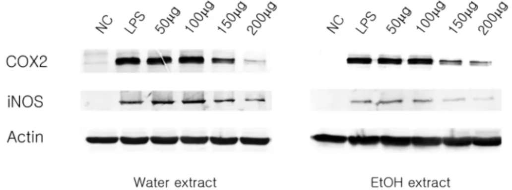
LPS-induced RAW 264.7 cells release NO by the action of the iNOS system. Therefore, we determined the expression levels of some common inflammatory molecules when macrophages were treated with the Paeoniae Radix Rubra extracts. COX-2 and iNOS expression were induced by LPS stimulation and were suppressed when treated with the extracts at a concentration of 150 and 200㎍/㎖ (Fig. 3). Similary, ERK1/2 becomes phosphorylated upon LPS stimulation and the phosphorylation can be inhibited by treatment with the extracts, especially at the higher concentrations (Fig. 4). Taken together, our data suggest that extracts prepared from Paeoniae Radix Rubra have anti- inflammatory effects by suppressing the expression and or
function of certain pro-inflammatory molecules. Also, it appears that both the ethanol-based and water-based extracts are nearly equal in their ability to inhibit these pro-inflammatory molecules.
DISCUSSION
Paeoniae Radix Rubra is a preparation of desiccated roots of Paeonia lactiflora PALL(belonging to Ranunculaceae). Our data demonstrate that Paeoniae Radix Rubra exhibits pharmacological activity via significant inhibitory effects on the LPS-induced inflammatory mediators and cytokines such as NO and PGE2 in RAW 264.7 mouse macrophages. These effects were mediated Fig. 2. Inhibitory effect of EtOH and water extracts from Paeoniae Radix on PGE2 production in LPS-stimulated RAW 264.7 cells. The cells (1.0× 10⁴ cells/well) were stimulated with 1㎍/㎖ of LPS or with LPS plus various concentrations (50, 100, 150, 200㎍/㎖) of ethanol-based or water- based Paeoniae Radix extract for 16 h. PGE2 production and release into the culture medium was assayed by ELISA. The data (n = 3) represent the mean±S.D. of experiments performed in triplicate (*p < 0.05).
through inhibition of iNOS activation and MAPK pathways.
Macrophages, located in most tissues and organs, have critical roles for immunity such as phagocytosis and secretion of pro- inflammatory cytokines (Adams and Hamilton, 1984; Lowenstein, et al., 1996). Macrophages are activated by bacteria and secrete inflammatory mediators including vascular amines, arachidonic acid metabolites, pro-inflammatory cytokines, PGE2 and reactive oxygen species (ROS). RAW 264.7 cells, a mouse macrophage cell line, generate NO when they are stimulated by cytokines or LPS. Therefore, this study examined the anti-inflammatory activities of Paeoniae Radix Rubra extracts using LPS-challenged RAW 264.7 macrophages as a model system.
Recent studies have reported that plant extracts inhibit the generation of inflammation mediators (NO, PGE2, iNOS and IL- 6) from macrophages such as Astragalus membranaceus, Ostericum koreanum and others (Clement-Kruzel et al., 2008;
Jung et al., 2010). Anti-inflammatory effect of leaves and stem from Paeonia lactiflora was reported, but the anti-inflammatory effect of root is yet to be reported (Kim et al., 2007). Other
studies suggest that LPS significantly activates macrophages and induces COX-2 expression, an enzyme that of converts arachidonic acid into PGE2. Studies have shown that increased COX-2 production is associated with cellular toxicity because inhibition of COX-2 expression and/or activity reduces brain injury after ischemia and slows the progression of Alzheimerís disease and Parkinsonís disease (Grunblatt et al., 2000; Selkoe, 2001; Calderon-Garciduenas et al., 2004). Nitric oxide is highly reactive as a free radical and has an important role physiologically for immune reactions in low concentrations (~nmol range) (Jeremy et al., 1999), but high concentrations (~µmol range) of NO results in many pathological processes including inflammation (Ridnour et al., 2006). It has been reported that iNOS is not normally expressed, but LPS up- regulates iNOS expression in RAW 264.7 cells. Exclusive release of NO from iNOS was correlated with several disorders (Ridnour et al., 2006). Generally, expression of iNOS is induced by immune reactions and plays a role in exacerbating inflammation.
In our study, Paeoniae Radix Rubra significantly suppressed Fig. 3. Inhibitory effects of EtOH and water extracts from Paeoniae Radix on iNOS and Cox2 expression in LPS-stimulated RAW 264.7 cells. RAW 264.7 cells (1.0× 10⁶ cells/㎖) were stimulated with LPS (1 ㎍/㎖) in the presence of extract (50, 100, 150, 200㎍/㎖). Whole cell lysates (50 ㎍) were prepared and subjected to 10% SDS-PAGE. Expression of iNOS, COX-2 and actin were determined by Western blot analysis. Actin was used as a loading control.
Fig. 4. Inhibitory effect of EtOH and water extracts from Paeoniae Radix on ERK1/2 Phosphorylation in LPS-stimulated RAW 264.7 cells. RAW 264.7 cells (1.0× 10⁶ cells/㎖) were stimulated with LPS (1 ㎍/㎖) in the presence of extract (50, 100, 150, 200㎍/㎖). Whole cell lysates (50 ㎍) were prepared and subjected to 10% SDS-PAGE. The expression of phosphorylated ERK1/
2 was determined by Western blot analysis and the total cellular ERK1/2 was used as the loading control.
LPS-induced NO production in RAW 264.7 cells in a concentration-dependent manner. This suppression did not show any cytotoxicity at low concentration (until 625㎍/㎖).
In addition, PGE2, an important mediator of the inflammatory response, is synthesized by COX-2 and excess PGE2 production causes inflammatory diseases such as Alzheimer's disease, Parkinson's disease and colon cancer. Suppression of PGE2, on the other hand, relieves inflammation and pain. Our results demonstrate that LPS-stimulated PGE2 production could be suppressed by both Paeoniae Radix Rubra extracts in RAW264.7 cells; however, the decrease was slight. Importantly, this inhibition was concentration-dependent and showed cell-toxicity only in high concentration.
A variety of intracellular signalling pathways are needed to induce and maintain the inflammatory process. MAPK signalling pathway is also involved in the inflammatory process and has been extensively investigated. Recently studies suggested that the MAPK pathways also play a role in regulating PGE2 expression (Guan et al., 1998; Rodriguez-Barbero et al., 2006). Therefore, we have demonstrated that activation of MAPK pathways induce COX-2 expression and PGE2 synthesis in LPS-stimulated RAW264.7 cells. Here, we demonstrated that extract prepared from Paeoniae Radix Rubra from 150㎍/㎖ inhibit the phosphorylation of ERK1/2, and therefore the MAPK pathway, in a concentration-dependent manner. COX-2 and PGE2 expression also decreased from 150㎍/㎖. These data indicates that the extracts prepared from Paeoniae Radix Rubra have anti- inflammation properties and exert their effects by inhibiting ERK1/2 phosphorylation in LPS-stimulated RAW264.7 cells. In the case of the water-based extract, however, excessive concentration can be cytotoxicity. Therefore, the ethanol-based extract prepared from Paeoniae Radix Rubra, which has a slightly different toxicity but similar efficiency on inflammatory activity, might be the preferred method of preparing extracts of Paeoniae Radix Rubra. Future studies might prove Paeoniae Radix Rubra to be an effective therapy for many other inflammatory diseases.
Acknowledgements
This work (Grant No. 000409150110) was supported by Business for Cooperative R&D between Industry, Academy, and Research Institute funded Korea Small and Medium Business Administration in 2010.
LITERATURE CITED
Adams DO and Hamiton TA. (1984). The cell biology of macrophage activation. Annual Review of Immunology. 2:283- 318.
Adams V, Yu J, Mobius-Winkler S, Linke A, Weigl C, Hibrich L, Schuler G and Hambrecht R. (1997). Increased inducible nitric oxide synthase in skeletal muscle biopsies from patients with chronic heart failure. Biochemical and Molecular Medicine. 61:152-160.
Aderem A. (2003). Phagocytosis and the inflammatory response.
The Journal of Infectious Diseases. 187:S340-S345.
Bredt DS and Snyder SH. (1989). Nitric oxide mediates glutamate-linked enhancement of cGMP levels in the cerebellum. Proceedings of the National Academy of Sciences of the United States of America. 86:9030-9033.
Bredt DS and Snyder SH. (1994). Transient nitric oxide synthase neurons in embryonic cerebral cortical plate, sensory ganglia, and olfactory epithelium. Neuron. 13:301-313.
Calderón-Garcidueñas L, Reed W, Maronpot RR, Henríquez- Roldán C, Delgado-Chavez R, Calderón-Garcidueñas A, Dragustinovis I, Franco-Lira M, Aragón-Flores M, Solt AC, Altenburg M, Torres-Jardón R and Swenberg JA. (2004).
Brain inflammation and Alzheimer`s-like pathology in individuals exposed to severe air pollution. Toxicologic Pathology. 32:650- 658.
Chen C, Zhang F, Xia ZY, Lin H and Mo AS. (2008).
Protective effect of pretreatment with Radix Paeoniae Rubra on acute lung injury induced by intestinal ischemia/reperfusion in rats. Chinese Journal of Traumatology. 11:37-41.
Chan SC, Kim JW, Hennerson WR Jr and Hanifin JM. (1993).
Altered prostaglandin E2 regulation of cytokine production in atopic dermatitis. Journal of Immunology. 151:3345-3352.
Clement-Kruzel S, Hwang SA, Kruzel MC, Dasgupta A and Actor JK. (2008). Immune modulation of macrophage pro- inflammatory response by goldenseal and Astragalus extracts.
Journal of Medicinal Food. 11:493-498.
Culbert AA, Skaper SD, Howlett DR, Evans NA, Facci L and Soden PE, Seymour ZM, Guillot F, Gaestel M and Richardson JC. (2006). MAPK-activated protein kinase 2 deficiency in microglia inhibits pro-inflammatory mediator release and resultant neurotoxicity. Relevance to neuroinflammati on in a transgenic mouse model of Alzheimer disease. The Journal of Biological Chemistry. 281:23658-23667.
Guan Z, Buckman SY, Miller BW, Springer LD and Morrison AR. (1998). Interleukin-1beta-induced cyclooxygenase-2 expression requires activation of both c-Jun NH2-terminal kinase and p38 MAPK signal pathways in rat renal mesangial cells. The Journal of Biological Chemistry. 273:28670-28676.
Grnblatt E, Mandel S and Youdim MB. (2000). Neuroprotective strategies in Parkinson`s disease using the models of 6- hydroxydopamine and MPTP. Annals of the New York Academy of Sciences. 889:262-273.
Hibbs JB, Taintor RR and Vavrin Z. (1987). Macrophage cytotoxicity: role of L-arginine deiminase and imino nitrogen oxidation to nitrite. Science. 235:473-476 .
Jeremy JY, Rowe D, Emsley AM and Newby AC. (1999). Nitric
oxide and the proliferation of vascular smooth muscle cells.
Cardiovascular Research. 43:580-594.
Jung HW, Mahesh R, Park JH, Boo YC, Park KM and Park YK. (2010). Bisabolangelone isolated from Ostericum kereanum inhibits the production of inflammatory mediators by down- regulation of NF-kappaB and ERK MAP kinase activity in LPS-stimulated RAW264.7 cells. International Immunopharma- cology. 10:155-162.
Kim SJ, Park JH, Choi SY, Son KH and Kim KU. (2007).
Isolated and identification of biological activity compounds from leaves and stem of Paeonia lactiflora Pallas. Korean Journal of Medicinal Crop Science. 15:6-11.
Kock A, Schwarz T, Kirnbauer R, Urbanski A, Perry P and Ansel JC. (1990). Human keratinocytes are a source for tumor necrosis factor-α: evidence for synthesis and release upon stimulation with endotoxin or ultraviolet light. Journal of Experimental Medicine. 172:1609-1614.
Lawrence T, Willoughby DA and Gilroy DW. (2002). Anti- inflammatory lipid mediators and insights into the resolution of inflammation. Nature Reviews Immunology. 2:787-795.
Lee SE, Lee JH, Kim JK, Kim GS, Kim YO, Soe JS, Choi JH, Lee, ES, Noh HJ and Kim SY. (2011). Anti-inflammatory activity of medicinal plant extracts. Korean Journal of Medicinal Crop Science. 19:217-226.
Lipton SA, Choi YB, Pan ZH, Lei SZ, Chen HS, Sucher NJ, Loscalzo J, Singel DJ and Stamler JS. (1993). A redox-based mechanism for the neuroprotective and neurodestructive effects of nitric oxide and related nitroso-compouds. Nature. 364:626-632.
Lowenstein CJ, Hill SL, Lafond-Walker A, Wu J, Allen G and Landavere M. (1996). Nitric oxide inhibits viral replication in murine myocarditis. The Journal of Clinical Investigation.
97:1837-1843.
Mahut B, Delclaux C, Tillie-Leblond I, Gosset P, Delacour C, Zerah-Lancner F, Harf A and de Blic J. (2004). Both inflammation and remodeling influence nitric oxide output in children with refractory asthma. The Journal of Allergy and Clinical Immunology. 113:252-256.
McGinty A, Foschi M, Chang YW, Han J, Dunn MJ and Sorokin A. (2000). Induction of prostaglandin endoperoxide synthase 2 by mitogen-activated protein kinase cascades. The Biochemical Journal. 352:419-424.
Nathan CF. (1987). Massive secretion of hydrogen peroxide in response to products of macrophages and lymphocytes. The Journal of Clinical Investigation. 80:1550-1560.
Puckett JL, Taylor RW, Leu SY, Guijon OL, Aledia AS, Galant SP and George SC. (2010). An elevated bronchodilator response predicts large airway inflammation in mild asthma. Pediatric Pulmonology. 45:174-181.
Palmer RM, Ashton DS and Moncada S. (1988). Vascular endothelial cells synthesize nitric oxide from L-arginine. Nature.
333:664-666.
Palmer RM and Moncada S. (1989). A novel citrulline-forming enzyme implicated in the formation of nitric oxide by vascular endothelial cells. Biochemical and Biophysical Research Communications. 158:348-352.
Ridnouur LA, Thomas DD, Donzelli S, Espey MG, Roberts DD, Wink DA and Isenberg JS. (2006). The biphasic nature
of nitric oxide responses in tumor biology. Antioxidants &
Redox Signaling. 8:1329-1337.
Rodriguez-Barbero A, Dorado F, Velasco S, Pandiella A, Banas B Lopez-Novoa JM. (2006). TGF-β1 induces COX-2 expression and PGE2 synthesis through MAPK and PI3K pathways in human mesangial cells. Kidney International.
70:901-909.
Schmidt H and Walter U. (1994). NO at work. Cell. 78:919-925.
Schneider A and Stahl RA. (1998). Cyclooxygenase-2 and the kidney: current status and potential perspectives. Nephrology, dialysis, transplantation:official publication of the European Dialysis and Transplant Association-European Renal Association.
13:10-12.
Selkoe DJ. (2001). Alzheimer`s disease: genes, protein, and therapy. Physiological Reviews. 81:741-766.
Sessa WC. (1994). The nitric oxide synthase family of proteins.
Journal of Vascular Research. 31:131-143.
Skidgel RA, Gao XP, Brovkovych V, Rahman A, Jho D, Predescu S, Standiford TJ and Malik AB. (2002). Nitric oxide stimulates macrophage inflammatory protein2 expression in sepsis. Journal of Immunology. 169:2093-2101.
Sun WY, Wei W, Gui SY, Wu L and Wang H. (2008).
Protective effect of extract from Paeonia lactiflora and Astragalus membranaceus against liver injury induced by Bacillus Calmette-Guerin and lipopolysaccharide in mice. Basic
& Clinical Pharmacology & Toxicology. 103:143-149.
Tetsuka T, Daphna-Iken D, Miller BW, Guan Z, Baier LD and Morrison AR. (1996). Nitric oxide amplifies interleukin 1- induced cyclooxygenase-1 expression in rat mesangial cells.
The Journal of Clinical Investigation. 97:2051-2056.
Torres SH, De Sanctis JB, de L Briceo M, Hernndez N and Finol HJ. (2004). Inflammation and nitric oxide production in skeletal muscle of type 2 diabetic patient. The Journal of Endocrinology. 181:419-427.
Williams CS and DuBois RN. (1996). Prostaglandin endoperoxide synthase: why two isoforms? The American Journal of Physiology. 270:G393-G400.
Wu GJ, Chen TL, Ueng YF and Chen RM. (2008). Ketamine inhibits tumor necrosis factor-α and interleukin-6 gene expressions in lipopolysaccharide-stimulated macrophages through suppression of toil-like receptor 4-mediated c-Jin N terminal kinase phosphorylation and activator protein-1 activation.
Toxicology and Applied Pharmacology. 228:105-113.
Wu KK. (1996). Cyclooxygenase 2 induction: molecular mechanism and pathophysiologic roles. The Journal of Laboratory and Clinical Medicine. 128:242-245.
Wu SH, Wu DG and Chen YW. (2010). Chemical constituents and bioactivities of plants from the genus Paeonia. Chemistry
& Biodiversity. 7:90-104.
Yoon T, Cheon MS, Kim SJ, Lee AY, Moon BC, Chun JM, Choo BK and Kim HK. (2010). Evaluation of solvent extraction on anti-inflammatory efficacy of Glycyrrhiza uralensis. Korean Journal of Medicinal Crop Science. 18:28-33.
Zhan LY, Xia ZY, Chen C and Wang XY. (2006). Effect of Radix Paeoniae Rubra on the expression of HO-1 and iNOS in rats with endotoxin-induced acute lung injury. Chinese Journal of Traumatology. 9:181-186.