• 검색 결과가 없습니다.

The preparation of skin analogue composition having the liquid crystalline structure and its cosmetic applications

N/A
N/A
Protected

Academic year: 2021

Share "The preparation of skin analogue composition having the liquid crystalline structure and its cosmetic applications"

Copied!
10
0
0

로드 중.... (전체 텍스트 보기)

전체 글

(1)

ISSN 2288-1069 (Online)

http://dx.doi.org/10.12925/jkocs.2013.30.2.215

The preparation of skin analogue composition having the liquid crystalline structure and its cosmetic applications

Dong-Kyu Lee

1

․Kwan-Young Jeong

2✝

1

Department of Industrial Engineering Chemistry, College of Engineering, Chungbuk National University, Cheong-ju 361-763, Korea

2

Skin research institute, Korea Kolmar Corporation, 170-7, Seojeong-Ri, Jeonui-Myun, Yeongi-Gun, Chung-Nam, Korea

(Received March 25, 2013 ; Revised June 25, 2013 ; Accepted June 25, 2013)

Abstract : Recently, many cosmetic researchers have been focused on the development of high functional cosmetics including anti-wrinkle and whitening. In these studies, they couldn’t afford to pay a deep attention to stable encapsulations for unstable materials and efficient drug deliveries for them. Particularly, in order to show a degree of instant effects as cosmetics, they can’t also ignore moisturizing effect enough to satisfy customers just after applying and its maintenance by improving the function of skin barrier as well as above two effects. Therefore, skin analogue systems have attracted considerable attention in the view of structural and compositional similarity to intercellular membrane in stratum corneum. And, some models for skin analogue composition were developed to improve the function of skin barrier, stably encapsulate unstable materials such as retinol, vitamin B, C, E, etc ., and control their skin penetration in order to show good effects as cosmetics. In this study, we suggest the new skin analogue model having the compositional similarity as well as conventional structural ones. Our skin analogue membrane(SAM) is mainly composed of ceramide/ cholesterol/phosphatidylcholin/fatty acids and its structural defects are compensated by including cholesterol amphiphile and controlling the ratio of ceramide/cholesterol.

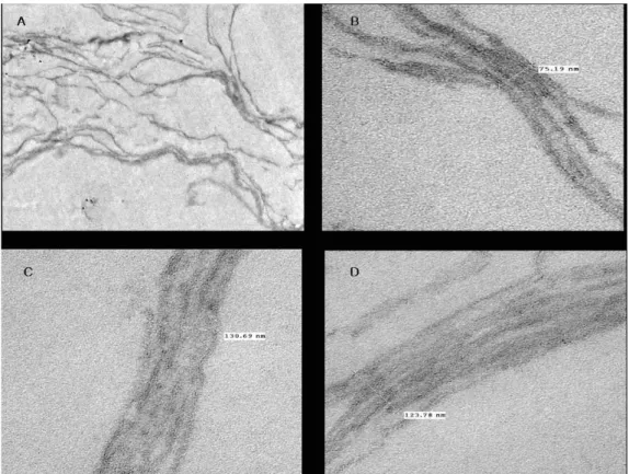
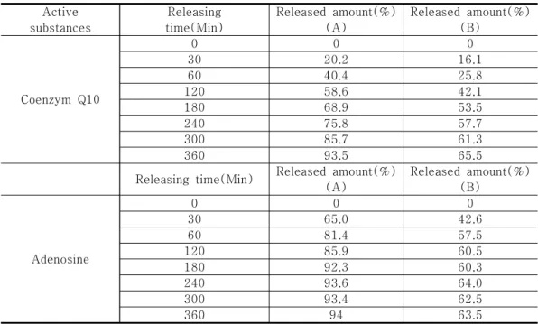
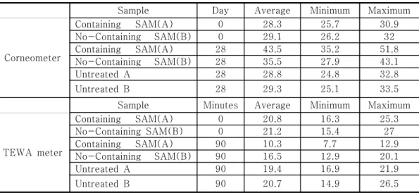
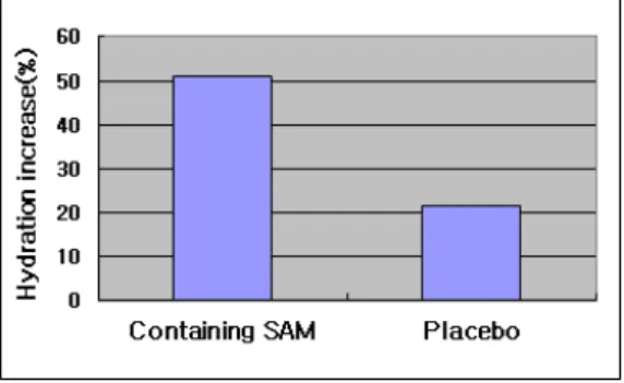
It was possible to confirm the formation of skin analogue membrane having highly-densed multilamella structure and compare them according to the change of each ratio with a polarized microscope, X-ray diffraction. More detaily, we observed their structures with a electron microscope(TEM). Finally, we dispersed them in excess of continuous water phase, observed the formation of maltese-cross liquid crystalline and measured the efficiency of drug deliveries and moisturizing effects.

Keywords : skin analogue membrane, phosphatidylcholin, Ceramide, Cholesterol amphiphile , multilamella structure, maltese-cross liquid crystalline



Corresponding author

(E-mail : [email protected])

(2)

1. Introduction

The natural function of the skin is to protect the body against the loss of endogenous substances. The main barrier for diffusion through the skin is the outermost layer, the stratum corneum(SC). This is composed of keratin-filled dead cells which are entirely surrounded by crystalline lamellar lipid regions having a very dense structure[1].

When most of active substances applied onto the skin diffuse along the crystalline lamella lipid layer in the intercellular region, this dense structure obstruct the diffusion of active substances, reduce the efficiency of drug delivery into skin, and decrease the desirable effects such as whitening, anti-wrinkle, or moisturizing[2]. Therefore, information on the relationship between lipid organization and composition is of great importance for improving these desirable cosmetic effects as well as skin barrier function. The lipid matrix in SC is composed of highly organized bilayers, mainly containing ceramides(CER), cholesterol(CHOL) and free fatty acid(FFA)[3-4], Fig. 1[5]. To make cosmetic formulations having a similar sturucture and composition to the intercellular membrane in SC, many efforts have been undertaken to characterize the lipid lamella region by electron microscopies(EM), Differntial scanning calorimetry(DSC), X-ray diffractometer(XRD), etc [6-8]. In this study, we will propose a new skin analogue membrane having a similar structure and composition to the intercellular membrane in SC. While it has a very stable encapsulation about unstable drugs, it is efficient to deliver these encapsulated drugs into skin and obtains desirable cosmetic effects mentioned above. To obtain these positive results, we modified the composition of lamella membrane to form a strong occlusive barrier, and stably maximized the encapsulating efficacy in polar drugs between bilayers or in its lipophilic portion. At first, our model membrane basically contains CER and CHOL,

since these two classes of lipids are very important for the formation and control of liquid crystalline(LC) structure similar to SC.

At second, Phospholipids(PL) have a crucial role of helping the formation of the narrow central layer which consists of polar liquid phases(polyols) due to the hydrogen bond of its strongly polar head group. At third, CHOL may play a role to form the occlusive barrier for the leak of encapsulated oil-soluble active substances as times go, and also to supply some fluidity to this rigid LC membrane. At fourth, in the case of transfersomes, a cholesterol amphiphile(CHOLA), and edge activators, may give LC structure a desirable elasticity to enhance the drug delivery and compensate the incompatibility of CHOL to be more packed with other lipids and help the function of PL binding with polar central layer[9]. Therefore, properly to cosmetic applications for the encapsulation and drug delivery, we will propose the elastic sandwich model which is composed of the occlusive bilayer having elasticity and the permeable liquid layer by means of modifying the sandwich structure of SC(Fig. 2.) suggested by Swarzendruber et al[10] and Kuempel et al[11]. Using these materials and controlling their composition, we designed a skin analogue model membrane to encapsulate a oil-soluble active substance(Coenzyme Q10) as well as a water-soluble one(Adenosine), enhance the skin penetration of these substances and restore defected or immature skin to a healthier skin.

To confirm this structure, we measured DSC

for the thermal property, XRDs for the

repeating distance of LC structure and the

interlayer length between bilayers, and EM for

monitoring the structure and matching the

analytical results. And, we used a new method

of enhancer cell to measure the efficiency of

drug deliveries after dispersing this LC in

excess of water. Finally, a corneometer and

tewameter were used to measure the effect

increasing the moist level in SC and decreasing

the TEWL(Transepidermal water loss) in order

(3)

to restore defected skin.

Fig. 1. A schematic drawing of a skin cross-section.

2. Experiments 2.1 Materials

A phospholipid(PL) was purchased from Lipoid(Germany). A ceramide(CER) was purchased from Doosan biotech(Korea), a cholesterol(CHOL) was purchased from Solvay (U.S.A), a cholesterol amphiphile(CHOLA) was purchased from Nikko chemical(Japan). These material were used without any pre-treatment.

All other ingredients were of cosmetic grade such polyols, capric/carplylic triglyceride(CCT), carbomers without any other before-treatment.

Active substances such as coenzyme Q10 and adenosine was purchased from Sigma aldrich as HPLC grade in order to analyze exactly.

2.2 Mehtods

2.2.1 Sample preparation

At first, all of the lipids, such as PL, CER, CHOL, CHOLA, were added to a polyol under a moderate mixing, and heated to 85℃

until being completely melted and lipid phase was prepared. Coenzyme Q10 was added to CCT at 50℃, mixed until being completely melted, and oil phase was prepared. Adenosine was added to polyol at 45℃, mixed until

being completely melted. Then, the lipid phase was cooled to 60℃, the oil phase and the melted adenosine phase was added to it one by one, and was well-mixed until being homogeneous. It was very slowly cooled to 3 5℃ and stored below 10℃ for 1 week before being analyzed. Also, this prepared LC was added to excess of water phase in order to make cosmetic samples and measure the efficiency of drug deliveries and the moisturizing effects.

2.2.2 DSC(Differential scanning calorimetry) analysis

Thermal analysis was made with a TA instrument(TA4100 model) from 10℃ to 70℃

at heating rate of 1℃/min after being cooled to a lower temperature. Sample quantities were about 15 mg, which was sealed in an aluminium sample cell. This analysis was done under a nitrogen gas and it measured the melting temperature and enthalpy to confirm the formation of LC structure and its phase transitions.

2.2.3. XRD

XRD spectra were taken with x-ray diffractometer(XDS 2000 model, SCINTAG INC., USA). During X-ray diffraction experiments the temperature of the samples deviated by maximum 1℃ from the adjusted temperature. XRD experiments were carried out with Ni-filtered CuKα-ray(λ=0.154nm) using photo dectection, operating 35 Kv, 50 mA under a room temperature.

2.2.4. EM(Electron microscope) and PM(Polarized microscope) analysis EM analysis was examined with a transmission electron microscope(TEM, Hitachi H-7600). At that time, we introduced RuO

4

as a post-fixation agent to preserve these lipid in LC structure by means of the same method as Hou et al and van den Bergh et al[12,13].

At first, a sample was slightly applied on the

inner region of a tube. And then, for RuO

4

(4)

post-fixation, the specimens were post-fixed in 0.25% RuO

4

(EMS), 0.1 M cacodylate buffer for 45min at room temperature in the dark.

All post-fixed tissues were rinsed in 0.1 M cacodylate buffer for 10 min, dehydrated in a graded ethanol series, respectively, and embedded in epon-epoxy resin. Ultra sections (Leica UCT) were cut, double stained with uranyl acetate and lead citrate, and then examined with TEM. PM strudy were performed with an Olympus BX-51 polarized microscope

2.2.5. Drug delivery

Drug delivery was examined with a dissolution tester(ERWEKA DT800, Germany) equipped by an enhancer cell(ERWEKA) and with a HPLC. The experiment was performed by the method similar to Rege, et al [14]. In a dissolution tester, 500 ml of round- bottomed vessel was equipped , 500 ml of a saline solution was added to it, this vessel was maintained at 37℃. A 1,000 mg of sample was homogeneously applied on the adjustment plate, an artificial cellulose-acetate membrane was placed on it, and they were fixed with washer. Then, they were assembled to the cell body with the retaining ring, put on the bottom of the dissolution vessel, and this test was performed under a moderate mixing(100 rpm). For each samples, released amounts of coenzyme Q10(275 nm) and adenosine(260 nm) were measured with a HPLC at 30, 60, 120, 180, 240, 300, 360 minutes, respectively.

2.2.6 Skin moisturizing tests

TEWL determination and corneometry were carried out on a sample of 10 healthy asian fermales having mean age(29±4) for 28 days.

Two test areas 2 cm in diameters were marked on the sides of all the subjects’

forearms and were measured under 22±2℃, relative humidity 41%. In an experiment for skin moisturizing effects, a corneometer(CM 825, CK electronic GmbH) which was mounted on a Multi- Probe Adapter MPA

5(CK electronics GmbH). Capacitance changes depending almost only upon the water content in the SC are detected and evaluated[16]. In another experiment, tewameter(TM 300, CK electronic GmbH)[17] was used to measure the TEWL from the skin in accordance with applicable guidelines after treating 5.0%

SLS(Sodium lauryl sulfate) solution[18].

TEWL is considered an important measurement of epidermal barrier function. Evaporimeter consists of applying a probe with two twin sensors directly to skin, with one sensor pair measuring humidity and the other temperature.

The acquired data are used by an integrated microcomputer to compute the water vapor partial pressures at the two parallel levels of each sensor pair and via the partial pressure gradient, the rate of evaporation.

3. Results & Discussion 3.1. The structure of skin analogue membrane from XRDs

Using XRD, we investigated the repeating distance of LC structure and the interlayer length between bilayers to analyze the detail LC structure. At first, the mean distance of interlayer was calculated from WAXD(Wide angle X-ray diffraction) results and was shown in Fig. 2. It showed only a reflection at 2 Theta(2Θ) = 21.49

o

for the lipid complex hydrated by a polar solvent and this reflection angle was characteristic for the α -modification in the region of lipid-bilayers [15]. From the diffraction angle the interlayer distance between the densely packed hydrophobic region is calculated: d=λ/(sinΘ)

= 2.11nm). Second, the repeating distance of

8.20 nm was derived from SAXD(Small angle

x-ray diffraction) at 2Θ = 10.77

o

. There

was only a reflection for a homogeneous

arrangement of LC similar to WAXD result.

(5)

(A)

(B)

Fig. 2. WAXD(A), SAXD(B) curves obtained.

The number at the maxima correspond with the diffraction angle 2θ.

3.2. Thermal behaviors from DSC

During heating, two endothermal effects were observed. As seen in Fig. 3., the lower temperature slope at 27.04℃ denotes an weak enthalpy change due to the transition from the β-modification to the α-modification[19].

The higher temperature peak at 41.59℃

denotes only an very strong enthalpy change due to the transition from the α-modification to the isotropic state. At this transition, the enthalpy change(ΔH) was 14.21 J/g and this only strong endothermal peak suggests that all

of the lipids are well-oriented to form a homogeneous LC structure and there aren’t other co-existing phases such as another separated LC or micelles. This result is in the good agreement with XRD results.

Fig. 3. DSC curve obtained in the system of skin analogue membrane.

3.3.3. Visualization for the structure of skin analogue membrane from PM and TEM.

With 1,000 magnification, the PM result showed that this skin analogue membrane had a very densed and regular lamella structure (Fig. 4(A).). Therefore, this showed an optical anisotropy behavior to form a good lamella structure. In TEM studies, a photograph(Fig.

5.) had a entirely good lamella structure in the

case of 3,500(x) magnification. In the case of

more 10x magnification to 30,000, it was

shown that the lamella thickness was ranged

from 75 nm to 130 nm and the structure was

also well-packed LC structure very similar to

the structure of intercellular membrane in SC

shown by Fig. 1. From SAXD result, because

the repeating distance was 8.20 nm, the

number of lipid bilayers packed in LC can be

calculated about as 9.1 to 15.8 layers. Adding

this LC to an excess of water phase, it formed

a very good maltese-cross LC structure

surrounding the oil phase and its particle size

was ranged about from 3.0 μm to 4.0 μ

m(Fig. 4(B).)

(6)

(A) (B)

Fig. 4. PM photographes of (A) Skin analogue membrane : Lamella LC structure (B) Maltese-cross LC structure after adding an excess of water.

Fig 5. TEM photograpes of skin analogue membrane having a lamella LC structure : A) Overall part of LC structure(3,500(x) Magnification),

B) 75.19 nm thickness of a partial LC structure(30,000(x) Magnification),

C) 130.69 nm thickness of a partial LC structure(30,000(x) Magnification),

D) 123.78 nm thickness of a partial LC structure(30,000(x) Magnification).

(7)

Active substances

Releasing time(Min)

Released amount(%) (A)

Released amount(%) (B)

Coenzym Q10

0 0 0

30 20.2 16.1

60 40.4 25.8

120 58.6 42.1

180 68.9 53.5

240 75.8 57.7

300 85.7 61.3

360 93.5 65.5

Releasing time(Min) Released amount(%) (A)

Released amount(%) (B)

Adenosine

0 0 0

30 65.0 42.6

60 81.4 57.5

120 85.9 60.5

180 92.3 60.3

240 93.6 64.0

300 93.4 62.5

360 94 63.5

Table 1. The Data of Released Amounts for Coenzyme Q10 and Adenosine with SAM(A) and without SAM(B).

3.3.4. Efficiency of drug deliveries

1000 mg of samples were loaded on adjustment plate, and were tested with the dissolution tester. In this experiment, the eluted solutions from the membrane were auto-sampled at each measuring time, and then were injected to HPLC in order to measure the released amount of coenzyme Q10 and adenosine. Table 1 and Fig. 6. show the profile of the released amounts for coenzyme Q10 and adenosine encapsulated into a sample A(with SAM) and sample B(a normal emulsion without SAM) in comparison to the original amounts, depending on each releasing time.

As shown in Fig. 6., coenzyme Q10 and adenosine showed a little different profile.

Coenzyme Q10 as an oil-soluble substance, showed a time-releasing profile to be penetrated with a relatively constant increasing slope and didn’t get to 80% released amount even after 4 hours, but adenosine as a water-soluble substance, showed a relatively fast-releasing profile and got to 80% released

amount only at 60 minutes, However, in the case of encapsulating them with SAM, both of coenzyme Q10 and adenosine ultimately reached to over 90% released amount at the end of measuring time and showed much higher efficiency for the drug deliveries of a water-soluble substance as well as an oil-soluble substance than a normal emulsion which showed nearby 60% released amounts.

Fig. 6. The time-released profiles of coenzyme

Q10 and adenosine depending on the

measuring times.

(8)

  Sample Day Average Minimum Maximum

Corneometer

Containing SAM(A) 0 28.3 25.7 30.9

No-Containing SAM(B) 0 29.1 26.2 32

Containing SAM(A) 28 43.5 35.2 51.8

No-Containing SAM(B) 28 35.5 27.9 43.1

Untreated A 28 28.8 24.8 32.8

Untreated B 28 29.3 25.1 33.5

  Sample Minutes Average Minimum Maximum

TEWA meter

Containing SAM(A) 0 20.8 16.3 25.3

No-Containing SAM(B) 0 21.2 15.4 27

Containing SAM(A) 90 10.3 7.7 12.9

No-Containing SAM(B) 90 16.5 12.9 20.1

Untreated A 90 19.4 16.9 21.9

Untreated B 90 20.7 14.9 26.5

Table 2. Descriptive Results for The Effects of Skin Moisturizing with The Corneometer and TEWA meter.

3.3.5. Skin moisturizing effect

The initial corneometer values were similar(Table 2.). At an area applied by skin analogue membrane, the average hydration level was increased from 28.3(min= 25.7, max=30.9) to 43.5(min=35.2, max=51.8) during the application period. In the case of placebo(No containing SAM), the average hydration level was slightly increased from 29.1(min= 26.2, max=32.0) to 35.5(min=27.9, max=43.1). The untreated controls remained almost unchanged, i.e 28.3, 29.1 on day 0 and 28.8, 29.3 on day 28. Variation analysis of the corneometer results is shown in Fig. 7.

This results are calculated from Eq.(1) below.

Parameter change(%)=(∑Q

ti

/∑Q

0i

–1)*100 (1) Where Q

ti

is the quotient after application time t of 28 days for each volunteer i, and Q

0i

is the quotient before application time for each volunteer i. In the case of applying this skin analogue membrane, average skin hydration was increased by 51.04%, and in the case of placebo it was increased by 21.16%. The use of SAM is significantly more effective than placebo no containing SAM.

One reason for this increase is expected due

to the excellent occlusivity of this skin analogue membrane and its capacity delivering moisturizers into skin.

The initial tewa values were very big different from 13.2 to 20.8 and 14.6 to 21.2 between before and after treating SLS solution(Table 2). In 60 minutes after treating SLS solution, each area was applied by a sample(A) containing SAM and a placebo sample(B). In 90 min after treating sample A, the average tewl level(g/cm

2

h) was decreased from 20.8(min=16.3, max=25.3) to 10.3 (min=7.7, max=12.9). In the case of sample B, the average tewl level(g/cm

2

h) was slightly decreased from 21.2(min=15.4, max=27.0) to 16.5(min=12.9, max=20.1). The untreated controls remained almost unchanged, i.e 19.4, 20.7 before treating SLS solution and 18.6, 20.1 at the final. Variation analysis of the tewameter results is shown in Fig. 8. This results are calculated from Eq.(2) below

Parameter change(%)=(∑Q

ti

/∑Q

0i

–1)*100 (2)

Where Qti is the quotient after application

time t of 90 minutes for each volunteer i, and

Q0i is the quotient before application time for

each volunteer i. In the case of applying

(9)

sample A(containing SAM), the average tewl level(g/cm

2

h) was decreased by 50.4%, and in the case of sample B(placebo) it was decreased by 22.17%. In terms of TEWL, the use of SAM is significantly more effective than placebo no containing SAM. One reason for this promotes to pack intercellular lipids more densely in SC and helps to repair a damaged and defected skin to healthier condition due to skin lipids such as CER, CHOL and promoters such as PL, CHOLA in this skin analogue membrane.

Fig. 7. Increasing corneometer values : 28 days after the application of sample A with SAM and sample B without SAM.

Fig. 8. Variances of TEWL values between before- and after- application of sample A with SAM and sample B without SAM.

4. Conclusion

In this study, we was successful to develop a skin analogue membrane having a highly densed LC structure mainly composed of CER, CHOL, PL and CHOLA without water. As results of XRDs, the mean distance length between bilayers was 2.11 nm from WAXD and the repeating distance of LC structure was 8.20 nm. Combining with TEM result, the number of lipid bilayers packed in LC can be calculated about as 9.1 to 15.8 layers. Also, only one reflection suggests that there is a regular LC structure being hydrated by a polar solvent containing excess of adenosine and being densely packed by lipid bilayers containing excess of coenzyme Q10. This LC structure was supported by DSC result having only one strong endothermal peak at 41.59℃.

In microscopic studies, this photo from PM showed that this LC formed a good lamella structure being very densed and these photo from TEM proved that this well-packed structure was very similar to the intercellular membrane one in SC as shown in Fig 1.

When this LC was applied on cosmetics, it

formed very homogeneous maltese-cross LC

particles which were ranged from 3.0 μm to

4.0 μm. As results of drug delivering and

skin moisturizing effects for this, we obtained

good results to significantly increase the

released amounts for both of coenzyme Q10

and adenosine in comparison to a normal

emulsion without SAM, and the skin

moisturizing effects were also much higher

than a placebo with both of corneometer and

tewameter. Therefore, this LC membrane is

very potential to various cosmetic

applications(encapsulation, drug delivery, skin

barrier repairing etc.).

(10)

References

1. P. W. Wertz and D. T. Dowing, "Stratum corneum: biological and biochemical considerations, in: J. Hadgraft, R. H. Guy,

"Transdermal Drug delivery", p. 1-17, Marcel Dekker, NewYork(1989).

2. R. O. Potts and R. H. Guy, Predicting skin permeability, Pharm. Res ., 9, 663-669(1992).

3. P. W. Wertz, The nature of the epidermal barrier: Biochemical aspects, Adv. Drug Del. Rev., 18(3), 283-294(1996).

4. P. W. Wertz and B. A. I. van den Bergh, The physical, chemical and functional properties of lipids in the skin and other biological barriers, C hem. Phys. Lipids., 91, 85-96(1998)

5. J. A. Bouwstra and P. L. Honeywell- Nguyen, Skin structure and mode of action of vesicles, Adv. Drug Delivery Rev ., 54 Suppl. 1, 41-55(2002).

6. D. C. Swartzendruber, Studies of epidermal lipids using electron microscopy, Semin.

Dermatol., 11, 157-161(1992).

7. B. F. van Duzee, Thermal analysis of human stratum corneum, J. Invest.

Dermatol ., 65, 404-408(1975).

8. S. H. White, D. Mirejovsky and G. I.

King, Structure of lamellar lipid domains and corneocytes envelopes of murine stratum corneum. An X-ray diffraction study, Biochemistry, 27, 3725-3732(1988).

9. G. Cevc and G. Blume, Lipid vesicles penetrate into intact skin owing to transdermal osmotic gradients and hydration force, Biochim. Biophys. Acta., 1104, 226-232(1992).

10. D. C. Swarzendruber, P. W. Wertz, D. J.

Kitko, K. C. Madison and D. T. Dowing, Molecular models of the intercellular lipid lamellae in mammalian stratum corneum, J.

Invest. Dermatol ., 92, 251-257(1989).

11. D. Kuempel, D. C. Swarzendruber, C. A.

Squier and P. W. Wertz, In vitro reconstitution of stratum corneum lipid

lamellae, Biochim. Biophys. Acta., 1372, 135-140(1998).

12. S. Y. Hou, A. K. Mitra, S. H. White, G.

K. Menon, R. Ghadially and P. Elias, Membrane structure in normal and essential fatty acid-deficient stratum corneum; characterization of ruthenium tetroxide stanning and X-ray diffraction, J.

Invest. Dermatol., 96, 215-223(1991).

13. B. A. I. van den Bergh, D. C.

Swarzendruber and A. Bos-van der Geest et al., Development od an optimal protocol for the ultrastructural examination of skin by transmission electron microscopey, J.

Microsc ., 187, 125-133(1997)

14. P. R. Rege, V. D. Vilivalam and C. C.

Collins, Development in release testing of topical dosage forms: use of the Enhancer cell with a sampling, J pharm and biochem. anal., 17, 1225-1233(1998).

15. T. de Vringer, J. G. H Joosten and H. E.

Junginger, A study of gel structure in a nonionic O/W cream by X-ray diffraction and microscopic methods, Colloid &

Polymer sci , 265,167-179(1987).

16. A. Sylvia, Wissing and Rainer H. Muller, The influence of solid lipid nanoparticles on skin hydration and viscoelasticity – in vivo study, Eur. J. Plarm. Biopharm. , 56, 67-72(2003).

17. L. Lehmann, S. Keipert and M. Gloor, Effects of microemulsion on the stratumcorneum and hydrocortisone penetration, Eur. J. Plarm. Biopharm., 52, 129-136(2001)

18. J. Pinnagoda, R. A. Tupker, T. Agner and J. Serup, Guidelines for transepidermal water loss(TEWL) measurement, Contact Dermatitis., 22, 164-178(1990).

19. T. de Vringer, J. G. H Joosten and H. E.

Junginger, A study of gel structure in a

nonionic O/W cream by differential

scanning calorimetry, Colloid & Polymer

sci. , 264, 691-700(1986).

수치

Fig.  1.  A  schematic  drawing  of  a  skin                        cross-section.
Fig. 2. WAXD(A),  SAXD(B)  curves  obtained.
Fig  5.  TEM  photograpes  of  skin  analogue  membrane  having  a  lamella  LC  structure  :          A)  Overall  part  of  LC  structure(3,500(x)  Magnification),
Fig. 6. The time-released profiles of coenzyme  Q10  and  adenosine  depending  on  the    measuring  times
+3

참조

관련 문서

• 이명의 치료에 대한 매커니즘과 디지털 음향 기술에 대한 상업적으로의 급속한 발전으로 인해 치료 옵션은 증가했 지만, 선택 가이드 라인은 거의 없음.. •

 The Dutch physicist Pieter Zeeman showed the spectral lines emitted by atoms in a magnetic field split into multiple energy levels...  With no magnetic field to align them,

Modern Physics for Scientists and Engineers International Edition,

Five days later, on 15 January 1975, the Portuguese government signed an agreement with the MPLA, FNLA and UNITA providing for Angola to receive its independence on 11

Usefulness of co-treatment with immunomodulators in patients with inflammatory bowel disease treated with scheduled infliximab maintenance therapy.. Oussalah A, Chevaux JB, Fay

Inclusion and Inclusiveness: Shared Vision of Youth for Local, National, and Global Village Inclusion at large stands for embracing populations with disabilities and

웹 표준을 지원하는 플랫폼에서 큰 수정없이 실행 가능함 패키징을 통해 다양한 기기를 위한 앱을 작성할 수 있음 네이티브 앱과

_____ culture appears to be attractive (도시의) to the