• 검색 결과가 없습니다.

Treatment Plan Delivery Accuracy of the ViewRay System in Two-Headed Mode

N/A
N/A
Protected

Academic year: 2021

Share "Treatment Plan Delivery Accuracy of the ViewRay System in Two-Headed Mode"

Copied!
6
0
0

로드 중.... (전체 텍스트 보기)

전체 글

(1)

http://dx.doi.org/10.14316/pmp.2016.27.3.169 pISSN 2508-4445, eISSN 2508-4453

This work was supported by the National Research Foundation of Korea (NRF) grant funded by the Korea government (MSIP) (No.

2015R1C1A1A01054192).

Received 22 September 2016, Revised 23 September 2016, Accepted 24 September 2016

Correspondence: Jung-in Kim ([email protected]) Tel: 82-2-2072-3573, Fax: 82-2-765-3317

cc This is an Open-Access article distributed under the terms of the Creative Commons Attribution Non-Commercial License (http://creativecommons.org/licenses/by-nc/4.0) which permits unrestricted non-commercial use, distribution, and reproduction in any medium, provided the original work is properly cited.

Treatment Plan Delivery Accuracy of the ViewRay System in Two-Headed Mode

Jong Min Park*

†‡§

, So-Yeon Park*

†‡

, Hong-Gyun Wu*

†‡║

, Jung-in Kim*

†‡

*Department of Radiation Oncology,

Biomedical Research Institute, Seoul National University Hospital,

Institute of Radiation Medicine, Seoul National University Medical Research Center, Seoul,

§

Center for Convergence Research on Robotics, Advanced Institutes of Convergence Technology, Suwon,

Department of Radiation Oncology, Seoul National University College of Medicine, Seoul, Korea

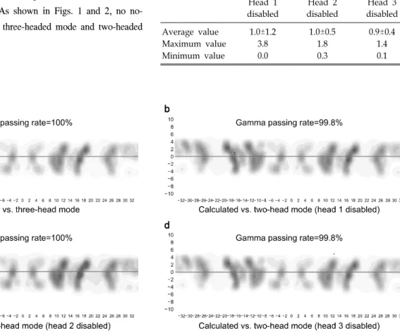
The aim of this study is to investigate the delivery accuracy of intensity-modulated radiation therapy (IMRT) plans in the two-headed mode of the ViewRay

TM

system in comparison with that of the normal operation treatment plan of the machine. For this study, a total of eight IMRT plans and corresponding verification plans were generated (four head and neck, two liver, and two prostate IMRT plans). The delivered dose distributions were measured using ArcCHECK

TM

with the insertion of an ionization chamber. We measured the delivered dose distributions in three-headed mode (normal operation of the machine), two-headed mode with head 1 disabled, two-headed mode with head 2 disabled, and two-headed mode with head 3 disabled. Therefore, a total of four measurements were performed for each IMRT plan. The global gamma passing rates (3%/3 mm) in three-headed mode, head 1 disabled, head 2 disabled, and head 3 disabled were 99.9±0.1%, 99.8±0.3%, 99.6±0.7%, and 99.7±0.4%, respectively. The difference in the gamma passing rates of the three- and two-headed modes was insignificant. With 2%/2 mm, the rates were 96.6±3.6%, 97.2±3.5%, 95.7±6.2%, and 95.5±4.3%, respectively. Between three-headed mode and head 3 disabled, a statistically significant difference was observed with a p-value of 0.02; however, the difference was minimal (1.1%). The chamber readings showed differences of approximately 1% between three- and two-headed modes, which were minimal. Therefore, the treatment plan delivery in the two-headed mode of the ViewRay

TM

system seems accurate and robust.

󰠏󰠏󰠏󰠏󰠏󰠏󰠏󰠏󰠏󰠏󰠏󰠏󰠏󰠏󰠏󰠏󰠏󰠏󰠏󰠏

Key Words: MRI-guided radiation therapy system, Two-headed mode, Co-60, Intensity modulated radiation therapy

Introduction

Magnetic resonance imaging (MRI) is advantageous for radi- ation therapy because of its superior soft tissue contrast capability.

1-3)

Therefore, if required in the clinic, MR images were acquired and registered to the CT images for accurate definition of the target volumes as well as organs at risk

(OARs).

4,5)

Recently, a magnetic resonance image-guided radia-

tion therapy (MR-IGRT) system, the ViewRay

TM

system

(ViewRay Inc., Cleveland, OH, USA), was introduced in the

field of radiotherapy.

6-10)

The ViewRay

TM

system facilitated the

delineation of target volumes, patient setup, adaptive radiation

therapy (ART), and respiratory gating based on MRI.

6)

The

ViewRay

TM

system can acquire volumetric MR images within

a few minutes for treatment planning and daily patient

positioning.

6,7)

The volumetric MR image can be acquired with

a 0.35 T static magnetic field, which is generated by a super-

conducting magnet.

6)

The ViewRay

TM

system can also acquire

a single sagittal planar cine MR image at 4 frames/s or three

sagittal planar cine images at 2 frames/s during treatment for

the respiratory gating.

10)

The rapid MR image acquisition, no

extra imaging dose requirement, and rapid optimization, as

well as the dose calculation ability of the ViewRay

TM

system

(2)

enabled ART with ease in the clinic.

6)

The respiratory gating of the ViewRay

TM

system based on patient anatomy, rather than on a surrogate placed on the patient’s skin, enabled more accurate delivery of the prescription dose to the target volume than did the conventional respiratory gating techniques.

To be compatible with the MR imaging system, the ViewRay

TM

system adopted a Co-60 radioisotope, rather than a linac, as the treatment beam delivery modality.

7)

To increase the dose rate, the ViewRay

TM

system uses a total of three Co-60 sources, which were located 120

o

apart in the ring-type gantry.

7)

The maximum capacity of the Co-60 sources for the ViewRay

TM

system is approximately 15,000 Ci. The inner diameter of the ring-type gantry is 70 cm and the source-to-axis distance (SAD) is 105 cm. Each source has its own multi-leaf collimator (MLC) system. The leaf width of each MLC system is 1.05 cm at the isoplane, i.e. 1 cm at a source-to-surface distance (SSD) of 100 cm. To reduce the large penumbra of the Co-60 source, the design of the MLC system is double-focused.

7)

As there are a total of three independent treatment heads with their own Co-60 sources and MLC systems, if one head is out of order, the other heads can deliver the prescription dose to the target volume and finish the treatment, which is called two-headed mode. In practice, therapists do not have to stop patient treatment owing to the malfunction of a single head. This is a unique and practical function of the ViewRay

TM

system, which prevents interruption of patient treatment in case one head has a problem, resulting in reduction of machine down time. This is quite a convenient and practical function; however, there is a concern about the accurate delivery of a treatment plan to a patient using two-headed mode, since the heads are not identical in reality. Therefore, in this study, we verified the de- livery accuracy of two-headed mode in comparison with three-headed mode, i.e. normal operation of the machine.

Materials and Methods

1. Generation of treatment plans and corresponding verification plans

We generated a total of eight step-and-shoot IMRT plans in the treatment planning system (TPS) of the ViewRay

TM

system and the MRIdian

TM

system (ViewRay Inc., Cleveland, OH, USA). Of the eight treatment plans, four IMRT plans were for

the treatment of head and neck cancer, two for the treatment of liver cancer, and two for the treatment of prostate cancer. A total of five beam groups (a total of 12∼15 fields) were used for the generation of the IMRT plans. The gantry angles of each field were modified patient-specifically, considering the patients’ anatomy, to reduce doses to OARs. The fields pass- ing through the corners of the patient couch were not used to eliminate the dosimetric uncertainty due to daily variations of the patient setup, because the attenuation of the treatment beam by the patient couch is considerable.

7,8)

Owing to the fact that the bore size is only 70 cm, the isocenter locations, relative to the patient body, were not moved laterally for the clearance of the patient setup. The value of the IMRT effi- ciency (degree of smoothing of the fluence map intensity pat- tern) was set to 0.5.

8,10)

The value of the level (the maximum value of the deliverable beam-on segments per field) was set to 20.

8,10)

The grid size of the dose calculation was 3 mm. The dose calculation algorithm was the Monte Carlo algorithm de- veloped by the manufacturer (ViewRay Inc., Cleveland, OH, USA). After treatment planning, a verification plan for each treatment plan was generated with the CT image set of the ArcCHECK

TM

(Sun Nuclear Corporation, Melbourne, FL, USA) in the MRIdian

TM

system. The reference dose dis- tributions were calculated in the MRIdian

TM

system.

2. Analysis of delivery accuracy

The delivered dose distributions of the verifications plans were measured using the MR-compatible ArcCHECK

TM

with the insertion of an ionization chamber. For each IMRT plan, we measured dose distributions in three-headed mode, i.e. nor- mal operation of the machine with full use of all three heads.

After that, we measured dose distributions three times in two-headed mode with head 1 disabled, head 2 disabled, and head 3 disabled. Therefore, we acquired a total of four meas- ured dose distributions for each IMRT plan.

The gamma-index method was applied for the analysis of the

delivery accuracy of the ViewRay

TM

system with the measured

and calculated dose distributions.

11)

We performed the global

gamma-index method with a 3%/3 mm gamma criterion as well

as a 2%/2 mm gamma criterion. The points with doses less

than 10% of the prescription dose were ignored as often cited

in the literature.

12,13)

The statistical significance of the differ-

(3)

Fig. 1. Measured dose distributions with the ArcCHECK

TM

of the intensity modulated radiation therapy (IMRT) plan for the treatment of head and neck cancer. The failed points by the gamma-index method with a 2%/2 mm gamma criterion are shown in red (measured value>calculated value) and blue (measured value<calculated value) colors. The distributions of the three-headed mode (a), two-headed mode with head 1 disabled (b), two-headed mode with head 2 disabled (c) and two-headed mode with head 3 disabled (d) are shown.

Table 1. Gamma passing rates of three-headed mode and two-headed mode.

Gamma passing rate Three-headed mode Head 1 disabled Head 2 disabled Head 3 disabled 2%/2 mm

Average value 96.6±3.6 97.2±3.5 95.7±6.2 95.5±4.3

Maximum value 99.7 100.0 99.9 98.8

Minimum value 89.1 88.1 80.1 85.8

3%/3 mm

Average value 99.9±0.1 99.8±0.3 99.5±0.8 99.6±0.6

Maximum value 100.0 100.0 100.0 100.0

Minimum value 99.6 99.0 97.6 98.3

ence in the gamma passing rates was investigated with paired a t-test between the three-headed mode and two-headed mode.

When measuring dose distributions with the ArcCHECK

TM

, we inserted an ionization chamber and measured the collected charges. Instead of measuring doses with the ionization cham- ber, we simply measured temperature- and pressure-corrected charges and compared those of the three- and two-headed modes. The percentage differences (%) of the charges in the two-headed mode compared to that of the three-headed mode were calculated as follows.

Percent difference=

(2 headed mode charge-3 headed mode charge)

×100 (1)

3 headed mode charge

Results

1. Difference in the gamma passing rates between three- and two-headed mode

The average values of the gamma passing rates with gamma

criteria of 2%/2 mm and 3%/3 mm for the three-headed mode

as well as two-headed modes are shown in Table 1. In the

(4)

Fig. 2. Measured dose distributions with the ArcCHECK

TM

of the intensity modulated radiation therapy (IMRT) plan for the treatment of head and neck cancer. The failed points by the gamma-index method with a 3%/3 mm gamma criterion are shown in red (measured value>calculated value) and blue (measured value<calculated value) colors. The distributions of the three-headed mode (a), two-headed mode with head 1 disabled (b), two-headed mode with head 2 disabled (c) and two-headed mode with head 3 disabled (d) are shown.

Table 2. The average, maximum, and minimum values of the percent difference in the measured charges with a chamber.

Head 1 disabled

Head 2 disabled

Head 3 disabled

Average value 1.0±1.2 1.0±0.5 0.9±0.4

Maximum value 3.8 1.8 1.4

Minimum value 0.0 0.3 0.1

case of gamma passing rates with 2%/2 mm, two-headed mode with head 1 disabled showed the highest gamma passing rate (97.2%); however, the difference was minimal, being less than approximately 1%. With the gamma criterion of 3%/3 mm, the gamma passing rate of three-headed mode was the highest (99.9%); however, the difference was again minimal, at less than 0.4%. No statistically significant difference was observed in the gamma passing rates between three-headed mode and two-headed mode, except in the gamma passing rates with 2%/2 mm of the two-headed mode with head 3 disabled (96.6% vs. 95.5% with p=0.020). However, the difference in the gamma passing rates between three-headed mode and the head 3 disabled was only 1.1%. This difference did not in- dicate any clinical significance.

13)

The representative dis- tributions of the failed points by gamma evaluation with 2%/2 mm using ArcCHECK

TM

of three-headed mode as well as two-headed modes are shown in Fig. 1. Those with 3%/3 mm are also shown in Fig. 2. As shown in Figs. 1 and 2, no no- ticeable difference between three-headed mode and two-headed mode was observed.

2. Difference in the measured values with an ioniza- tion chamber

The average, maximum, and minimum values of the per-

centage difference in the measured charges with a chamber are

shown in Table 2. As shown in Table 2, the average differ-

ence in charges between three-headed mode and two-headed

mode was approximately 1%. The maximum difference ob-

served was 3.8%, when head 1 was disabled. In this case,

there was a considerable dose gradient in the vicinity of the

chamber location, which rendered the measured values vulner-

able to setup errors. Therefore, this relatively large difference

seemed to be induced by the setup error of the ArcCHECK

TM

,

(5)

rather than by the two-headed mode beam delivery. The meas- ured charges showed no noticeable difference between three- headed mode and two-headed mode, similar to the results of the gamma evaluation.

Discussion

In this study, the delivery accuracy of the two-headed mode of the ViewRay

TM

system was verified with a total of eight IMRT plans. As shown in the results, gamma passing rates were lowest when head 3 was disabled, while the deviation in the chamber measurements from those of normal operation were the highest when head 1 was disabled. Although these were inconsistent results, the differences in both gamma pass- ing rates and chamber measurements between three- and two-headed modes were minimal. Despite the tight gamma cri- terion of 2%/2 mm, there was no considerable difference in the gamma passing rates between three-headed mode and two-headed modes, which showed approximately 1% difference.

The results of the ionization chamber also showed an accurate delivery of the treatment plan in two-headed mode. The aver- age difference of the chamber readings between three-headed mode and two-headed mode was approximately 1%. Therefore, the treatment plan delivery in two-headed mode of the ViewRay

TM

system was accurate and clinically acceptable.

In order to make the two-headed mode delivery accurate, the characteristics of the beam delivery system of each head should be identical as far as possible. In other words, the char- acteristics of the beam delivery system of each head, such as the beam output of each Co-60 source, geometry of each Co-60 source, shapes of the beam profiles, output factors of each head, and characteristics of the MLC system, should be identical among the heads. We verified that the difference in the reference outputs of the Co-60 sources was less than 0.5%, following the protocol of American Association of Physicists in Medicine (AAPM) Task Group 51 (TG-51). Moreover, dur- ing commissioning we tuned the delivery system of each head to be identical to one another as far as possible. Therefore, the beam deliveries in two-headed mode were similar to those in three-headed mode in this study. Since the mechanical per- formance of the machine would change as machine use in- creases, periodic checks should be performed to ensure an ac-

curate beam delivery in two-headed mode.

Conclusion

We verified the IMRT plan delivery accuracy in the two- headed mode of the ViewRay

TM

system by gamma evaluation with gamma criteria of 2%/2 mm and 3%/3 mm. The results showed highly accurate beam delivery performance of two- headed mode in comparison with three-headed mode. To guar- antee and maintain the delivery accuracy in two-headed mode, periodic quality assurance and verification should be performed.

References

1. Mera Iglesias M, Aramburu Nunez D, Del Olmo Claudio JL, et al: Multimodality functional imaging in radiation therapy planning: relationships between dynamic contrast-enhanced MRI, diffusion-weighted MRI, and 18F-FDG PET. Comput Math Methods Med 2015:103843 (2015)

2. Ghose S, Mitra J, Rivest-Henault D, et al: MRI-alone ra- diation therapy planning for prostate cancer: Automatic fiducial marker detection. Med Phys 43(5):2218 (2016)

3. Sun J, Dowling J, Pichler P, et al: MRI simulation: end-to- end testing for prostate radiation therapy using geometric pelvic MRI phantoms. Phys Med Biol 60(8):3097-109 (2015) 4. Ireland RH, Woodhouse N, Hoggard N, et al: An image

acquisition and registration strategy for the fusion of hyper- polarized helium-3 MRI and x-ray CT images of the lung. Phys Med Biol 53(21):6055-63 (2008)

5. Sarkar A, Santiago RJ, Smith R, et al: Comparison of manual vs. automated multimodality (CT-MRI) image registration for brain tumors. Med Dosim 30(1):20-4 (2005).

6. Mutic S, Dempsey JF: The ViewRay system: magnetic reso- nance-guided and controlled radiotherapy. Semin Radiat Oncol 24(3):196-9 (2014)

7. Park JM, Park S, Wu H, et al: Commissioning experience of tri-cobalt-60 MRI-guided radiation therapy system. Prog Med Phys 26(4):193-200 (2015)

8. Wooten HO, Green O, Yang M, et al: Quality of Intensity Modulated Radiation Therapy Treatment Plans Using a

60

Co Magnetic Resonance Image Guidance Radiation Therapy System. Int J Radiat Oncol Biol Phys 92(4):771-8 (2015) 9. Wooten HO, Rodriguez V, Green O, et al: Benchmark

IMRT evaluation of a Co-60 MRI-guided radiation therapy system. Radiother Oncol 114(3):402-5 (2015)

10. Park JM, Park SY, Kim HJ, et al: A comparative planning

study for lung SABR between tri-Co-60 magnetic resonance

image guided radiation therapy system and volumetric modu-

lated arc therapy. Radiother Oncol 120(2):279-85 (2016)

11. Low DA, Harms WB, Mutic S, et al: A technique for the

(6)

quantitative evaluation of dose distributions. Med Phys 25(5):

656-61 (1998)

12. Fredh A, Scherman JB, Fog LS, et al: Patient QA sys- tems for rotational radiation therapy: a comparative experimental

study with intentional errors. Med Phys 40(3):031716 (2013) 13. Heilemann G, Poppe B, Laub W: On the sensitivity of com-

mon gamma-index evaluation methods to MLC misalignments

in Rapidarc quality assurance. Med Phys. 40(3):031702 (2013)

수치

Fig.  1.  Measured  dose  distributions  with  the  ArcCHECK TM   of  the  intensity  modulated  radiation  therapy  (IMRT)  plan  for  the  treatment  of  head  and  neck  cancer
Table 2. The average, maximum, and minimum values of the  percent difference in the measured charges with a chamber.

참조

관련 문서

we performed 3D conformal radiation therapy (3D-CRT), intensity-modulated radiation therapy (IMRT), and tomotherapy on 10 patients with brain tumors in the frontal

Keywords : radiation therapy, intensity modulated radiotherapy, volumetric radiation arctherapy, dynamic conformal therapy, dose distribution.. 두경부 방사선 치료

In particular, arc therapy was applied to reduce irradiated volume and spread of low-dose during intensity modulated radiation therapy and volumetric intensity

[1] 대표적으로 세기변조 방사선치료(Intensity Modulated Radiation Therapy, IMRT), 영상유도방사선치료(Image Guided Radiation Therapy, IGRT),

IMRT: intensity-modulated radiation therapy; LAD: left anterior descending artery; LN: lymph node; OAR: organ at risk; PTV: planning target volume; RNI: regional node irradiation;

The aim of this study was to evaluate the patient specific quality assurance (QA) results of intensity modulated radiation therapy (IMRT) and volumetric

Landry JC, Yang GY, Ting JY, et al.: Treatment of pancreatic cancer tumors with intensity-modulated radiation therapy (IMRT) using the volume at risk approach (VARA):

Influence of Parotid from Various Dose Rate in Intensity Modulated Radiation Therapy Planning for Head and Neck Cancer. Joo Wan Hong, Yun Ju Jeong, Hui Su Won, Nam Jun Chang,