http://dx.doi.org/10.14316/pmp.2014.25.2.79
Evaluation of Dose Distributions Recalculated with Per-field Measurement Data under the Condition of
Respiratory Motion during IMRT for Liver Cancer
Ju-Young Song
†, Yong-Hyeob Kim*, Jae-Uk Jeong*, Mee Sun Yoon
†, Sung-Ja Ahn
†, Woong-Ki Chung
†, Taek-Keun Nam
†*Department of Radiation Oncology, Chonnam National University Hwasun Hospital, Hwasun,
†
Department of Radiation Oncology, Chonnam National University Medical School, Gwangju, Korea
The dose distributions within the real volumes of tumor targets and critical organs during internal target volume-based intensity-modulated radiation therapy (ITV-IMRT) for liver cancer were recalculated by applying the effects of actual respiratory organ motion, and the dosimetric features were analyzed through comparison with gating IMRT (Gate-IMRT) plan results. The ITV was created using MIM software, and a moving phantom was used to simulate respiratory motion. The doses were recalculated with a 3 dose-volume histogram (3DVH) program based on the per-field data measured with a MapCHECK2 2-dimensional diode detector array. Although a sufficient prescription dose covered the PTV during ITV-IMRT delivery, the dose homogeneity in the PTV was inferior to that with the Gate-IMRT plan. We confirmed that there were higher doses to the organs-at-risk (OARs) with ITV-IMRT, as expected when using an enlarged field, but the increased dose to the spinal cord was not significant and the increased doses to the liver and kidney could be considered as minor when the reinforced constraints were applied during IMRT plan optimization. Because the Gate-IMRT method also has disadvantages such as unsuspected dosimetric variations when applying the gating system and an increased treatment time, it is better to perform a prior analysis of the patient's respiratory condition and the importance and fulfillment of the IMRT plan dose constraints in order to select an optimal IMRT method with which to correct the respiratory organ motional effect.
Key Words: Respiratory organ motion, IMRT, Internal target volume (ITV), Gating system, 3DVH
Received 8 May 2014, Revised 3 June 2014, Accepted 9 June 2014 Correspondence: Taek-Keun Nam ([email protected])
Tel: 82-61-379-7224, Fax: 82-61-379-7249
cc This is an Open-Access article distributed under the terms of the Creative Commons Attribution Non-Commercial License (http://creativecommons.org/licenses/by-nc/3.0) which permits unrestricted non-commercial use, distribution, and reproduction in any medium, provided the original work is properly cited.Introduction
Intensity-modulated radiation therapy (IMRT) has been used effectively in complicated clinical cases that require improved dose distribution to tumor targets and reduced doses to normal tissues and critical organs near the tumor.
Although IMRT has been most often performed in cases of head and neck and prostate cancer in which the tumor motion is very small and within a limited range, this technique has al- so been applied to tumors located in the abdomen and chest,
including liver cancer and lung cancer which are affected by respiratory organ motion. Many studies on dosimetric errors during IMRT beam delivery consequent to respiratory organ motion have been conducted, and some practical methods have been devised to resolve these errors.
1-8)An active breathing coordinator (ABC) system and real-time
position management (RPM) respiratory gating system are typ-
ically applied to clinical cases in order to reduce the respira-
tory organ motional effects. These methods often require the
patient's cooperation with controlling their regulatory respira-
tion pattern, and the motional correlation between the external
marker and internal organ should be uniform in the RPM gat-
ing system. These requirements are very difficult to achieve in
actual clinical practice, which can introduce more errors in
cases involving IMRT, given the complicated dose distribu-
tions delivered through the accurate movements of a multi-leaf
collimator (MLC). IMRT administered through sliding win- dow-type MLC movements can lead to substantial errors in the gating method due to the possible dosimetric inconstancy and inaccuracy of the MLC position during the repeated-beam on-off process.
9)Another conventional way to consider the organ motional effect without using a beam on-off gating system is to apply the entire range of respiratory organ motion as a margin when delineating the planning target volume (PTV). The accurate motion range can be applied to the internal target volume (ITV) using 4-dimensional computed tomography (4DCT), which can minimize unnecessary margins when delineating the PTV.
10-13)When creating an IMRT plan based on the 4DCT- contoured ITV, there is no need to apply the gating method and stable IMRT can be performed without dosimetric error, due to the beam on-off process. However, a disadvantage of the ITV method is that the dose to the organ at risk (OAR) near the PTV might be increased as a result of the treatment field, which is larger than that used in the gating method. It is also difficult to analyze the accurate dosimetric distributions in the real PTV and OARs using ITV because the IMRT plan is created using an inflated PTV and OARs, which differ from the actual volumes.
In this study, we devised a method for calculating the dose distributions in the real PTV and OARs when an ITV-based IMRT (ITV-IMRT) plan was delivered under the condition of actual respiratory organ motion, and analyzed the calculated dosimetric data through comparison with a gating IMRT (Gate-IMRT) plan. The ITV-IMRT plans for this study were created from 4DCT scans of liver cancer patients, and an ac- curate ITV that applied the entire motion range was delineated.
The doses in the patients' CT data were recalculated based on the measured ITV-IMRT doses delivered under the condition of respiratory motion in each patient. The calculated dose dis- tributions in the actual volumes of the PTV and OARs were compared with the results of the Gate-IMRT plan and ana- lyzed for the application of the proper IMRT method for treat- ing liver tumors, which includes a variable motional aspect due to the differences in the patients' respiratory conditions.
Materials and Methods
The 4DCT data for 10 patients who had been treated with Gate-IMRT for liver cancer were selected to create ITV-IMRT plans. The 4DCT data were acquired with a BrightSpeed CT scanner (GE Medical Systems, Milwaukee, WI), and retro- spectively reconstructed 10-phase CT data were transferred to the MIM program (MIM Software Inc., Cleveland, OH) to generate the ITV and planning OAR volume (PRV). The CTV and OAR contours in 50%-phase CT for the Gate-IMRT plan were propagated to the other phase CT using a deformable registration algorithm, and all contours from each phase CT were combined into a final ITV and PRVs.
The PTV was generated by adding a 0.5-cm setup margin to the ITV, and all ITV-IMRT plans were made using 7 fixed 10-MV X-ray beams from a Clinac iX linear accelerator (Varian Medical Systems, Palo Alto, CA); these plans used the same gantry angles as did Gate-IMRT plans. The Eclipse radi- ation treatment planning (RTP) system (Varian Medical Systems, Palo Alto, CA) was employed, and the same dosi- metric constraints were used during the optimization process for IMRT planning. The prescription dose to the PTV was 50 Gy, applied in 25 fractions, and the optimization constraints for the PTV were that the 95% isodose (prescription) surface had to cover 95% of the PTV and that no portion of the PTV could receive more than 110% of the prescription dose. The OARs considered during the optimization were the liver, kid- ney, and spinal cord. The liver constraint was that the volume irradiated with >30 Gy should comprise <30% of the total volume. The kidney constraint was that the volume irradiated with >20 Gy should comprise <20% of the total volume.
The dose limit to the spinal cord was 45 Gy.
The Dynamic Platform Model 008PL (CIRS Inc., Norfolk,
VA), which can simulate respiratory motion, was used to ap-
ply the same respiratory motional effects in each patient dur-
ing ITV-IMRT beam delivery. The period and range of respi-
ratory motion in the region of tumor were measured in all pa-
tients from 4DCT-generated movie data, and the same period
and range were applied when operating the dynamic phantom
to realize coincident respiratory conditions in each patient. In
this study, single motional range in the superior-inferior direc-
Fig. 1. MapCHECK2 laid on the dynamic moving phantom for the per-field measurement of ITV-IMRT under the condition of respiratory motion.
Fig. 2. Diagram of the overall study process.
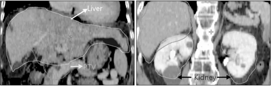
Fig. 3. Example of PTV and OARs in ITV-IMRT plan delineated in MIM program reflecting the respi- ratory motional effect.
tion was measured and applied to the phantom simulation be- cause the dynamic phantom could move in only 1-direction, and the greatest changes in respiratory motion usually occurred in the superior-inferior direction.
The per-field measurement for verifying the dose distribution in each IMRT filed under the condition of respiratory motion was performed using a MapCHECK2 2-dimensional diode de- tector array (SunNuclear, Melbourne, FL), which was laid on the dynamic phantom as shown in Fig. 1. The delivered dose distribution in each patient's CT was recalculated using a 3 dose-volume histogram program (3DVH; SunNuclear, Melbourne, FL) and applying the per-field measurement data. The 3DVH was used because the program is the only verified method that can calculate internal dose distribution of patient with the 2D data measured by MapCHECK2. The DVHs of the actual PTV and OARs during ITV-IMRT beam delivery under the con- dition of respiratory organ motion were calculated with a 3DVH and compared with the DVHs calculated for the Gate-IMRT plan to analyze the organ motional effect on dosi- metric changes during ITV-IMRT delivery. The overall process
of this study is summarized in Fig. 2.
Results
An example of ITV delineation with the MIM program is
shown in Fig. 3, and proper contour expansion can be verified
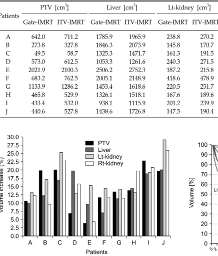
Table 1. Volume comparison of PTV and OARs between ITV-IMRT plan and Gate-IMRT plan with information on the period and range of respiratory motion in each patient.
Patients
PTV [cm
3] Liver [cm
3] Lt-kidney [cm
3] Rt-kidney [cm
3] Respiration period
[sec]
Respiratory motional range [cm]
Gate-IMRT ITV-IMRT Gate-IMRT ITV-IMRT Gate-IMRT ITV-IMRT Gate-IMRT ITV-IMRT
A 642.0 711.2 1785.9 1965.9 238.8 270.2 196.8 221.2 9.0 1.7
B 273.8 327.8 1846.3 2073.9 145.8 170.7 141.9 155.7 5.0 1.8
C 49.5 58.7 1325.3 1471.7 161.3 191.5 153.0 174.5 6.0 1.2
D 573.0 612.5 1053.3 1261.6 240.3 271.5 185.6 214.8 7.0 1.5
E 2021.9 2100.3 2506.2 2752.3 187.2 215.8 175.1 183.1 7.0 1.3
F 683.2 762.5 2005.1 2148.9 418.6 478.9 213.5 238.7 4.0 1.4
G 1133.9 1286.2 1453.4 1618.6 220.5 251.7 231.7 258.4 5.0 1.5
H 465.8 529.9 1326.1 1518.1 167.6 189.6 154.4 184.8 5.0 1.5
I 433.4 532.0 938.1 1115.9 201.2 239.9 165.2 199.5 7.0 1.8
J 440.6 527.8 1438.6 1726.8 147.5 190.4 156.2 196.8 6.0 1.2
Fig. 4. Volume increase rate of PTV and OARs in ITV-IMRT
plan compared with the volume in Gate-IMRT plan. Fig. 5. Example of the DVH comparison between the recal- culated dose in the ITV-IMRT beam delivery and the DVH of Gate-IMRT plan.
based on the shapes of the kidney and liver. The ITV-IMRT plan volumes of the PTV and OARs of 10 patients are listed in Table 1 along with the respiration periods and tumor mo- tion ranges of each patient. The rates of increase in the PTV and OARs, compared with the volumes obtained in the Gate- IMRT plans, are shown in a graph in Fig. 4. The ITV-IMRT volume increases due to the respiratory motion range were the same for both the PTV and OARs, but the correlation trends were different. Although Pearson's coefficient value was pos- itive when calculated for the correlation between the motion range and the PTV increase, a negative value was obtained for the OARs, indicating that, in contrast to the PTV, the volume increase in the OARs was not proportional to the motion range.
An example of a DVH produced from the recalculated dose
obtained from a patient's CT and based on the measured per-
field data under the condition of respiratory motion is shown
in Fig. 5, along with a comparison with the DVH from a Gate-
IMRT plan. Results of comparisons between the 2 DVHs at
the specific points are provided in Tables 2∼5. The DVH dif-
ferences in the PTV are verified in Fig. 6, which shows that
the PTV doses increased more with the ITV-IMRT beam de-
livery than with the Gate-IMRT plan. The average dose in-
creases were 3.85±1.79 Gy for the maximum dose (Dmax) to
the PTV and 1.30±0.96 Gy for the mean dose (Dmean). The
average rate of increase in the PTV volume that received >47.5
Table 2. Comparison of PTV's DVH on the specific points between the beam delivery of ITV-IMRT and Gate-IMRT plan.
Patients PTV mean-dose [Gy] PTV max-dose [Gy] PTV V
47.5Gy[%vol]
Gate-IMRT ITV-IMRT Gate-IMRT ITV-IMRT Gate-IMRT ITV-IMRT
A 51.19 50.95 53.21 55.14 99.66 99.40
B 50.78 52.84 52.62 55.46 99.94 100.00
C 51.92 55.04 53.53 56.89 100.00 100.00
D 50.53 51.24 52.86 54.49 99.82 99.29
E 49.61 50.61 53.67 59.21 98.33 97.63
F 49.56 51.74 53.52 57.74 88.95 98.87
G 48.95 49.81 52.22 55.26 94.88 95.75
H 50.13 50.75 52.37 55.22 99.02 90.23
I 49.64 51.19 54.83 61.36 67.32 73.57
J 51.28 52.47 53.29 56.83 99.88 99.70
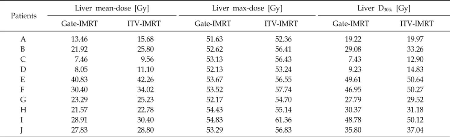
Table 3. Comparison of liver's DVH on the specific points between the beam delivery of ITV-IMRT and Gate-IMRT plan.
Patients Liver mean-dose [Gy] Liver max-dose [Gy] Liver D
30%[Gy]
Gate-IMRT ITV-IMRT Gate-IMRT ITV-IMRT Gate-IMRT ITV-IMRT
A 13.46 15.68 51.63 52.36 19.22 19.97
B 21.92 25.80 52.62 56.41 29.08 33.26
C 7.46 9.56 53.13 56.43 7.43 12.90
D 8.05 11.10 52.13 53.24 9.23 14.83
E 40.83 42.26 53.67 56.55 49.61 50.64
F 30.40 34.02 53.52 57.74 46.95 50.27
G 23.29 25.23 52.17 54.70 27.79 29.52
H 21.57 22.78 54.43 55.14 30.37 31.18
I 28.91 30.40 54.83 61.36 48.78 50.12
J 27.83 28.80 53.29 56.83 35.80 37.04
Gy, (95% of the prescribed dose) was 0.66±4.85%. Fig. 7 sh- ows the dose homogeneity within a PTV, which was calcu- lated as a homogeneity index (HI) using equation (1).
14)The average HI of the PTV during ITV-IMRT delivery was 13.66±
9.32% which was slightly inferior to the average HI value of, 10.10±6.98%, for the Gate-IMRT plan.
HI = (D
2%−D
98%)
×100% (1)
(D
prescription)
D
2%: the dose to the 2% of the target volume, as displayed on the DVH
D
98%: the dose to the 98% of the volume as displayed on the cumulative DVH
D
prescription: the prescription dose
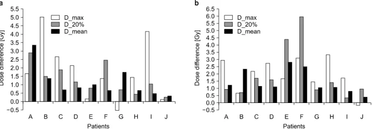
The results of a comparison of the OAR DVH differences are shown in Fig. 8, 9, and 10. Fig. 8 shows the results of a
liver comparison, in which a higher dose was irradiated during
ITV-IMRT delivery than with the Gate-IMRT plan. The aver-
age dose increases were 2.93±1.80 Gy for the D
max, 2.47±1.16
Gy for the D
mean, and 2.31±1.66 Gy for the dose to 30% of
the liver volume on the cumulative DVH (D
30%). Fig. 9 shows
that the results of a kidney comparison, in which higher doses
were also observed during ITV-IMRT delivery. The average
dose increases to the left kidney were 1.66±2.09 Gy for the
D
max, 1.12±0.89 Gy for the D
mean, and 1.33±0.87 Gy for the
dose to 20% of the kidney volume (D
20%). The average dose
increases to the right kidney were 1.97±1.13 Gy for the D
max,
1.45±0.81 Gy for the D
mean, and 1.90±1.82 Gy for the D
20%.
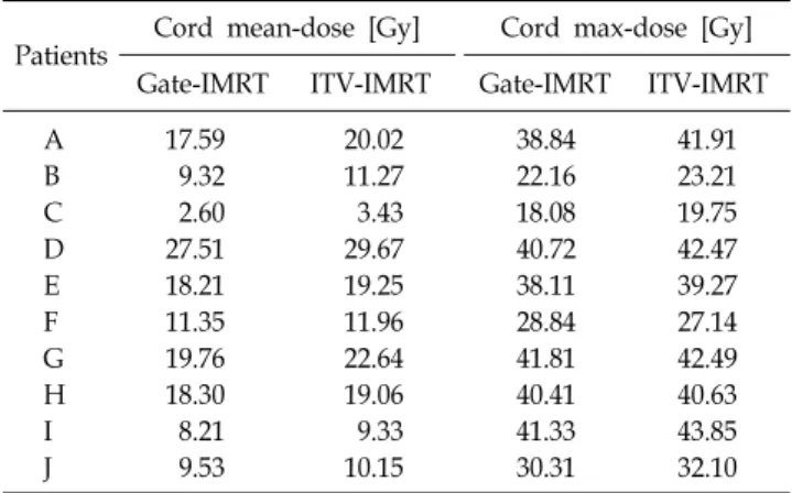
Higher irradiation doses were also delivered to the spinal cord
during ITV-IMRT delivery, as shown in Fig. 10; the average
dose increases were 1.22±1.32 Gy for the D
maxand 1.44±0.84
Gy for the D
mean.
Fig. 6. Differences of PTV's DVH values between the ITV-IMRT beam delivery and Gate-IMRT plan at the major points.
Table 5. Comparison of cord's maximum dose and mean dose between the beam delivery of ITV-IMRT and Gate- IMRT plan.
Patients Cord mean-dose [Gy] Cord max-dose [Gy]
Gate-IMRT ITV-IMRT Gate-IMRT ITV-IMRT
A 17.59 20.02 38.84 41.91
B 9.32 11.27 22.16 23.21
C 2.60 3.43 18.08 19.75
D 27.51 29.67 40.72 42.47
E 18.21 19.25 38.11 39.27
F 11.35 11.96 28.84 27.14
G 19.76 22.64 41.81 42.49
H 18.30 19.06 40.41 40.63
I 8.21 9.33 41.33 43.85
J 9.53 10.15 30.31 32.10
Table 4. Comparison of kidney's DVH on the specific points between the beam delivery of ITV-IMRT and Gate-IMRT plan.
Patients Lt-kidney mean-dose [Gy] Lt-kidney max-dose [Gy] Lt-kidney D
20%[Gy]
Gate-IMRT ITV-IMRT Gate-IMRT ITV-IMRT Gate-IMRT ITV-IMRT
A 22.72 26.07 52.52 54.18 39.48 42.39
B 5.54 6.92 24.17 29.21 7.58 9.09
C 1.52 2.24 14.14 16.81 1.12 3.03
D 15.71 16.54 44.53 46.69 18.86 20.05
E 7.09 8.09 24.38 24.55 18.22 19.05
F 0.79 1.45 5.42 6.80 0.90 3.38
G 12.57 14.31 43.48 46.17 19.35 20.07
H 7.57 8.25 29.53 31.00 13.46 13.91
I 0.74 1.22 7.11 11.28 0.89 1.93
J 2.58 2.94 19.14 19.28 5.16 5.43
Patients Rt-kidney mean-Dose [Gy] Rt-kidney max-Dose [Gy] Rt-kidney D
20%[Gy]
Gate-IMRT ITV-IMRT Gate-IMRT ITV-IMRT Gate-IMRT ITV-IMRT
A 15.98 17.10 28.68 31.63 19.33 20.27
B 11.87 14.21 34.53 35.21 16.92 17.85
C 5.27 6.40 20.96 23.14 14.65 16.36
D 14.40 15.51 46.97 49.73 19.07 20.67
E 10.56 13.38 51.32 53.00 19.14 23.55
F 5.81 8.33 31.05 34.17 12.64 18.60
G 14.88 15.94 50.80 52.25 18.79 19.70
H 12.44 13.52 48.35 51.68 19.03 20.45
I 6.75 7.55 25.16 26.89 17.34 17.70
J 2.58 3.00 35.47 35.29 2.24 3.21
Discussion
The PTV and OARs have virtually enlarged volumes that differ from those of real tumors and organs when the entire
motional range is considered during ITV-IMRT planning. This
phenomenon makes it difficult to analyze the exact dose dis-
tributions in the actual PTV and OARs, although increased
doses to the OARs can be assumed with ITV-IMRT, given the
enlarged field size. In this study, the real dose distributions in
Fig. 8. Differences of liver's DVH values between the ITV-IMRT beam delivery and Gate-IMRT plan at the major points.
Fig. 7. Comparison of homogeneity index (HI) between the beam delivery of ITV-IMRT and Gate-IMRT plan.
Fig. 9. Differences of kidney's DVH values between the ITV-IMRT beam delivery and Gate-IMRT plan at the major points. (a) Left kidney. (b) Right kidney.
Fig. 10. Differences of maximum dose and mean dose of spinal cord between the ITV-IMRT beam delivery and Gate-IMRT plan.
the actual volumes of the PTV and OARs during ITV-IMRT
for liver cancer could be calculated based on the measured
doses during beam delivery under respiratory motional condi-
tions. We analyzed the recalculated doses through comparisons
with the Gate-IMRT doses and verified that the delivered
doses to the actual volumes of the PTV and OARs in ITV-
IMRT were somewhat higher than those calculated in the
Gate-IMRT plan. Generally, the increased respiratory motional
range caused the volume increases in the PTV and OARs, and
we were able to confirm that higher actual doses were deliv-
ered to the PTV and OARs that moved within the enlarged
field area.
The correlation between the volume increases of the OARs and the respiratory motional range was negative, which might have been a result of the selection of a motional range in the area of the PTV's center that was not in the region of OARs.
Although the exact 3-dimensional movements of the organs could not be simulated in this study, the motional effect could be evaluated sufficiently well by applying the largest motional change in the superior-inferior direction.
In this study, the accuracy of the dosimetric data calculated with the 3DVH program could be considered reliable because the dose calculation algorithm that incorporates the per-field dose measurement data using 3DVH has already been verified in many studies.
15,16)The average increase in the D
meanof the PTV was 1.30±
0.96 Gy, and the D
maxand V
95%of the PTV were also slightly elevated with ITV-IMRT delivery. Although the sufficient pre- scription dose coverage of the PTV could be considered a ben- efit of ITV-IMRT, the dose homogeneity in the PTV, eval- uated using the HI, was inferior to that of the Gate- IMRT plan. This double-sided nature of the dose distribution to the PTV should be considered in ITV-IMRT cases.
Although the potential disadvantage of ITV-IMRT with re- spect to higher dose distribution to the OARs was again con- firmed in this study, the dose increase rate was modest com- pared with the constraint criteria, and these increases should be evaluated from a biological point of view, rather than simply from a numerical dose increase. In other words, the dose in- creases should be analyzed with regard to the impact on the increased probability of complications in the OARs. The aver- age increase in the D
maxof the spinal cord with ITV-IMRT was 1.22±1.32 Gy, which could be considered a meaningless increase if the maximum dose constraint for the spinal cord (45 Gy) was fulfilled. The important constraint value in the kidney, the D
20%, increased with ITV-IMRT, and the average dose increase to both kidneys was 1.61±1.42 Gy. The D
30%liver constraint also showed an increase with ITV-IMRT, and the average dose increase was 2.31±1.66 Gy. The dose in- creases to the kidney and liver might be significant factors in failures of constraint fulfillment, and therefore the criteria for OAR constraints should be further restricted in the opti- mization process of ITV-IMRT plans.
The respiratory motional direction simulated in this study
was only the superior-inferior direction, which could not simu- late the real 3-dimensional movement of tumor. This limitation can be solved in the further study by the application of 3-di- mensional moving phantom which can simulate tumor motion similarly with a real condition.
The problem of dose increases to the OARs during ITV- IMRT can be solved by applying the Gate-IMRT method, which has a theoretical advantage with regard to the ideal dose distribution without a respiratory motional effect. However, Gate-IMRT also presents difficulties when attempting to real- ize the theoretical treatment conditions in actual clinical sit- uations, including accurate MLC operation, gating system pre- cision, stable patient respiration, and consistent organ motional correlation with the respiratory signals. When considering the increased treatment time consequent to the gating system ap- plication, Gate-IMRT cannot be considered a superior method with which to correct respiratory motional effects in actual clinical circumstances.
Therefore, it would be better to perform an analysis of the patient's respiratory patterns and constraints during the opti- mization process in order to select the proper IMRT method for areas of motion such as the liver and lung.
Conclusions
Dose distributions within the real volumes of the PTV and
OARs during ITV-IMRT for liver cancer while under respira-
tory motional conditions could be acquired, and these data
confirmed that higher irradiation doses were delivered to the
OARs when compared to those calculated in the Gate-IMRT
plan. However, the increased doses could be considered as mi-
nor risk factors if constraints were reinforced during the IMRT
optimization process. Because Gate-IMRT cannot always be
considered an ideal method with which to correct the respira-
tory motional effect, given the dosimetric variations in the gat-
ing system application and the increased treatment time, a pri-
or analysis for optimal IMRT method selection should be per-
formed while considering the patient's respiratory condition
and IMRT plan results.
References
1. Duan J, Shen S, Fiveash JB, et al: Dosimetric effect of respiration-gated beam on IMRT delivery. Med Phys 30(8):2241- 2252 (2003)
2. Ahmed RS, Shen S, Ove R, et al: Intensity modulated with respiratory gating for radiotherapy of the pleural space. Med Dosim 32(1):16-22 (2007)
3. van der Geld YG, van Triest B, Verbakel WF, et al:
Evaluation of four-dimensional computed tomography-based intensity-modulated and respiratory-gated radiotherapy techni- ques for pancreatic carcinoma. Int J Radiat Oncol Biol Phys 72(4):1215-1220 (2008)
4. Seco J, Sharp GC, Wu Z, et al: Dosimetric impact of mo- tion in free-breathing and gated lung radiotherapy: a 4D Monte Carlo study of intrafraction and interfraction effects. Med Phys 35(1):356-366 (2008)
5. Chen H, Wu A, Brandner ED, et al: Dosimetric evaluations of the interplay effect in respiratory-gated intensity-modulated radiation therapy. Med Phys 36(3):893-903 (2009)
6. Kang H, Yorke ED, Yang J, et al: Evaluation of tumor mo- tion effects on dose distribution for hypofractionated intensity- modulated radiotherapy of non-small-cell lung cancer. J Appl Clin Med Phys 11(3):78-89 (2010)
7. Cheong KH, Kang SK, Lee M, et al: Evaluation of delivered monitor unit accuracy of gated step-and-shoot IMRT using a two-dimensional detector array. Med Phys 37(3):1146-1151 (2010)
8. Yoganathan SA, Maria Das KJ, Agarwal A, et al:
Performance evaluation of respiratory motion-synchronized dy- namic IMRT delivery. J Appl Clin Med Phys 14(3):39-51 (2013) 9. Hugo GD, Agazaryan N, Solberg TD: An evaluation of gating window size, delivery method, and composite field dosim- etry of respiratory-gated IMRT. Med Phys 29(11):2517-2525 (2002)
10. Xi M, Liu MZ, Deng XW, et al: Defining internal target vol- ume (ITV) for hepatocellular carcinoma using four-dimensional CT. Radiother Oncol 84(3):272-278 (2007)
11. Xi M, Liu MZ, Zhang L, et al: How many sets of 4DCT im- ages are sufficient to determine internal target volume for liver radiotherapy? Radiother Oncol 92(2):255-259 (2009)
12. Reitz B, Parda DS, Colonias A, et al: Investigation of sim- ple IMRT delivery techniques for non-small cell lung cancer pa- tients with respiratory motion using 4DCT. Med Dosim 34(2):
158-169 (2009)
13. Speight R, Sykes J, Lindsay R, et al: The evaluation of a deformable image registration segmentation technique for semi-automating internal target volume (ITV) production from 4DCT images of lung stereotactic body radiotherapy (SBRT) patients. Radiother Oncol 98(2):277-283 (2011)
14. Wu Q, Mohan R, Morris M, et al: Simultaneous integrated boost intensity modulated radiotherapy for locally advanced head-and-neck squamous cell carcinomas. I: dosimetric results.
Int J Radiat Oncol Biol Phys 56(2):573-585 (2003)
15. Olch AJ: Evaluation of the accuracy of 3DVH software esti- mates of dose to virtual ion chamber and film in composite IMRT QA. Med Phys 39(1):81-86 (2012)
16. Carrasco P, Jornet N, Latorre A, et al: 3D DVH-based
metric analysis versus per-beam planar analysis in IMRT pre-
treatment verification. Med Phys 39(8):5040-5049 (2012)
간암 환자의 세기조절방사선치료 시 호흡에 의한 움직임 조건에서 측정된 조사면 별 선량결과를 기반으로 재계산한 체내 선량분포 평가