493
Intrahepatic portosystemic shunt with a second degree atrioventricular block fixed by transvenous coil embolization in a dog
Seung-Gon Lee
1, So-Jeong Nam
1, Hyun-Wook Kim
2, Changbaig Hyun
1,*
1
School of Veterinary Medicine, Kangwon National University, Chuncheon 201-100, Korea
2
Haemaru Animal Medical Centre, Sungnam 463-824, Korea (Accepted: December 3, 2008)
Abstract : A 2-year-old female Pekingese dog was presented with primary complaints including exercise intolerance and neurological sign associated with hepatic encephalopathy. The major findings in clinical examination included an intermittent seizure, a slow heart rate with pulse deficit, leukocytosis and anemia in hemogram, elevated pre- and post-prandial serum bile acid and hepatic enzymes, hypoproteinemia, coagulopathy, ammonium urate crystaluria and bilirubinuria. Diagnostic tests revealed an intrahepatic portosystemic shunt complicated with a second degree atrioventricular block and QT prolongation. The case was successfully treated with a transvenous coil embolization. Clinical signs were gradually improved and cardiac bradyarrhythmia disappeared. This case is a rare case of intrahepatic portosystemic shunts complicated with a cardiac bradyarrhythmia in a small breed dog fixed by a transvenous coil embolization.
Keywords : atrioventricular block, IPSS, portosystemic shunt, transvenous coil embolization
Introduction
A portosystemic shunt (PSS) is an abnormal connection between the portal vein and the systemic circulation. The PSS can be subdivided into extraheptic (shunts located outside the liver) and intrahepatic (shunts located inside the liver) PSS, depending on the location of the shunted vessel. The PSS can be also subdivided into congenital and acquired, depending on the etiology. Either incomplete or botched closure of patent ductus venosus after birth is the main cause of PSS in dogs [17]. In the small breed dogs, extrahepatic portosystemic shunts (EPSS) are more common, while intrahepatic portosystemic shunts (IPSS) are more common in large breed dogs. To date, the exact mode of inheritance for this disorder has not been determined, although PSS is over-presented in certain dog breeds such as Havanese, Yorkshire Terriers, Miniature Schnauzers, and Maltese [11, 24].
Most dogs with PSS will develop clinical signs early in life. The common clinical presentations associated with PSS are a history of doing poorly, the smallest size in littermates, waxing and waning neurologic
signs, polyuria/polydipsia, microhepatica and urate urolithiasis [17]. The characteristic hemogram is a microcytic, normochromic nonregenerative anemia with target cells and poikilocytes, resulted from abnormal lipid metabolism, iron sequestration, or iron deficiency [3, 10, 27].
Liver enzymes are often not significantly elevated in dogs with PSS. The more common biochemical changes in PSS include hypoalbuminemia, hypocho- lesterolemia, hypoglycemia (especially after fasting), and decreased BUN. Low urine specific gravity and ammonium biurate crystals are the most common abnormalities observed in urinalysis of dogs with PSS.
Coagulation abnormalities are gradually apparent, when the disease progresses. Both serum bile acids (fasting and 2-h postprandial) and ammonia tolerance tests are more accurate method for diagnosing PSS in dogs, although they may be increased by other causes of hepatic dysfunction.
For definitive diagnosis, a visualization of shunting vessel is required using an exploratory laparotomy, contrast radiography (portography), computed tomography, ultrasound, or nuclear scintigraphy. Medical manage-
*Corresponding author: Changbaig Hyun
School of Veterinary Medicine, Kangwon National University, Chuncheon 201-100, Korea
[Tel: +82-33-250-8681, Fax: +82-33-244-2367, E-mail: [email protected]]
ment for PSS predominantly aims to control signs of hepatic encephalopathy using a low-protein diet and oral ammonia absorption blockers. However, the medical management alone is usually not very successful and most animals are euthanized because of inability to adequately control signs.
Ultimately, all dogs with PSS are required either surgical or non-surgical occlusion of shunted vessel(s).
Many different surgical procedures of PSS have been described in veterinary literatures [6, 24, 26]. Gradual ligation (for preventing portal hypertension) using cellophane tape and ameroid constrictors is the most commonly used method for EPSS. However, for IPSS, this surgical ligation alone is often difficult to occlude this shunt. Transvenous coil embolization (TCE) has been recently developed and successfully applied to occlude the shunted vessel located inside the liver [5, 22]. However, overzealous occlusion of a shunt vessel can result in acute portal hypertension, which can cause ascites, bowel ischemia, and endotoxemic shock [17].
Cirrhotic cardiomyopathy (CC) is a relatively new clinical entity characterized by inconstant and often subclinical series of heart abnormalities induced by
chronic hepatic failure (e.g. hepatic cirrhosis) [18]. In humans with CC, a significant prolongation of QT interval on basal ECG has been well recognized [13, 15, 16], presumably due to an impaired electrical ventricular recovery. The abnormally prolonged QT is associated with sudden cardiac arrest in dogs and humans [7, 8, 20]. In humans, about 30-60% of cirrhotic patients have prolonged QT interval [1, 2].
Furthermore, the extent and magnitude of long QT were closely correlated with the severity of liver disease [1, 2]. Although the long QT disappeared in most human patients after liver transplantation, the clinical relation between QT prolongation and hepatic cirrhosis is still debatable [4].
This case study described a rare case of IPSS complicated with a cardiac bradyarrhythmia and long QT in a small breed dog fixed by a TCE.
Case history
A 2-year-old female Pekingese dog (body weight 4.1 kg) was referred to the Veterinary Teaching Hospital, Kangwon National University with primary
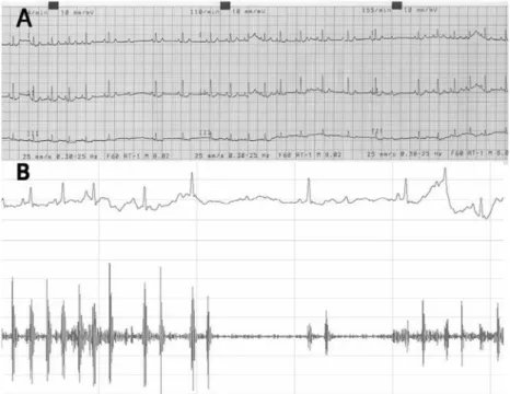
Fig. 1. The electrocardiogram (A) and phonocardiogram (B) of this case. The electrocardiogram showed a second degree atrioventricular heart block (atrial rate: 125 bpm, ventricular rate 120 bpm) with QT prolongation (QTc = 269
×10.8 msec).
The phonocardiogram showed an irregular interval and intensity of heart sound. There is also intermittent absence of heart
sound.
complaints including exercise intolerance and neurological sign associated with hepatic encephalopathy. Before the presentation, the dog was received mediations to reduce the absorption and production of ammonia (i.e.
metronidazol, 7.5 mg/kg PO q 8 h; lactulose, 0.5 ml/kg PO q 8 h with a protein-restricted diet).
In physical examination, the dog was anorexic and lethargic. Femoral pulsation was intermittently deficient with slow heart rate. Pulse deficit was more obvious in the phonocardiogram and electrocardiogram (ECG;
Fig. 1). The 12 lead- surface ECG and digital ECG recordings revealed a second degree atrioventricular (AV) heart block with QT prolongation (269
±10.8 msec; Fig. 1A). A subsequent atropine response test revealed second degree Mobitz type I-AV block.
Complete blood count revealed a leukocytosis (23.78
×
10
3/
µl; reference range: 6-17
×10
3/
µl) with neutrophilia (13.30
×10
3/
µl; reference range: 3.0-11.8
×10
3/
µl) and mild anemia (5.1
×10
12/
µl; reference range: 5.5-7.5
×10
12/
µl). Serum biochemistry revealed reduced hepatic
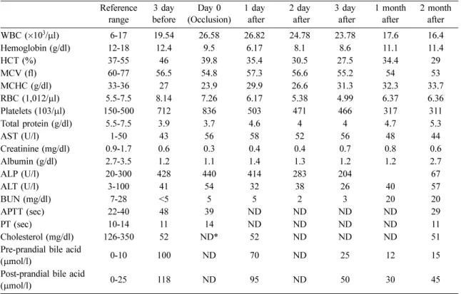
function with an increased pre- and post-prandial serum bile acid levels (Table 1). Blood coagulation profiles were also prolonged (Table 1). Urinalysis revealed ammonium urate crystaluria and bilirubinuria.
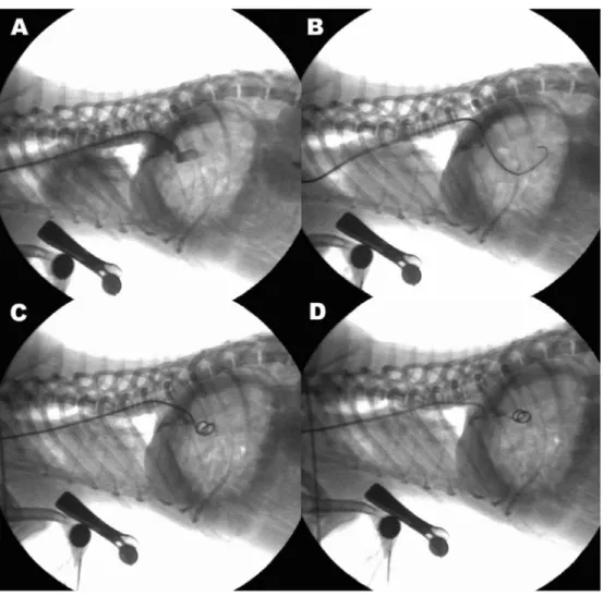
Diagnostic imaging studies revealed a cranial gastric axis deviation indicating small size liver, abdominal fluid accumulation, decreased number of portal veins in the liver and generalized enhancement of the hepatic echogenicity (Fig. 2). The portal vein was not tapered in the hepatic parenchyma (Fig. 2). The portography clearly visualized a single straight shunted portal vein to the caudal vena cava (Fig. 3). The ultrasonography revealed the maximal diameter of the shunted vessel was 5.0 mm in diameter.
Based on diagnostic findings, the case was diagnosed as IPSS complicated with second degree atrioventricular (AV) block and QT prolongation. With the consent of owner, we decided to occlude IPSS by lodging transvenous embolization coils. Since the shunt diameter was 5 mm, we decided to initially lodge a Table 1. Hemogram and blood chemistry of this case
Reference
range 3 day
before Day 0
(Occlusion) 1 day
after 2 day
after 3 day
after 1 month
after 2 month after
WBC (
×10
3/
µl) 6-17 19.54 26.58 26.82 24.78 23.78 17.6 16.4
Hemoglobin (g/dl) 12-18 12.4 9.5 6.17 8.1 8.6 11.1 11.4
HCT (%) 37-55 46 39.8 35.4 30.5 27.5 34.4 29
MCV (fl) 60-77 56.5 54.8 57.3 56.6 55.2 54 53
MCHC (g/dl) 33-36 27 23.9 29.9 26.6 31.3 32.3 33.7
RBC (1,012/
µl) 5.5-7.5 8.14 7.26 6.17 5.38 4.99 6.37 6.36
Platelets (103/
µl) 150-500 712 836 503 471 466 317 311
Total protein (g/dl) 5.5-7.5 3.9 3.7 4.6 4 4 4.7 5.3
AST (U/l) 1-50 43 56 58 52 56 48 44
Creatinine (mg/dl) 0.9-1.7 0.6 0.3 0.4 0.4 0.7 0.8 0.6
Albumin (g/dl) 2.7-3.5 1.2 1.1 1.4 1.3 1.2 1.2 2.7
ALP (U/l) 20-300 428 440 414 283 204 67
ALT (U/l) 3-100 41 54 32 38 26 40 57
BUN (mg/dl) 7-28 <5 5 5 2 3 20 20
APTT (sec) 22-40 48 39 ND ND ND ND 29
PT (sec) 10-14 11 14 ND ND ND ND 11
Cholesterol (mg/dl) 126-350 52 ND* 52 ND ND ND 51
Pre-prandial bile acid
(
µmol/l) 0-10 100 ND 70 ND 25 12 15
Post-prandial bile acid
(
µmol/l) 0-25 118 ND 95 ND 50 30 45
WBC, white blood cell; HCT, hematocrit; MCV, mean corpuscular volume; MCHC, mean corpuscular hemoglobin concen- tration, RBC, red blood cell; AST, aspirate transaminase; ALP, alkaline phosphatase; ALT, alanine transaminase; BUN, blood urea nitrogen; APTT, activated partial thrombin time; PT, prothrombin time.
*