Introduction
Carbon monoxide (CO) is a colorless, odorless, and nonirritating toxic gas commonly encountered in daily life. CO poisoning is particularly damaging to organs with high oxygen requirements, such as the brain
1). Therefore, CO can cause various neuropsy-
의식이 명료한 일산화탄소 중독환자를 대상으로 응급실에서 시행한 간이정신상태검사의 임상적 의의
연세대학교 원주의과대학 응급의학교실1, 재활의학과학교실2
육@현1∙차용성1∙김@현1∙김성훈2∙김지현2 김오현1∙김형일1∙차경철1∙이강현1∙황성오1
Incidence and Features of Cognitive Dysfunction Identified by Using Mini-mental State Examination at the Emergency Department among
Carbon Monoxide-poisoned Patients with an Alert Mental Status
Hyun Youk, M.D.1, Yong Sung Cha, M.D.1, Hyun Kim, M.D., Ph.D.1, Sung Hoon Kim, M.D., Ph.D.2, Ji Hyun Kim, M.D.2, Oh Hyun Kim, M.D., Ph.D.1, Hyung Il Kim, M.D.1, Kyoung Chul Cha, M.D., Ph.D.1,
Kang Hyun Lee, M.D., Ph.D.1, Sung Oh Hwang, M.D., Ph.D.1
Department of Emergency Medicine1,
Department of Rehabilitation Medicine2, Wonju College of Medicine, Yonsei University, Korea
Purpose: Because carbon monoxide (CO)-intoxicated patients with an alert mental status and only mild cognitive dysfunction may be inadequately assessed by traditional bedside neurologic examination in the emergency depart- ment (ED), they may not receive appropriate treatment.
Methods: We retrospectively investigated the incidence and features of cognitive dysfunction using the Korean ver- sion of the Mini-Mental State Examination (MMSE-K) in ED patients with CO poisoning with alert mental status. We conducted a retrospective review of 43 consecutive mild CO poisoned patients with a Glasgow Coma Scale score of 15 based on documentation by the treating emergency physician in the ED between July 2014 and August 2015.
Results: Cognitive dysfunction, defined as a score of less than 24 in the MMSE-K, was diagnosed in six patients (14%) in the ED. In the MMSE-K, orientation to time, memory recall, and concentration/calculation showed greater impairments. The mean age was significantly older in the cognitive dysfunction group than the non-cognitive dysfunc- tion group (45.3 yrs vs. 66.5 yrs, p<0.001). Among the initial symptoms, experience of a transient change in mental sta- tus before ED arrival was significantly more common in the cognitive dysfunction group (32.4% vs. 100%, p=0.003).
Conclusion: Patients with CO poisoning and an alert mental status may experience cognitive dysfunction as assessed using the MMSE-K during the early stages of evaluation in the ED. In the MMSE-K, orientation to time, memory recall, and concentration/calculation showed the greatest impairment.
Key Words: Carbon monoxide, Cognitive disorder, Emergency care 원
원 저저
책임저자: 차 용 성
강원도 원주시 일산로 20
연세대학교 원주의과대학 응급의학교실 Tel: 033) 741-1615, Fax: 033) 742-3030 E-mail: [email protected]
투고일: 2016년 6월 4일 1차 심사일: 2016년 7월 25일 게재 승인일: 2016년 8월 12일
chological symptoms, including impairment in cogni- tive function, mood disorders, irritability and coma
2-5). Additionally, unique and problematic complications of CO poisoning can lead to delayed neuropsycho- logical sequelae (DNS), including impairment in cog- nitive functions, akinetic mutism, anxiety and depres- sion, sphincter incontinence, temperament changes, dystonia, parkinsonism, and amnesia, which may develop in some patients after a period of apparent recovery from acute symptoms
3-5). DNS develops in 3% to 23% of cases of CO poisoning
6,7)and appears 1 to 4 weeks after poisoning
7,8).
Although the neurotoxic effect of moderate to severe CO poisoning is well-established
9-11), CO poi- soning in patients with an alert mental status has received relatively little attention and the extent of neuropsychological damage is controversial
12). Although traditional methods of assessment such as the bedside neurologic examination can readily identi- fy unconscious or severely affected patients, many CO-poisoned patients with only mild cognitive impair- ment are inadequately assessed by such methods
13). Failure to assess the cerebral functioning of patients exposed to CO may result in neuropsychological sequelae. To the best of our knowledge, there has not been a report on the evaluation of cognitive dysfunc- tion using neuropsychological screening tests in
patients with CO poisoning and an alert mental status in an emergency department (ED) setting in Korea.
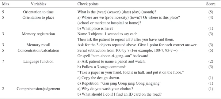
Since July 2014, our ED has been investigating cogni- tive dysfunction using the Korean version of the Mini- Mental State Examination (MMSE-K), which was devel- oped for use in a Korean population (Table 1)
14,15).
We retrospectively investigated the incidence and features of cognitive dysfunction using the MMSE-K in ED patients with CO poisoning with alert mental status.
Methods
1. Study design and data
This was a retrospective observational study that collected data from consecutive patients diagnosed with acute CO poisoning in an ED between July 2014 and August 2015. The ED was located in a single urban, tertiary-care hospital, which experiences more than 45,000 annual visits and is staffed 24 hours per day by board-certified emergency physicians.
All patients with the word “CO poisoning”in ED discharge codes registered in the computerized hospi- tal records were initially considered. For selected patients, a diagnosis of CO poisoning was made according to medical history and carboxyhemoglobin
Table 1. The Korean version of the mini-mental state examination (MMSE-K)
Max Variables Check points Score
5 Orientation to time What is the (year) (season) (date) (day) (month)? (5)
5 Orientation to place a) Where are we (province/city) (town)? Or where is this place? (4) (school or market or hospital or home)?
b) What place is here? (1)
3 Memory registration Name 3 objects: 1 second to say each. (3)
Then ask the patient to repeat all 3 after you have said them.
3 Memory recall Ask for the 3 objects repeated above. Give 1 point for each correct answer. (3) 5 Concentration/calculation Serial subtraction from 100 by 7 (For example, 100-7, 93-7…) (5)
Or spell “sam-cheon-ri-gang-san” backward.
7 Language function a) Ask patient to name a pencil and watch. (2)
b) Follow a 3-stage command: (3)
“Take a paper in your hand, fold it in half, and put it on the floor.”
c) Copy the design shown. (1)
d) Repetition: “Gan jang Gong jang Gong jangjang” (1)
2 Comprehension/judgement a) Why do you wash your clothes? (1)
b) What should I do if I find an ID card on the road?
(CO-Hb) level of >5% (>10% in smokers). Mild CO poisoning was defined as patients with a Glasgow Coma Scale (GCS) score of 15, which relied on the documentation by the treating emergency physician, upon arrival to the ED. All patients complaining of acute CO poisoning were treated with 100 % high- flow oxygen therapy and, if indicated
16), hyperbaric oxygen therapy (HBOT). Exclusion criteria were a his- tory of a previous stroke or dementia, inability to per- form MMSE-K for unavoidable reasons, and a GCS less than 15. The reasons for these exclusions were as fol- lows: 1) in patients with a history of stroke or demen- tia, it was not possible to prove whether the cognitive dysfunction occurred as a result of the current event.
2) Patients with a GCS less than 15 were excluded because we wanted to investigate the results of cogni- tive dysfunction using the MMSE-K in patients with CO poisoning and no deficits in mental status.
Data were collected by retrospectively reviewing the electronic medical records. Data collection was conducted by two emergency physicians blinded to the study objectives and hypothesis; if there was inter-observer disagreement in the interpretation of the clinical data, the two emergency physicians reviewed the case together to come to a conclusion.
Training of the abstractors was conducted prior to data collection to reduce bias. The clinical parame- ters assessed were age, gender, intentionality of CO poisoning, duration of CO exposure, ED arrival time, medical history, initial symptoms, and vital signs. The duration of CO exposure was ascertained from patient history via the patients themselves or through interviews with guardians and was thus recorded as an estimated maximum duration of CO exposure, measured from the time of normal consciousness to the time of detection or reported CO exposure. The laboratory parameters assessed were the initial CO- Hb level from pre-hospital sources or the level obtained at our hospital, creatinine kinase (CK) to evaluate muscle injury, high sensitivity troponin I (hs-TnI) to evaluate myocardial injury, base excess (BE), and lactate to evaluate tissue hypoperfusion.
Patients available for testing underwent the MMSE- K administered in the ED to measure the presence
and severity of cognitive function. The MMSE-K includes 7 subsets that evaluate higher cognitive functions, including orientation to time, orientation to place, memory registration, memory recall, con- centration/calculation, language function, and com- prehension/judgement (Table 1)
14,15). The cognitive dysfunction group included those with an MMSE-K score less than 24 in test performed in the ED. Two experienced rehabilitation physicians (S.H.K and J.H.K) interpreted the test findings while blinded to the patients’clinical data.
The primary outcome of this study was to investi- gate the early incidence and features of cognitive dysfunction in patients with CO poisoning and an alert mental status using the MMSE-K performed in an ED. This study was approved by the Institutional Review Board of Wonju College of Medicine, Yonsei University.
2. Statistical analysis
Data are expressed according to the properties of the variable. Continuous variables are presented as the mean and standard deviation, median and interquartile range, or range after assessment for nor- mality using the Shapiro-Wilk test. Categorical vari- ables are presented as frequency and percentage.
The Chi-square test or Fisher’s exact test was used for comparison of categorical variables, while the two-sample t-test or Mann-Whitney U-test was used to compare continuous variables. P values less than 0.05 were considered statistically significant, and all statistical analyses were performed using SPSS 20 Ver. (IBM, Armonk, NY, USA).
Results
1. Characteristics of the study subjects
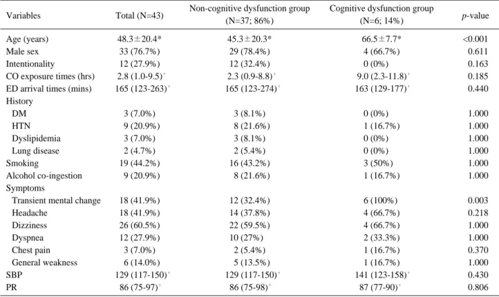
Between July 2014 and August 2015, a total of 47,005 patients visited our hospital ED. A total of 55 consecutive patients with acute CO poisoning and a GCS score 15 was identified during the study period.
The following patients were excluded: those unable
to undergo the MMSE-K for unavoidable reasons including refusal to undergo the examination and dif- ficulties associated with test performance (6 patients) and a history of stroke or dementia (6 patients).
Therefore, we ultimately included 43 of the 55 total patients with CO poisoning and an alert mental status.
Of the 43 patients analyzed, 76.7% were men, and the mean age was 48.3 years (range, 12-82 years).
Intentional CO poisoning accounted for 27.9% of cases, and the median duration of CO exposure was 2.8 hours. Hypertension (20.9%) was the most com-
mon finding in the medical history. The most com- mon symptom at initial presentation was dizziness (60.5%). The median initial CO-Hb level was 19.8%, and the median serum lactate levels was 1.82 mmol/L (Table 2, 3).
2. The incidence and features of cognitive dysfunction identified in the ED
Acute cognitive dysfunction, defined as an abnor- mal score on MMSE-K, was diagnosed in 6 patients
Table 3. Laboratory findings according to the presence of cognitive dysfunction in patients with CO poisoning and an alert mental status Variables Total (N=43) Non-cognitive dysfunction group Cognitive dysfunction group
p-value
(N=37; 86%) (N=6; 14%)
CO-Hb (%) 19.8 (11.4-36.7)� 20.3 (10.4-36.7)� 17.2 (15.6-41.6)� 0.483
Creatinine kinase (U/L) 119 (78-169)�0. 119 (80-186)�0. 97 (65-195)�. 0.335 Troponin I (ng/mL) 00.015 (0.015-0.047)� 00.015 (0.015-0.025)� 00.131 (0.015-0.280)� 0.085 Base excess (mmol/L) -1.8 (-3.7- -1.0)� -2.0 (-4.2- -0.8)� -1.3 (-3.4- -0.9)� 0.587 Lactate (mmol/L) 1.82 (1.15-3.07)� 1.82 (1.13-2.97)� 1.65 (1.21-3.59)� 0.902
* mean±standard deviation, �Median (interquartile range) CO-Hb: carboxyhemoglobin
Table 2. General characteristics according to the presence of cognitive dysfunction in patients with CO poisoning and an alert mental status Variables Total (N=43) Non-cognitive dysfunction group Cognitive dysfunction group
p-value
(N=37; 86%) (N=6; 14%)
Age (years) 48.3±20.4* 45.3±20.3* 66.5±7.7* <0.001
Male sex 33 (76.7%) 29 (78.4%) 4 (66.7%) <0.611
Intentionality 12 (27.9%) 12 (32.4%) 0 (0%)00. <0.163
CO exposure times (hrs) 02.8 (1.0-9.5)� 02.3 (0.9-8.8)� 09.0 (2.3-11.8)� <0.185 ED arrival times (mins) 0.165 (123-263)� 0.165 (123-274)� .163 (129-177)� <0.440 History
DM 3 (7.0%) 3 (8.1%) 0 (0%)00. <1.000
HTN 09 (20.9%) 08 (21.6%) 1 (16.7%) <1.000
Dyslipidemia 3 (7.0%) 3 (8.1%) 0 (0%)00. <1.000
Lung disease 2 (4.7%) 2 (5.4%) 0 (0%)00. <1.000
Smoking 19 (44.2%) 16 (43.2%) 3 (50%)0. <1.000
Alcohol co-ingestion 09 (20.9%) 08 (21.6%) 1 (16.7%) <1.000
Symptoms
Transient mental change 18 (41.9%) 12 (32.4%) 6 (100%). <0.003
Headache 18 (41.9%) 14 (37.8%) 4 (66.7%) <0.218
Dizziness 26 (60.5%) 22 (59.5%) 4 (66.7%) <1.000
Dyspnea 12 (27.9%) 10 (27%)0. 2 (33.3%) <1.000
Chest pain 3 (7.0%) 2 (5.4%) 1 (16.7%) <0.370
General weakness 06 (14.0%) 05 (13.5%) 1 (16.7%) <1.000
SBP 0.129 (117-150)� 0.129 (117-150)� .141 (123-158)� <0.430
PR .86 (75-97)� .86 (75-98)� 87 (77-90)�. <0.806
* mean±standard deviation, �Median (interquartile range)
CO: carbon monoxide, ED: emergency department, DM: diabetes mellitus, HTN: hypertension, SBP: systolic blood pressure, PR: pulse rate
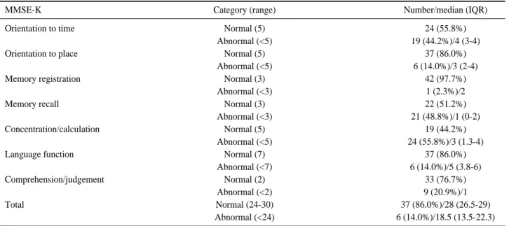
(14%). In the MMSE-K, orientation to time, memory recall, and concentration/calculation showed the greatest impairment (Table 2-4). All of the 6 patients received once HBOT at the ED. They were follow-up test before discharge and normalized score of MMSE- K. For the 6 patients with cognitive dysfunction, we attempted to contact each patient or their guardian to inquire about the patient’s current condition, includ- ing newly developed DNS symptoms after discharge.
All of these 6 patients were successfully contacted and verified that they did not experience any DNS symptoms. A comparison of the baseline characteris- tics of the study participants is shown in Table 2. The mean age was significantly older in the cognitive dys- function group than in the non-cognitive dysfunction group (45.3 yrs vs. 66.5 yrs, p<0.001). Among the ini- tial symptoms, experience of a transient change in mental status before ED arrival was significantly more common in the cognitive dysfunction group (32.4%
vs. 100 %, p=0.003). There was no statistical differ- ence in terms of intentionality, CO exposure time, and past medical history. Initial CO-Hb, CK, hs-TnI, and lactate levels were not significantly different between the cognitive dysfunction group and the non-cognitive dysfunction group (Table 3).
Discussion
It would be easy for emergency physicians to con- clude that patients have no neuropsychiatric abnor- malities, such as cognitive dysfunction, if the patient is alert as assessed using traditional assessment meth- ods such as bedside neurologic examination.
Although bedside neurologic examination can readily identify unconscious or severely affected patients, such methods may inadequately assess many CO- poisoned patients with only mild cognitive impair- ment
13). In this study, we included patients with a GCS score of 15 as assessed by the treating emer- gency physician. However, the incidence of cogni- tive dysfunction as assessed by tests performed in the ED was as high as 14% in patients with CO poisoning and an alert mental status, with orientation to time, memory recall, and concentration/calculation show- ing the greatest impairment. There was a large frac- tion of patients with disorientation to time and/or place, which would automatically reduce their GCS to 14 (losing 1 point for a disoriented verbal response). In other words, this means that the pre- liminary neurologic examination was relatively insensitive and inconsistent. Therefore, cognitive function tests such as the MMSE-K may be required
Table 4. Result of cognitive function tests (MMSE-K)
MMSE-K Category (range) Number/median (IQR)
Orientation to time Normal (5) 24 (55.8%)
Abnormal (<5) 19 (44.2%)/4 (3-4)
Orientation to place Normal (5) 37 (86.0%)
Abnormal (<5) 6 (14.0%)/3 (2-4)
Memory registration Normal (3) 42 (97.7%)
Abnormal (<3) 1 (2.3%)/2
Memory recall Normal (3) 22 (51.2%)
Abnormal (<3) 21 (48.8%)/1 (0-2)
Concentration/calculation Normal (5) 19 (44.2%)
Abnormal (<5) 24 (55.8%)/3 (1.3-4)
Language function Normal (7) 37 (86.0%)
Abnormal (<7) 6 (14.0%)/5 (3.8-6)
Comprehension/judgement Normal (2) 33 (76.7%)
Abnormal (<2) 9 (20.9%)/1
Total Normal (24-30) 37 (86.0%)/28 (26.5-29)
Abnormal (<24) 6 (14.0%)/18.5 (13.5-22.3)
MMSE-K: Korean version of the Mini-Mental State Examination, IQR: interquartile range
to identify patients with cognitive dysfunction.
Although various areas of the brain can be dam- aged in acute CO poisoning, the temporal lobe and the hippocampus are particularly vulnerable areas
17)and are responsible for many of the aspects of cogni- tive function evaluated by the MMSE, including ori- entation to time and place, memory recall, and lan- guage
18). Therefore, the MMSE may be one of useful methods for assessing cognitive function. Amitai et al. reported that the cognitive dysfunction resulting from low-level exposure to CO may be extensive and may involve memory, concentration, temporal and spatial orientation, construction skills, visuomotor coordination, and visuospatial function
19). The authors used the Carbon Monoxide Neuropsychological Screening Battery (CONSB), which includes 6 subsets that evaluate higher cognitive functions including memory, visuomotor coordination, visuospatial plan- ning and processing, temporospatial orientation, and constructive skills. Although one advantage of the CONSB is that it can be conducted in a short amount of time
20), Rottman et al. reported that the CONSB does not appear to be as useful in cases of low-level CO poisoning
21).
In this study, age differed between the patient groups, with the cognitive dysfunction group being older than the non-cognitive dysfunction group (p<0.001). This result may be explained by the lack of physiologic functional reserve in older patients. A history of transient mental status change before arrival at the ED was significantly more common in the cognitive dysfunction group than in the group without cognitive dysfunction (p=0.003) (Table 2).
Even if patients only lose consciousness for a short period of time, their brains may have already been injured by the exposure and the recovery of con- sciousness may not positively correlate with the level of brain injury resulting from the CO poisoning.
There were some limitations to this study. First, the study was conducted using a retrospective design at a single hospital. As a result, not all relevant assess- ment parameters were included. Second, we did not compare the usefulness of the MMSE-K with that of other tests such as the CONSB, because the length of
time required for testing makes it difficult to conduct cognitive function tests in CO-poisoned patients.
Third, we did not evaluate the long-term outcomes of patients with mild CO poisoning with only cognitive dysfunction. Long term outcomes, such as DNS, in mild CO-poisoned with only reversible cognitive dys- function should be investigated. Fourth, our exclu- sion criteria may have resulted in bias and we did not administer the MMSE-K to all patients with mild CO poisoning. Additional prospective studies with a larg- er sample size are required to validate our results.
Conclusion
Our study demonstrated that patients with CO poi- soning and an alert mental status may experience cognitive dysfunction as assessed using the MMSE-K during the early stages of evaluation in the ED. In the MMSE-K, orientation to time, memory recall, and concentration/calculation show the greatest impair- ment.
Conflict of interests
The authors report no declarations of interest.
REFERENCES
01. Ernst A, Zibrak JD. Carbon monoxide poisoning. N Engl J Med 1998;339:1603-8.
02. Katirci Y, Kandis H, Aslan S, Kirpinar I. Neuropsychiatric disorders and risk factors in carbon monoxide intoxication.
Toxicol Ind Health 2011;27:397-406.
03. Prockop LD, Chichkova RI. Carbon monoxide intoxica- tion: an updated review. J Neurol Sci 2007;262:122-30.
04. Chambers CA, Hopkins RO, Weaver LK, Key C.
Cognitive and affective outcomes of more severe com- pared to less severe carbon monoxide poisoning. Brain Inj 2008;22:387-95.
05. Jasper BW, Hopkins RO, Duker HV, Weaver LK.
Affective outcome following carbon monoxide poisoning:
a prospective longitudinal study. Cogn Behav Neurol 2005;18:127-34.
06. Choi S. Delayed neurologic sequelae in carbon monoxide intoxication. Arch Neurol 1983;40:433-5.
07. Thom SR, Taber RL, Mendiguren, II, Clark JM, Hardy
KR, Fisher AB. Delayed neuropsychologic sequelae after carbon monoxide poisoning: prevention by treatment with hyperbaric oxygen. Ann Emerg Med 1995;25:474-80.
08. Choi IS, Cheon HY. Delayed movement disorders after carbon monoxide poisoning. Eur Neurol 1999;42:141-4.
09. Ellenhorn MJ, Barceloux DG. Diagnosis and treatment of human poisoning. Med Toxicol 1997:609-10.
10. Garland H, Pearce J. Neurological complications of car- bon monoxide poisoning. Q J Med 1967;36:445-55.
11. Ginsburg R, Romano J. Carbon monoxide encephalopa- thy: need for appropriate treatment. Am J Psychiatry 1976;
133:317-20.
12. Seger D, Welch L. Carbon monoxide controversies: neu- ropsychologic testing, mechanism of toxicity, and hyper- baric oxygen. Ann Emerg Med 1994;24:242-8.
13. Heckerling PS, Leikin JB, Terzian CG, Maturen A. Occult carbon monoxide poisoning in patients with neurologic ill- ness. J Toxicol Clin Toxicol 1990;28:29-44.
14. Park JH, Kwon YC. Modification of the mini-mental state examination for use in the elderly in a non-western soci- ety. Part 1. Development of korean version of mini-mental state examination. Int J Geriatr Psychiatry 1990;5:381-7.
15. Park JH, Park YN, Ko HJ. Modification of the mini-mental state examination for use with the elderly in a non-western
society. Part II: Cutoff points and their diagnostic validi- ties. Int J Geriatr Psychiatry 1991;6:875-82.
16. Tibbles PM, Edelsberg JS. Hyperbaric-oxygen therapy. N Engl J Med 1996;334:1642-8.
17. O’Donnell P, Buxton PJ, Pitkin A, Jarvis LJ. The magnet- ic resonance imaging appearances of the brain in acute carbon monoxide poisoning. Clin Radiol 2000;55:273-80.
18. Folstein MF, Folstein SE, McHugh PR. “Mini-mental state”. A practical method for grading the cognitive state of patients for the clinician. J Psychiatr Res 1975;12:189- 98.
19. Amitai Y, Zlotogorski Z, Golan-Katzav V, Wexler A, Gross D. Neuropsychological impairment from acute low- level exposure to carbon monoxide. Arch Neurol 1998;
55:845-8.
20. Rottman SJ, Kaser-Boyd N, Cannis T, Alexander J. Low- level carbon-monoxide poisoning: inability of neuropsy- chological testing to identify patients who benefit from hyperbaric oxygen therapy. Prehosp Disaster Med 1995;10:276-82.
21. Messier LD, Myers RA. A neuropsychological screening battery for emergency assessment of carbon-monoxide- poisoned patients. J Clin Psychol 1991;47:675-84.