Increased Risk of Exacerbation in Asthma Predominant Asthma–Chronic Obstructive Pulmonary Disease Overlap Syndrome
Jisoo Park, M.D.
1, Eun-Kyung Kim, M.D., Ph.D.
1, Mi-Ae Kim, M.D., Ph.D.
1, Tae-Hyung Kim, M.D., Ph.D.
2, Jung Hyun Chang, M.D., Ph.D.
3, Yon Ju Ryu, M.D., Ph.D.
3, Sei Won Lee, M.D., Ph.D.
4, Yeon- Mok Oh, M.D., Ph.D.
4, Suk Joong Yong, M.D., Ph.D.
5, Won-Il Choi, M.D., Ph.D.
6, Kwang Ha Yoo, M.D., Ph.D.
7and Ji-Hyun Lee, M.D., Ph.D.
11
Division of Pulmonary, Allergy and Critical Care Medicine, Department of Internal Medicine, CHA Bundang Medical Center, CHA University, Seongnam,
2Division of Pulmonary and Critical Care Medicine, Department of Internal Medicine, Hanyang University Guri Hospital, Guri,
3Division of Pulmonary and Critical Care Medicine, Department of Medicine, Ewha Womans University College of Medicine, Seoul,
4Department of Pulmonary and Critical Care Medicine, Asan Medical Center, University of Ulsan College of Medicine, Seoul,
5Department of Internal Medicine, Wonju Severance Christian Hospital, Yonsei University Wonju College of Medicine, Wonju,
6Department of Internal Medicine, Keimyung University Dongsan Medical Center, Keimyung University School of Medicine, Daegu,
7Department of Pulmonary and Critical Care Medicine, Konkuk University School of Medicine, Seoul, Korea
Background: Obstructive airway disease patients with increased variability of airflow and incompletely reversible airflow obstruction are often categorized as having asthma–chronic obstructive pulmonary disease (COPD) overlap syndrome (ACOS). ACOS is heterogeneous with two sub-phenotypes: asthma-ACOS and COPD-ACOS. The objective of this study was to determine the difference in risk of exacerbation between the two sub-phenotypes of ACOS.
Methods: A total of 223 patients exhibiting incompletely reversible airflow obstruction with increased variability (spirometrically defined ACOS) were enrolled. These patients were divided into asthma-ACOS and COPD-ACOS according to their physician’s diagnosis and smoking history of 10 pack-years. Within-group comparisons were made for asthma-ACOS versus COPD-ACOS and light smokers versus heavy smokers.
Results: Compared to patients with COPD-ACOS, patients with asthma-ACOS experienced exacerbation more often despite their younger age, history of light smoking, and better lung function. While the light-smoking group showed better lung function, they made unscheduled outpatient clinic visits more frequently. On multivariate analysis, asthma-ACOS and poor inhaler compliance were significantly associated with more than two unscheduled clinic visits during the previous year.
Conclusion: Spirometrically defined ACOS includes heterogeneous subgroups with different clinical features.
Phenotyping of ACOS by physician’s diagnosis could be significant in predicting future risk of exacerbation.
Keywords: Asthma; Pulmonary Disease, Chronic Obstructive; Phenotype
Copyright © 2018
The Korean Academy of Tuberculosis and Respiratory Diseases.
Address for correspondence: Ji-Hyun Lee, M.D., Ph.D.
Division of Pulmonary, Allergy and Critical Care Medicine, Department of Internal Medicine, CHA Bundang Medical Center, CHA University, 59 Yatap-ro, Bundang-gu, Seongnam 13496, Korea
Phone: 82-31-780-6140, Fax: 82-31-780-5170, E-mail: [email protected]
Received: May. 23, 2017, Revised: Oct. 23, 2017, Accepted: Jan. 2, 2018, Published online: Mar. 7, 2018
cc It is identical to the Creative Commons Attribution Non-Commercial License (http://creativecommons.org/licenses/by-nc/4.0/).
Introduction
Asthma and chronic obstructive pulmonary disease (COPD) are common among the general population, and a significant proportion of patients present with characteristics of both
1,2. However, the estimated prevalence of asthma–
COPD overlap syndrome (ACOS) varies depending on how it is defined. There is no general consensus regarding the definition, although several have been suggested. The main context for any definition is recognition of the coexistence of increased variability of airflow and incompletely reversible airway obstruction
1-5.
Patients with ACOS have more rapid disease progression
6, worse health-related quality of life
7, more frequent exacerba- tions
7-10, and more comorbidities and healthcare utilization than do patients with either disease alone
10-13. However, rec- ommendations for the management of ACOS are vague and extrapolated from the guidelines for either asthma or COPD alone
5,6,14,15.
ACOS is considered heterogeneous but can be divided into two clinical phenotypes
2,5: asthma with fixed airflow limitation and COPD accompanied by reversible airway obstruction.
Either phenotype may have distinct clinical features.
Characterization of different phenotypes in ACOS must be addressed to individualize and optimize phenotype-guided treatment and thus achieve the best outcome with the fewest side effects for the patient. Therefore, distinguishing the differ- ent ACOS phenotypes and their respective characteristics is clinically worthwhile.
We investigated the difference in risk of exacerbation be- tween clinical phenotypes of ACOS.
Materials and Methods
1. Study subjects
This was a multicenter (seven institutes), cross-sectional study in which clinical data were collected by physicians via patient interviews and reviews of their medical records. Pa- tients with ACOS were enrolled on a day-to-day basis when they met the following criteria: age ≥40 years, >1 year of in- clinic follow-up, and demonstrable incompletely reversible airflow obstruction with increased variability on spirometry during the previous year as suggested by Gibson and Simp- son
1. Incompletely reversible airflow obstruction was defined as a forced expiratory volume in 1 second (FEV
1)/forced vital capacity (FVC) of <70% and an FEV
1of <80% after inhalation of a bronchodilator, while increased airflow variability was established if patients met at least one of the following criteria:
increased diurnal variability in peak expiratory flow rates (PE- FRs; maximum-minimum/average >10%), increased response to a bronchodilator (>200 mL and >12% improvement in FEV
1from baseline after immediate bronchodilator inhalation or
>20% increase in FEV
1from baseline after treatment), or in- creased airway responsiveness (methacholine provocation concentration [PC
20] of <8 mg/mL). Patients were excluded if they experienced acute exacerbations or respiratory infec- tions within 4 weeks or other comorbid obstructive airway diseases, such as bronchiectasis and sequelae of tuberculosis.
Patients with terminal cancer or other severe diseases that would affect the clinical manifestation or prognosis were also excluded.
2. Measurements of clinical parameters
Patients were asked about the following parameters: age at onset of respiratory symptoms, diagnosis of asthma before the age of 40 years, history of other allergic diseases, comor- bidities, smoking history, duration of respiratory disease treatment, compliance with medication, and history of acute exacerbations (unscheduled visits to the outpatient clinic, emergency room attendance, hospitalization, and intensive care unit [ICU] admission). Each patient’s medication history and the total amount of inhalers or systemic corticosteroids prescribed over the previous 6 months were also checked. In- haler compliance of patient was indirectly calculated accord- ing to the amount of inhalers. In addition, we used the Korean version of the Asthma Control Test (ACT), COPD Assessment Test (CAT), and Patient Health Questionnaire (PHQ-9). The best spirometry result was selected for patients who had un- dergone pulmonary function testing several times during the previous year.
3. Categorization and determination of ACOS phenotype The physicians who enrolled patients in the study also made the final diagnosis for each patient according to the weight of evidence and their experience. The final diagnosis comprised five categories: asthma, COPD, asthma-dominant ACOS (A-ACOS), COPD-dominant ACOS (C-ACOS), and asthma=COPD ACOS. After categorization, the ACOS phe- notypes were determined based on the physicians’ diagno- ses. There were two phenotypes: the asthma predominant ACOS, which included asthma or A-ACOS, and the COPD predominant ACOS, which included COPD or C-ACOS. The asthma=COPD ACOS category was excluded. We compared the clinical characteristics and frequency of exacerbations be- tween the two phenotypes.
We then divided the patients with ACOS into two groups in
terms of their smoking history (in pack-years): light smokers
(<10 pack-years) versus heavy smokers (≥10 pack-years). We
also compared the clinical parameters and frequency of exac-
erbations between the two groups.
4. Statistics
Descriptive statistics are presented as mean and standard deviation for continuous variables and number and percent- age for categorical variables. To compare the two groups in terms of demographic and baseline characteristics, Student’s
t test and the chi-square test were used for continuous and categorical variables, respectively. The parameters associated with exacerbations leading to more than two unscheduled outpatient clinic visits during the previous year were com- pared by calculating the odds ratios and their 95% confidence intervals. Univariate and multivariate linear regression analy- ses were also performed. All statistical analyses were under- taken by a two-sided test at the conventional 5% significance level using the statistical software SPSS version 11 (SPSS Inc., Chicago, IL, USA).
5. Ethics
This study was approved by the institutional review board of each individual hospital. Informed consent was obtained from each patient prior to enrollment.
Results
1. Baseline characteristics
In total, 223 patients were enrolled in the study between May 2013 and April 2014. The demographic profiles of all pa- tients are presented in Table 1. Overall mean age was 66 years, and 19.7% had never smoked. Average age of symptom onset was 53 years, and mean treatment duration was 8 years. Ap- proximately 17.9% had been diagnosed with asthma before age 40 years, and 24.2% had a history of other allergic diseases.
The most common comorbidity was hypertension (38.6%).
The mean post-bronchodilator FEV
1was 59.6% of the pre- dicted value. More than 80.0% had an immediate response to bronchodilation. Some patients showed an increase of >20%
in the FEV
1from baseline after treatment (18.4%), PEFR vari- ability (20.2%), or positive methacholine provocation test (4.5%).
According to the physicians’ diagnoses, 22 patients (9.9%) were diagnosed with asthma, 45 (20.2%) with COPD, 72 (32.3%) with A-ACOS, and 84 (37.7%) with C-ACOS. Only one patient was diagnosed with asthma=COPD ACOS. After the patient with asthma=COPD ACOS was excluded, asthma predominant phenotype (94 patients, 42.1%) and COPD pre- dominant phenotype (129 patients, 57.8%) were divided.
2. Comparisons of clinical characteristics between the asthma and COPD predominant ACOS
Patients with the asthma predominant ACOS were younger, more likely to be female, and had a history of light smoking.
They were also younger at the onset of respiratory symptoms, had more accompanying allergic diseases, and better lung function compared with patients with the COPD predomi- nant ACOS. However, the proportion of patients diagnosed
Table 1. Baseline characteristics of study populationCharacteristic Value
Age, yr 66.4±9.5
Male sex 174 (78.0)
Smoking status
Pack-years 34.2±35.4
Current/Ex-/Never smoker 72 (32.3)/107 (48.0)/44 (19.7)
Age at symptom onset, yr 52.9±15.0
Duration of treatment, yr 7.7±6.8
Asthma diagnosis before age 40 40 (17.9)
Other allergic diseases 54 (24.2)
Post-bronchodilator FEV1, % predicted 59.6±13.7 Diagnosis of airflow variability
Immediate BDR 184 (82.5)
>20% FEV1 after treatment 41 (18.4)
PEFR variability 45 (20.2)
Methacholine provocation test 10 (4.5) Comorbidity
Rhinosinusitis 23 (10.3)
Gastroesophageal reflux 28 (12.6)
Hypertension 86 (38.6)
Ischemic heart disease 23 (10.3)
Heart failure 13 (5.8)
Arrhythmia 11 (4.9)
Diabetes mellitus 33 (14.8)
Osteoporosis 27 (12.1)
Aspirin sensitivity 8 (3.6)
Depression 24 (10.8)
Physician’s diagnosis
Asthma 22 (9.9)
COPD 45 (20.2)
Asthma-dominant ACOS 72 (32.3)
COPD-dominant ACOS 84 (37.7)
Values are presented as mean±SD or number (%) unless otherwise indicated.
FEV1: forced expiratory volume in 1 second; BDR: bronchodilator response; PEFR: peak expiratory flow rate; COPD: chronic obstruc- tive pulmonary disease; ACOS: asthma-COPD overlap syndrome.
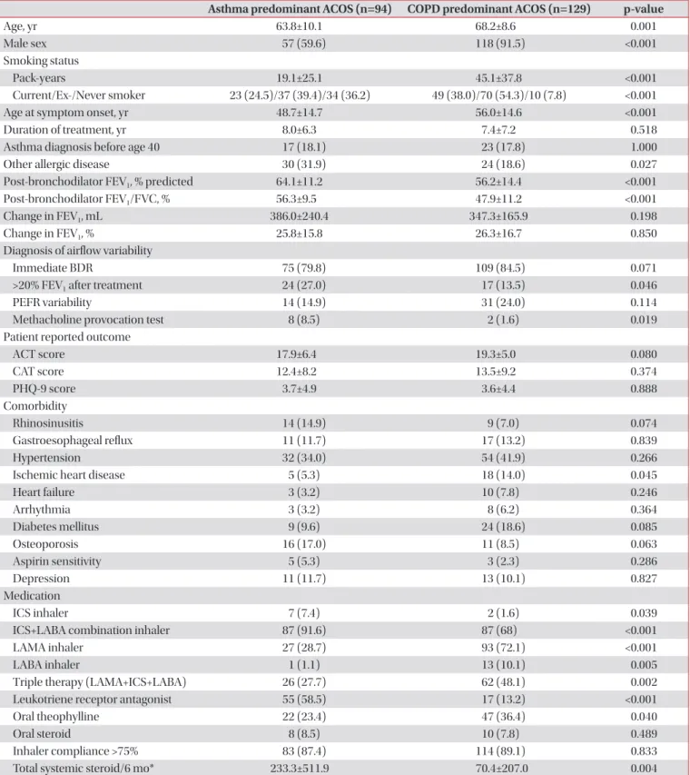
Table 2. Demographic and baseline characteristics of asthma and COPD phenotype groups
Asthma predominant ACOS (n=94) COPD predominant ACOS (n=129) p-value
Age, yr 63.8±10.1 68.2±8.6 0.001
Male sex 57 (59.6) 118 (91.5) <0.001
Smoking status
Pack-years 19.1±25.1 45.1±37.8 <0.001
Current/Ex-/Never smoker 23 (24.5)/37 (39.4)/34 (36.2) 49 (38.0)/70 (54.3)/10 (7.8) <0.001
Age at symptom onset, yr 48.7±14.7 56.0±14.6 <0.001
Duration of treatment, yr 8.0±6.3 7.4±7.2 0.518
Asthma diagnosis before age 40 17 (18.1) 23 (17.8) 1.000
Other allergic disease 30 (31.9) 24 (18.6) 0.027
Post-bronchodilator FEV1, % predicted 64.1±11.2 56.2±14.4 <0.001
Post-bronchodilator FEV1/FVC, % 56.3±9.5 47.9±11.2 <0.001
Change in FEV1, mL 386.0±240.4 347.3±165.9 0.198
Change in FEV1, % 25.8±15.8 26.3±16.7 0.850
Diagnosis of airflow variability
Immediate BDR 75 (79.8) 109 (84.5) 0.071
>20% FEV1 after treatment 24 (27.0) 17 (13.5) 0.046
PEFR variability 14 (14.9) 31 (24.0) 0.114
Methacholine provocation test 8 (8.5) 2 (1.6) 0.019
Patient reported outcome
ACT score 17.9±6.4 19.3±5.0 0.080
CAT score 12.4±8.2 13.5±9.2 0.374
PHQ-9 score 3.7±4.9 3.6±4.4 0.888
Comorbidity
Rhinosinusitis 14 (14.9) 9 (7.0) 0.074
Gastroesophageal reflux 11 (11.7) 17 (13.2) 0.839
Hypertension 32 (34.0) 54 (41.9) 0.266
Ischemic heart disease 5 (5.3) 18 (14.0) 0.045
Heart failure 3 (3.2) 10 (7.8) 0.246
Arrhythmia 3 (3.2) 8 (6.2) 0.364
Diabetes mellitus 9 (9.6) 24 (18.6) 0.085
Osteoporosis 16 (17.0) 11 (8.5) 0.063
Aspirin sensitivity 5 (5.3) 3 (2.3) 0.286
Depression 11 (11.7) 13 (10.1) 0.827
Medication
ICS inhaler 7 (7.4) 2 (1.6) 0.039
ICS+LABA combination inhaler 87 (91.6) 87 (68) <0.001
LAMA inhaler 27 (28.7) 93 (72.1) <0.001
LABA inhaler 1 (1.1) 13 (10.1) 0.005
Triple therapy (LAMA+ICS+LABA) 26 (27.7) 62 (48.1) 0.002
Leukotriene receptor antagonist 55 (58.5) 17 (13.2) <0.001
Oral theophylline 22 (23.4) 47 (36.4) 0.040
Oral steroid 8 (8.5) 10 (7.8) 0.489
Inhaler compliance >75% 83 (87.4) 114 (89.1) 0.833
Total systemic steroid/6 mo* 233.3±511.9 70.4±207.0 0.004
Values are presented as mean±SD or number (%) unless otherwise indicated.
*The amount of total systemic steroid used for the last 6 months is described as the equivalent dose of prednisolone.
COPD: chronic obstructive pulmonary disease; ACOS: asthma-COPD overlap syndrome; FEV1: forced expiratory volume in 1 second; FVC:
forced vital capacity; BDR: bronchodilator response; PEFR: peak expiratory flow rate; ACT: asthma control test; CAT: COPD Assessment Test;
PHQ-9: Patient Health Questionnaire; ICS: inhaled corticosteroid; LABA: long acting β2 agonist; LAMA: long acting muscarinic antagonist.
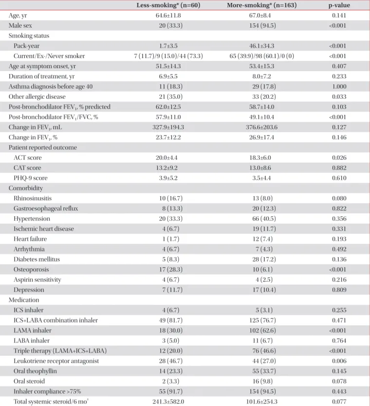
Table 3. Demographic and baseline characteristics of the less-smoking and the more-smoking groups
Less-smoking* (n=60) More-smoking* (n=163) p-value
Age, yr 64.6±11.8 67.0±8.4 0.141
Male sex 20 (33.3) 154 (94.5) <0.001
Smoking status
Pack-year 1.7±3.5 46.1±34.3 <0.001
Current/Ex-/Never smoker 7 (11.7)/9 (15.0)/44 (73.3) 65 (39.9)/98 (60.1)/0 (0) <0.001
Age at symptom onset, yr 51.5±14.3 53.4±15.3 0.407
Duration of treatment, yr 6.9±5.5 8.0±7.2 0.233
Asthma diagnosis before age 40 11 (18.3) 29 (17.8) 1.000
Other allergic disease 21 (35.0) 33 (20.2) 0.033
Post-bronchodilator FEV1, % predicted 62.0±12.5 58.7±14.0 0.103
Post-bronchodilator FEV1/FVC, % 57.9±11.0 49.1±10.4 <0.001
Change in FEV1, mL 327.9±194.3 376.6±203.6 0.127
Change in FEV1, % 23.7±12.2 26.9±17.4 0.146
Patient reported outcome
ACT score 20.0±4.4 18.3±6.0 0.026
CAT score 13.2±9.2 13.0±8.6 0.882
PHQ-9 score 3.9±5.2 3.5±4.4 0.610
Comorbidity
Rhinosinusitis 10 (16.7) 13 (8.0) 0.080
Gastroesophageal reflux 8 (13.3) 20 (12.3) 0.822
Hypertension 20 (33.3) 66 (40.5) 0.356
Ischemic heart disease 4 (6.7) 19 (11.7) 0.331
Heart failure 1 (1.7) 12 (7.4) 0.193
Arrhythmia 4 (6.7) 7 (4.3) 0.492
Diabetes mellitus 5 (8.3) 28 (17.2) 0.136
Osteoporosis 17 (28.3) 10 (6.1) <0.001
Aspirin sensitivity 4 (6.7) 4 (2.5) 0.216
Depression 7 (11.7) 17 (10.4) 0.809
Medication
ICS inhaler 4 (6.7) 5 (3.1) 0.255
ICS+LABA combination inhaler 49 (81.7) 125 (76.7) 0.471
LAMA inhaler 18 (30.0) 102 (62.6) <0.001
LABA inhaler 3 (5.0) 11 (6.7) 0.764
Triple therapy (LAMA+ICS+LABA) 12 (20.0) 76 (46.6) <0.001
Leukotriene receptor antagonist 28 (46.7) 44 (27.0) 0.006
Oral theophyllin 14 (23.3) 55 (33.7) 0.145
Oral steroid 2 (3.3) 16 (9.8) 0.078
Inhaler compliance >75% 55 (91.7) 154 (94.5) 0.443
Total systemic steroid/6 mo† 241.3±582.0 101.6±254.3 0.077
Values are presented as mean±SD or number (%) unless otherwise indicated.
*The less-smoking group represents the patients group with a smoking history of less than 10 pack-years and the more-smoking group repre- sents the patients group with a smoking history of equal or more than 10 pack-years. †The amount of total systemic steroid used for the last 6 months is described as the equivalent dose of prednisolone.
FEV1: forced expiratory volume in 1 second; FVC: forced vital capacity; ACT: Asthma Control Test; CAT: COPD Assessment Test; PHQ-9: Pa- tient Health Questionnaire; ICS: inhaled corticosteroid; LABA: long acting β2 agonist; LAMA: long acting muscarinic antagonist.
with asthma before 40 years of age did not differ significantly between the two phenotypes, nor did ACT, CAT, or PHQ-9 scores. Ischemic heart disease was more frequent in patients with the COPD predominant ACOS.
As expected, patients with the asthma predominant ACOS underwent more frequent treatment with inhaled cortico- steroids (ICS) or a combination of ICS+long-acting beta- agonists (LABA) or leukotriene receptor antagonists (LTRA), while patients with the COPD predominant ACOS were more commonly treated with long-acting muscarinic antagonists (LAMA), inhaled LABA, oral theophylline, or triple inhaler therapy (LAMA+ICS+LABA).
The total amount of systemic steroids prescribed over the previous 6 months was much higher in the asthma pre- dominant ACOS than in the COPD predominant ACOS (233.3±511.9 vs. 70.4±207.0 mg of an equivalent dose of pred- nisolone, respectively; p=0.004) (Table 2).
3. Comparisons of clinical characteristics between the light- and heavy-smoking groups
The heavy-smoking group was predominantly male and had a mean smoking history of 46 pack-years. The light-smok- ing group had more incidences of other allergic diseases and
showed a higher post-bronchodilator FEV
1/FVC percentage and ACT score. Osteoporosis was more common in the light- smoking group.
Patients in the light-smoking group were more frequently treated with LTRA, while patients in the heavy-smoking group were more frequently treated with LAMA or triple inhaler therapy (LAMA+ICS+LABA) (Table 3).
Approximately three-quarters (73.3%) of the patients in the light-smoking group were categorized as the asthma predomi- nant ACOS and 69.3% of those in the heavy-smoking group were categorized as the COPD predominant ACOS.
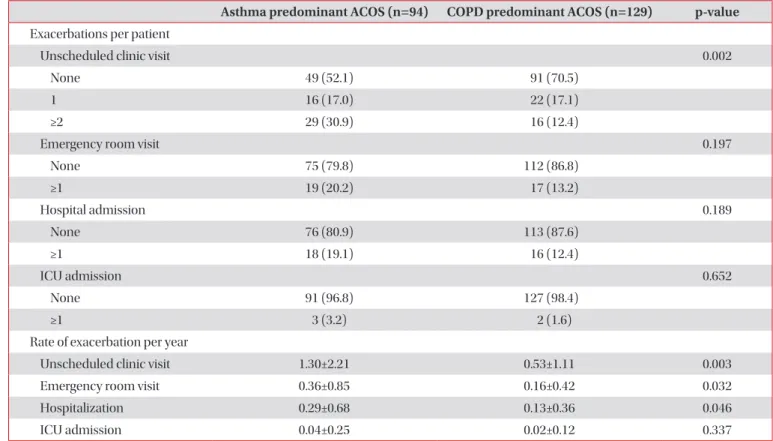
4. Comparison of exacerbation between the asthma and COPD predominant ACOS
During the previous year, 52.1% of patients with the asthma predominant ACOS and 70.5% of those with the COPD pre- dominant ACOS had no unscheduled clinic visits, while 30.9%
of the patients with the asthma predominant ACOS and 12.4%
of those with the COPD predominant ACOS had more than two unscheduled clinic visits (p=0.002). Additionally, patients with the asthma predominant ACOS had approximately twice the number of unscheduled outpatient clinic visits, emer- gency room visits, and hospitalizations during the previous
Table 4. Exacerbation history of asthma phenotype and COPD phenotype groups
Asthma predominant ACOS (n=94) COPD predominant ACOS (n=129) p-value Exacerbations per patient
Unscheduled clinic visit 0.002
None 49 (52.1) 91 (70.5)
1 16 (17.0) 22 (17.1)
≥2 29 (30.9) 16 (12.4)
Emergency room visit 0.197
None 75 (79.8) 112 (86.8)
≥1 19 (20.2) 17 (13.2)
Hospital admission 0.189
None 76 (80.9) 113 (87.6)
≥1 18 (19.1) 16 (12.4)
ICU admission 0.652
None 91 (96.8) 127 (98.4)
≥1 3 (3.2) 2 (1.6)
Rate of exacerbation per year
Unscheduled clinic visit 1.30±2.21 0.53±1.11 0.003
Emergency room visit 0.36±0.85 0.16±0.42 0.032
Hospitalization 0.29±0.68 0.13±0.36 0.046
ICU admission 0.04±0.25 0.02±0.12 0.337
Values are presented as number (%) or mean±SD.
COPD: chronic obstructive pulmonary disease; ACOS: asthma-COPD overlap syndrome; ICU: intensive care unit.
year compared with the COPD predominant ACOS group, with only the rate of ICU admission having no statistical sig- nificance (Table 4).
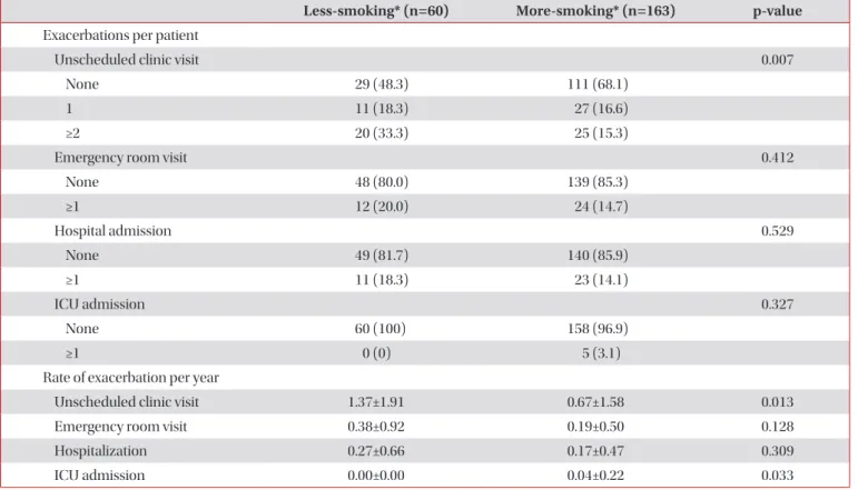
5. Comparison of exacerbation between the light- and heavy-smoking groups
The proportion of patients with more than two unscheduled
outpatient clinic visits, as well as the rate of unscheduled out- patient clinic visits during the previous year, was significantly higher in the light than heavy-smoking group. However, ICU admission was more common in the heavy-smoking group (Table 5).
Table 5. Exacerbation history of the less-smoking and the more-smoking groups
Less-smoking* (n=60) More-smoking* (n=163) p-value Exacerbations per patient
Unscheduled clinic visit 0.007
None 29 (48.3) 111 (68.1)
1 11 (18.3) 27 (16.6)
≥2 20 (33.3) 25 (15.3)
Emergency room visit 0.412
None 48 (80.0) 139 (85.3)
≥1 12 (20.0) 24 (14.7)
Hospital admission 0.529
None 49 (81.7) 140 (85.9)
≥1 11 (18.3) 23 (14.1)
ICU admission 0.327
None 60 (100) 158 (96.9)
≥1 0 (0) 5 (3.1)
Rate of exacerbation per year
Unscheduled clinic visit 1.37±1.91 0.67±1.58 0.013
Emergency room visit 0.38±0.92 0.19±0.50 0.128
Hospitalization 0.27±0.66 0.17±0.47 0.309
ICU admission 0.00±0.00 0.04±0.22 0.033
Values are presented as number (%) or mean±SD.
*The less-smoking group represents the patients group with a smoking history of less than 10 pack-years and the more-smoking group repre- sents the patients group with a smoking history of equal or more than 10 pack-years.
ICU: intensive care unit.
Table 6. Association of clinical parameters with exacerbation (≥2 unscheduled outpatient clinic visits last year)
Univariate Multivariate
Odds ratio (95% CI) p-value Odds ratio (95% CI) p-value
Age 0.911 (0.948–1.015) 0.939
Female sex 3.605 (1.773–7.332) <0.001 2.423 (0.929–6.315) 0.070
Less-smoking group (smoking <10 pack-years) 2.760 (1.386–5.490) 0.003 1.029 (0.390–2.715) 0.954 Post-bronchodilator FEV1, % predicted 0.972 (0.951–0.994) 0.158
Poor inhaler adherence (<75%) 6.198 (2.029–18.930) <0.001 4.321 (1.746–10.695) 0.002 ACOS phenotype (asthma phenotype) 3.151 (1.593–6.234) 0.001 2.433 (1.117–5.302) 0.025 CI: confidence interval; FEV1: forced expiratory volume in 1 second; ACOS: asthma‒chronic obstructive pulmonary disease overlap syndrome.
6. Parameters associated with more than two unscheduled clinic visits during the last year
On univariate analysis, female sex, light smoking, a poor in- haler compliance of <75%, and a diagnosis of the asthma pre- dominant ACOS were all significantly associated with exacer- bation (i.e., more than two unscheduled clinic visits). However, only poor inhaler compliance and the asthma predominant ACOS were found to be significant on multivariate analysis. In particular, the risk of exacerbation for patients with the asthma predominant ACOS was 2.4 times higher than that for patients with the COPD predominant ACOS. The post-bronchodilator FEV
1was not associated with exacerbation (Table 6).
Discussion
This study is meaningful to support the clinical heterogene- ity of patients with ACOS with increased airflow variability and incompletely reversible airway obstruction on spirom- etry.
The clinical characteristics of physician-diagnosed asthma and COPD predominant ACOS among patients with ACOS differed: patients with the asthma predominant ACOS were younger, more likely to be female, more commonly had never smoked, had a smoking history of fewer pack-years, and had better lung function. More importantly, patients with the asthma predominant ACOS of ACOS experienced more exac- erbations than did those with the COPD predominant ACOS.
Although some patients were clinically diagnosed with pure asthma or pure COPD rather than ACOS, they could be regarded as having asthma with fixed airflow limitation or COPD with airflow variability, respectively, and could there- fore have a distinct clinical phenotype of asthma or COPD.
We included these patients in the analysis because we were attempting to identify clinical heterogeneity among the group of patients who showed fixed airflow limitation with airflow variability.
Asthmatics who smoke frequently show fixed airway ob- struction and have more severe symptoms of asthma
16,17, an accelerated decline in lung function
6,18, an increased risk of frequent exacerbations
19, and an increased risk of death
20.
In this study, phenotyping using smoking history proved less definitive than the physician’s diagnosis. Interestingly, when patients with ACOS were divided according to smoking history, patients in the light-smoking group had more frequent unscheduled clinic visits despite having better lung function and higher ACT scores. The more frequent occurrence of exacerbation in the light-smoking group may be related to the larger number of patients with the asthma predominant ACOS in this group, because the light-smoking group was as- sociated with frequent exacerbation only on univariate analy- sis and not on multivariate analysis. However, smoking history
is easily obtainable from patients and would be a straightfor- ward guide for choosing the initial treatment, particularly in primary-care clinics when physicians are confronted with complex cases of ACOS.
Exacerbation of asthma and COPD is associated with poor health status and accelerated lung-function decline
21-23, and patients with ACOS experience exacerbations more frequent- ly than do those with either disease alone
11-13. A major target in managing patients with obstructive airway disease is reduc- tion of acute exacerbation. Therefore, it is important to know which patients with ACOS are susceptible to exacerbation.
In our patient population, other parameters related to frequent exacerbation in obstructive lung disease, such as poor lung function
24and female sex
25-27, were not significantly associated with exacerbation. However, poor inhaler compli- ance was strongly related to frequent exacerbation, as in other studies
28,29. In asthmatic patients in particular, inadequate use of ICS is the primary risk factor for frequent and fatal exacer- bation
30,31. However, in our study almost all patients with the asthma predominant ACOS used ICS, and inhaler compliance did not differ between the asthma and COPD predominant ACOS.
It is important to note that the asthma predominant ACOS, rather than the degree of airflow limitation, was related to fre- quent exacerbation. Airflow variability and symptom variabil- ity are the most important characteristics of asthma. Airway hyperresponsiveness in patients with COPD is also an inde- pendent predictor of exacerbation and mortality
32,33. Symp- tom variability is also associated with increased frequency of COPD exacerbation
34. Given the above, the variability of either airflow or symptoms may partly explain the high frequency of exacerbation in patients with ACOS.
This study has several limitations. First, we defined ACOS using only spirometry results. Although several criteria for the definition of ACOS exist, no definite ACOS criteria have been established, and most suggested criteria are relatively complex. In this situation, simplicity may offer the best solu- tion. Second, we used the criteria of a positive bronchodilator response of >12% and a 200-mL improvement in FEV
1after inhalation of a bronchodilator when we enrolled patients, although a higher positive bronchodilator response has been suggested by some recently published ACOS criteria
4,5. How- ever, due to the retrospective observational nature of this study, we could not control bronchodilator use during the bronchial reversibility test, and most of the hospitals used 200 g of salbu- tamol, a smaller dose than used in Western studies. Therefore, we considered that the criterion of an increase in FEV
1would be sufficient to observe a bronchodilator response. Third, the criteria for diagnosis used by our physicians appeared to be arbitrary. However, considering that each patient had been treated by their physician for several years, the physicians’
diagnosis based on the weight of evidence that the physicians
encountered in each patient was likely relatively accurate. In
the situation where neither biomarkers nor spirometry has any value in distinguishing between asthma and COPD pre- dominant ACOS, the physician’s diagnosis would be impor- tant. In addition, a physician’s diagnosis is a widely accepted diagnostic criterion in clinical studies of ACOS or asthma. It is also important to note that only one of 223 patients was diag- nosed with asthma=COPD ACOS. Lastly, we could not evalu- ate the relationship between the two phenotypes and blood eosinophil counts. In a recent study, phenotypes of ACOS, classified according to blood eosinophil counts and smoking history of the patients, showed difference in the proportion of patients free of severe exacerbation
35. It could be confounding bias.
In conclusion, we found that patients who met the spiromet- ric ACOS criteria could be divided into two clinically distinct groups: those with the asthma predominant ACOS and those with the COPD predominant ACOS. The most striking result was that the asthma predominant ACOS group showed more frequent exacerbation despite their history of light smoking and better lung function. Smoking history could, therefore, be another diagnostic criterion when differentiation between the asthma and COPD predominant ACOS proves difficult.
Future studies are needed to further our understanding of the heterogeneity of ACOS and to establish an appropriate thera- peutic intervention.
Authors' Contributions
Conceptualization: Park J, Lee JH. Methodology: Park J, Kim EK, Kim MA, Lee JH. Formal analysis: Park J, Lee JH. Data cu- ration: all authors. Investigation: all authors. Writing - original draft preparation: Park J, Lee JH. Writing - review and editing:
Park J, Lee JH. Approval of final manuscript: all authors.
Conflicts of Interest
No potential conflict of interest relevant to this article was reported.
References
1. Gibson PG, Simpson JL. The overlap syndrome of asthma and COPD: what are its features and how important is it?
Thorax 2009;64:728-35.
2. Zeki AA, Schivo M, Chan A, Albertson TE, Louie S. The asth- ma-COPD overlap syndrome: a common clinical problem in the elderly. J Allergy (Cairo) 2011;2011:861926.
3. Louie S, Zeki AA, Schivo M, Chan AL, Yoneda KY, Avdalovic M, et al. The asthma-chronic obstructive pulmonary disease overlap syndrome: pharmacotherapeutic considerations. Ex-
pert Rev Clin Pharmacol 2013;6:197-219.
4. Miravitlles M, Soler-Cataluna JJ, Calle M, Molina J, Almagro P, Quintano JA, et al. Spanish COPD Guidelines (GesEPOC):
pharmacological treatment of stable COPD. Spanish Society of Pulmonology and Thoracic Surgery. Arch Bronconeumol 2012;48:247-57.
5. Global Initiative for Asthma. Diagnosis of diseases of chronic airflow limitation: asthma, COPD and asthma-COPD overlap syndrome (ACOS), 2014 [Internet]. Bethesda: Global Initia- tive for Asthma; 2014 [cited 2014 Jul 27]. Available from:
http://www.ginasthma.org/documents/14.
6. Lange P, Parner J, Vestbo J, Schnohr P, Jensen G. A 15-year follow-up study of ventilatory function in adults with asthma.
N Engl J Med 1998;339:1194-200.
7. Kauppi P, Kupiainen H, Lindqvist A, Tammilehto L, Kilpe- lainen M, Kinnula VL, et al. Overlap syndrome of asthma and COPD predicts low quality of life. J Asthma 2011;48:279-85.
8. Hardin M, Silverman EK, Barr RG, Hansel NN, Schroeder JD, Make BJ, et al. The clinical features of the overlap between COPD and asthma. Respir Res 2011;12:127.
9. Menezes AM, Montes de Oca M, Perez-Padilla R, Nadeau G, Wehrmeister FC, Lopez-Varela MV, et al. Increased risk of exacerbation and hospitalization in subjects with an overlap phenotype: COPD-asthma. Chest 2014;145:297-304.
10. Sin DD. Asthma-COPD overlap syndrome: what we know and what we don’t. Tuberc Respir Dis 2017;80:11-20.
11. Rhee CK, Yoon HK, Yoo KH, Kim YS, Lee SW, Park YB, et al.
Medical utilization and cost in patients with overlap syn- drome of chronic obstructive pulmonary disease and asthma.
COPD 2014;11:163-70.
12. Shaya FT, Dongyi D, Akazawa MO, Blanchette CM, Wang J, Mapel DW, et al. Burden of concomitant asthma and COPD in a Medicaid population. Chest 2008;134:14-9.
13. Soriano JB, Visick GT, Muellerova H, Payvandi N, Hansell AL.
Patterns of comorbidities in newly diagnosed COPD and asthma in primary care. Chest 2005;128:2099-107.
14. Kim DK, Park YB, Oh YM, Jung KS, Yoo JH, Yoo KH, et al.
Korean asthma guideline 2014: summary of major updates to the Korean asthma guideline 2014. Tuberc Respir Dis 2016;79:111-20.
15. Yoon HK, Park YB, Rhee CK, Lee JH, Oh YM; Committee of the Korean COPD Guideline 2014. Summary of the chronic obstructive pulmonary disease clinical practice guideline revised in 2014 by the Korean Academy of Tuberculosis and Respiratory Disease. Tuberc Respir Dis 2017;80:230-40.
16. Siroux V, Pin I, Oryszczyn MP, Le Moual N, Kauffmann F. Re- lationships of active smoking to asthma and asthma severity in the EGEA study: epidemiological study on the genetics and environment of asthma. Eur Respir J 2000;15:470-7.
17. Polosa R, Russo C, Caponnetto P, Bertino G, Sarva M, Antic T, et al. Greater severity of new onset asthma in allergic sub- jects who smoke: a 10-year longitudinal study. Respir Res 2011;12:16.
18. James AL, Palmer LJ, Kicic E, Maxwell PS, Lagan SE, Ryan GF, et al. Decline in lung function in the Busselton Health Study:
the effects of asthma and cigarette smoking. Am J Respir Crit Care Med 2005;171:109-14.
19. Kupczyk M, ten Brinke A, Sterk PJ, Bel EH, Papi A, Chanez P, et al. Frequent exacerbators: a distinct phenotype of severe asthma. Clin Exp Allergy 2014;44:212-21.
20. Althuis MD, Sexton M, Prybylski D. Cigarette smoking and asthma symptom severity among adult asthmatics. J Asthma 1999;36:257-64.
21. Seemungal TA, Donaldson GC, Paul EA, Bestall JC, Jeffries DJ, Wedzicha JA. Effect of exacerbation on quality of life in patients with chronic obstructive pulmonary disease. Am J Respir Crit Care Med 1998;157(5 Pt 1):1418-22.
22. Quint JK, Baghai-Ravary R, Donaldson GC, Wedzicha JA. Re- lationship between depression and exacerbations in COPD.
Eur Respir J 2008;32:53-60.
23. Donaldson GC, Seemungal TA, Bhowmik A, Wedzicha JA.
Relationship between exacerbation frequency and lung func- tion decline in chronic obstructive pulmonary disease. Tho- rax 2002;57:847-52.
24. Garcia-Aymerich J, Serra Pons I, Mannino DM, Maas AK, Miller DP, Davis KJ. Lung function impairment, COPD hospi- talisations and subsequent mortality. Thorax 2011;66:585-90.
25. Singh AK, Cydulka RK, Stahmer SA, Woodruff PG, Camargo CA Jr. Sex differences among adults presenting to the emer- gency department with acute asthma. Multicenter Asthma Research Collaboration Investigators. Arch Intern Med 1999;159:1237-43.
26. Mullerova H, Shukla A, Hawkins A, Quint J. Risk factors for acute exacerbations of COPD in a primary care popula- tion: a retrospective observational cohort study. BMJ Open 2014;4:e006171.
27. Kilic H, Kokturk N, Sari G, Cakir M. Do females behave differ- ently in COPD exacerbation? Int J Chron Obstruct Pulmon Dis 2015;10:823-30.
28. Vestbo J, Anderson JA, Calverley PM, Celli B, Ferguson GT, Jenkins C, et al. Adherence to inhaled therapy, mortality and hospital admission in COPD. Thorax 2009;64:939-43.
29. Stern L, Berman J, Lumry W, Katz L, Wang L, Rosenblatt L, et al. Medication compliance and disease exacerbation in patients with asthma: a retrospective study of managed care data. Ann Allergy Asthma Immunol 2006;97:402-8.
30. O’Byrne PM, Pedersen S, Lamm CJ, Tan WC, Busse WW;
START Investigators Group. Severe exacerbations and de- cline in lung function in asthma. Am J Respir Crit Care Med 2009;179:19-24.
31. Hasegawa K, Bittner JC, Nonas SA, Stoll SJ, Watase T, Gabriel S, et al. Children and adults with frequent hospitalizations for asthma exacerbation, 2012-2013: a multicenter observational study. J Allergy Clin Immunol Pract 2015;3:751-8.e1.
32. Hospers JJ, Postma DS, Rijcken B, Weiss ST, Schouten JP.
Histamine airway hyper-responsiveness and mortality from chronic obstructive pulmonary disease: a cohort study. Lan- cet 2000;356:1313-7.
33. Scichilone N, Battaglia S, La Sala A, Bellia V. Clinical implica- tions of airway hyperresponsiveness in COPD. Int J Chron Obstruct Pulmon Dis 2006;1:49-60.
34. Kessler R, Partridge MR, Miravitlles M, Cazzola M, Vogelmei- er C, Leynaud D, et al. Symptom variability in patients with severe COPD: a pan-European cross-sectional study. Eur Respir J 2011;37:264-72.
35. Joo H, Han D, Lee JH, Rhee CK. Heterogeneity of asthma- COPD overlap syndrome. Int J Chron Obstruct Pulmon Dis 2017;12:697-703.