• 검색 결과가 없습니다.

Fat Embolism Syndrome - Three Case Reports and Review of the Literature

N/A
N/A
Protected

Academic year: 2021

Share "Fat Embolism Syndrome - Three Case Reports and Review of the Literature"

Copied!
5
0
0

로드 중.... (전체 텍스트 보기)

전체 글

(1)

Received: June 15, 2017 Revised: August 31, 2017 Accepted: September 7, 2017 TRAUMA AND INJURY

Correspondence to

Leonidas Grigorakos, M.D., Ph.D.

Faculty of Nursing, National and Kapodistri- an University of Athens, 2 Nikis Str., Kifissia, Athens 14561, Greece

Tel: +30-210-3709522 Fax: +30-210-3709520 E-mail: [email protected]

Fat Embolism Syndrome – Three Case Reports and Review of the Literature

Leonidas Grigorakos, M.D., Ph.D.

1,2

, Ioannis Nikolopoulos, M.D.

3

,

Stamatina Stratouli, M.D.

2

, Anastasia Alexopoulou, M.D.

2

, Eleftherios Nikolaidis, M.D.

2

, Eleftherios Fotiou, M.D.

2

, Daria Lazarescu, Ph.D.

2

, Ioannis Alamanos, M.D.

2

1

Faculty of Nursing, National and Kapodistrian University of Athens, Athens, Greece

2

Intensive Care at Trauma, Hospital of Athens, KAT, Kifissia, Athens, Greece

3

Center of Respiratory Insufficiency, Sotiria Chest Hospital, Athens, Greece

The fat embolism syndrome (FES) represents a condition, usually with traumatic etiology, which may pose challenges to diagnosis while its treatment usually requires supportive measures in the intensive care units (ICUs). The clinical criteria, including respiratory and cerebral dysfunction and a petechial rash, along with imaging studies help in diagnosis. Here we present three case reports of young male who developed FES and were admitted to our ICUs after long bones fractures emerging after vehi- cle crashes and we briefly review FES literature. All patients' treatment was directed towards: 1) the restoration of circulating volume with fresh blood and/or plasma; 2) the correction of acidosis; and 3) immobilization of the affected part. All patients re- covered and were released to the orthopedic wards. The incidence of cases of patients with FES admitted in our ICUs records a significant decrease. This may be explained in terms effective infrastructure reforms in Greece which brought about significant improvement in early prevention and management.

Keywords: Embolism, Fat; Intensive Care Units; Long bone fractures; Fat embolism prevention; Fat embolism management

INTRODUCTION

Since it was first described, more than 150 years ago, the fat embolism syndrome (FES)

has been considered a diagnostic enigma, which still poses challenges to diagnosis. This

difficulty is based on the fact that it can complicate an array of clinical presentation

with a variable severity of illness. Although simple fat embolism may be a pathologic

finding with little clinical significance, patients with FES have fat emboli in multiple

organs, which show extensive damage from this embolization [1]. While most patients

(2)

with FES fully recover, there is still an estimated 5 to 20%

mortality risk [2] while care is generally supportive.

We report three cases of young males with FES in the setting of traumatic long bones fractures. Although rare, FES is more common at level I trauma centers, where polytrauma patients are often transferred for specialized care. Early diagnosis, high-pressure positive end-expira- tory pressure, and supportive treatment are the mainstays of treatment [3]. Major and minor diagnostic criteria for FES were proposed by Gurd and Wilson (Table 1).

Using their system, a diagnosis of FES could be made if one major feature, four minor features, and fat macro- globulinemia were present [4] Schonfeld proposed the fat embolism index to aid in diagnosing FES (Table 2) [5].

A cumulative score of five or more over the first three days of hospitalization corresponds with a diagnosis of FES. However, given the complex nature of polytrauma patients, it is often difficult to accurately diagnose FES as these patients have multiple injuries and are often intu- bated upon arrival. Here, we present three case reports of young male who developed FES after long bones fractures emerging after vehicle crashes.

CASE REPORT

We present the cases of three patients who were admitted to our intensive care units (ICUs) three to five days upon their previous admissions to orthopedic clinics after ve-

hicle crashes. Their duration of stay before release to the orthopedic clinics ranges from 5 to 17 days (mean average 10 days). All patients were male and their age ranged from twenty to twenty-four years. All cases followed multiple fractures with one or more long bones involved (Table 3).

In all cases there was a latent period between injury and the onset of symptoms with an average time of seven- ty-two hours after surgery for remanipulation of fractures.

No significant correlation was found between the time of onset and the severity of the subsequent course. There was marked variation in the clinical presentation (Table 4).

In one case the earliest recorded symptoms were cerebral, usually drowsiness or confusion. In two cases unexplained tachycardia and pyrexia heralded more specific signs.

However, respiratory dysfunction was observed first in all patients with dyspnoea, tachypnoea and/or haemoptysis.

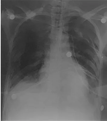
One patient was intubated in the emergency department due to respiratory insufficiency - shunt FiO

2

100%, Sat O

2

78-82%, tachypnea up to 40 breaths/min, tachycardia 120-140 beats/min. Before intubation the patient main-

Table 1. Gurd and Wilson’s major and minor criteria for fat embolism

Major features Minor features

Petechial rash Tachycardia

Respiratory symptoms plus bilat- eral signs with positive radio- graphic changes

Pyrexia

Cerebral signs unrelated to head injury

Retinal fat or petechiae

Urinary fat globules or oligoanuria Sudden drop in Hg-level Sudden thrombocytopenia High erythrocyte sedimentation

rate

Fat globules in sputum

Table 2. Schonfeld’s fat embolism index (FEI) score

Feature Points

a

Diffuse petechiae 5

Alveolar infiltrates 4

Hypoxemia (<70 mm Hg) 3

Confusion 1

Fever >38℃ 1

Heart rate >120 beats/min 1

Respiratory rate >30/min 1

a

Five or more points are needed to diagnose FES. FES: embolism syn- drome

Table 3. Nature of the injury

Patient 1 Patient 2 Patient 3

Segmental right femur fracture

Left femur fracture

Right femur fracture Fracture of right femur left

condyle

Extra articular wirst fracture Mandibule fracture Open rupturing trauma of

chin and lower lip

(3)

tained good level of communication. Petechial rash was a presenting sign in all our cases.

Upon admission in our ICUs, respiratory involvement was predominant in all three patients. Two of them pre- sented hypoxemia, diffuse pulmonary infiltrates, mainly median type, of middle and low pulmonary areas, bi- lateral ground glass. One patient presented hypocapnia, dyspnoea, tachypnoea and respiratory failure. Cyanosis was uncommon even when arterial hypoxia was marked, presumably because of concomitant anaemia. The arterial oxygen tension was monitored in two cases and the mini- mum PO

2

level recorded ranged from 51 to 80.

All patients had radiographic examination of the chest and the films showed typical bilateral diffuse patchy areas of consolidation (Fig. 1). In one case veins’ triplex was performed and it revealed pathological findings (bilateral fine non-muscular sprains of pulmonary bases). In other case, chest computed tomography (CT) showed a bilateral ground-glass image while limb and cardiac ultrasound ex- amination did not reveal any pathological findings. There was mild cerebral involvement in one patient associated with head injury. One patient was fully orientated on ad- mission, while the other two became confused. Petechial rash were observed in all patients.

Pyrexia of 38.5 degrees Celsius or above and tachycar- dia of 120 per minute or more were noted in two cases.

Ophthalmoscopy was recorded in two patients and was normal. Some renal involvement was manifested in two patients, as they became oliguric. Daily haemoglobin es- timations were recorded and a drop of more than 25%

was found in all patients. Daily platelet counts were also monitored with a recorded drop of 50% in only one pa- tient. The erythrocyte sedimentation rate was raised, with values of 30 to 50 mm. Fat globules larger than 8 microns were found circulating in all cases, which resulted in me- chanical obstruction of lung capillaries. The amount of circulating fat did not appear to correlate with the clinical severity of the condition. Fat globules were also found in the urine on all three cases.

In all cases, treatment was directed towards: 1) the restoration of circulating volume with fresh blood and/

or plasma; 2) the correction of acidosis; and 3) immo- bilization of the affected part. Additional treatment was primarily concerned with respiratory support. All patients required full respiratory care with assisted ventilation and oxygen while one patient who remained under suppres- sion and analgesia for 7 days received all the routine care of the unconscious patient with Pentothal, Remifentanil Table 4. Patients’ recorded symptoms and complications

Patient 1 Patient 2 Patient 3

Drowsiness and confusion Confusion Fully orientated

Tacycardia and pyrexia - Tachycardia and pyrexia

Respiratory dysfunction (dyspnoea, tachypnoea, haemoptysis)

Respiratory dysfunction (dyspnoea, tachypnoea, haemoptysis)

Respiratory dysfunction (dyspnoea, tachypnoea, haemoptysis)

Petechial rash Petechial rash Petechial rash

Lung typical bilateral diffuse patchy areas of consolidation

Lung typical bilateral diffuse patchy areas of con- solidation

Lung typical bilateral diffuse patchy areas of con- solidation

Hypocapnia and respiratory failure Hypoxemia Hypoxemia

Intubation - -

Pathological CT findings suggesting mild cere- bral involvement (bilateral grand glass image)

- -

Fat globules >8 microns present, resulting in result in mechanical obstruction of lung capil- laries

Fat globules >8 microns present, resulting in result in mechanical obstruction of lung capillaries

Fat globules >8 microns present, resulting in result in mechanical obstruction of lung capil- laries

Fat globules in the urine Fat globules in the urine Fat globules in the urine

Oliguria Oliguria -

CT: computed tomography.

(4)

and Midazolam.

One patient was transfused with red blood cells and fresh frozen plasma due to low hematocrit. Antibiotics were given to two patients with moderate or severe lung involvement. Esmolol was required in one case with un- controllable tachycardia. Two patients were given Lasix or Furosemide for diuresis while one patient was given hydrocortisone sodium succinate. All patients recovered and were released to the orthopedic wards.

DISCUSSION

FES, which was first described by Zenker in 1861, is com- monly associated with long bone fractures and frequently presents a constellation of neurological, pulmonary, dermatological, and hematological symptoms. Actually, cerebral complications are considered to be the most se- vere complications of fat embolism [5]. In our case, the petechial rash was a presenting sign in all our cases which proved to be determinant factor for early diagnosis of FES.

Since its first description, two main theories were pro-

posed to explain the origin of FES: the mechanical and biochemical theories. The former states that three condi- tions are necessary for the development of fat embolism:

injury to adipose tissue, rupture of veins within the zone of injury, and a mechanism that causes the passage of free fat into the open ends of blood vessels. The latter theory argues that plasma mediators mobilize fat from body stores and cause the coalescence of larger droplets.

The presence of fat within various tissues (i.e., lungs and brain) initiates an inflammatory cascade causing injury [6]. However, these mechanisms are not necessarily mu- tually exclusive The former presumes that fractures of marrow containing bone have the highest incidence of FES due to disrupted venules in the marrow which are tethered open by osteous attachments that allows easy entry of marrow contents into the circulation [7]. From there on, it can enter the arterial circulation via a patent foramen ovale (PFO) causing increases in pulmonary artery and right heart pressures or by micro embolism via the lung demonstrated even in the absence of a PFO;

the latter could explain the neurologic disease and pe- techiae associated with FES [8]. The biochemical theory or mechanism explains that destabilization of a plasma chylomicronemulsion lead stocoalescence off at stores.

Subsequently, degradation of embolized fat into toxic intermediaries (free fatty acids and Creactive protein) re- sults in the development of FES [9].

The incidence of FES ranges from <1 to 29% and it varies considerably according to the cause. The actual in- cidence of FES is not known, as mild cases often go unno- ticed. In patients with long bone fractures FES incidence has been reported to occur in 0.5 to 11% [10]. However, it is seen more frequently in closed fractures than open frac- tures and risk increases in proportion with the number of bone structures involved [11].

The early fixation of long-bone fracture (early immo- bilization of fractures and operative correction rather than conservative management) is important to prevent or to decrease the severity of FES. One report estimates a 70% reduction in pulmonary complications from this intervention alone [12]. Further, two surgical techniques are debated as possible means of preventing fat embolism syndrome. However, these two techniques are considered but not used routinely by surgeons. The first is “venting,”

Fig. 1. Patient 2 - chest X-ray.

(5)

in which a hole is made distal to the site of intramedul- lary nail placement. This reduces intramedullary pressure elevation and, therefore, extravasation of fat into the circulation. The second technique is the use of a reamer, irrigator, aspirator (RIA) device. Reaming before intra- medullary nail placement can release fat deposits into the circulation. The RIA device irrigates and aspirates resident fat deposits as it reams the canal, releasing fewer deposits into the circulation.

Preoperative use of methylprednisolone may prevent the occurrence of FES [13]. The use of corticosteroid prophylaxis is controversial and few studies have shown decrease in the incidence and severity of FES. However, the administration to one of the three patients of hydro- cortisone sodium succinate (125 gr/6 h for three days) proved to be effective for his treatment. Smaller-diameter nails and unreamed nailing have been mentioned as being useful in the prevention of FES. Both in polytraumatism patients and in those being submitted to surgery, it is cru- cial to avoid hypovolemia and hypoxia, because these are factors that much worsen a FES prognosis. Thus, in both situations, the close monitoring of the blood pressure and the PaO

2

is recommended, as well as correcting their de- viations as soon as they are detected.

To conclude, we have to stress out that during the last years, in Greece, we record a serious decrease not only in mortality rates of patients with FES but also in the cases of patients with FES admitted in our ICUs, when compared to the same units 15-20 years ago [14]. This may be ex- plained in terms of infrastructure reforms which induced:

1) a significant reduction of road accidents, 2) the mod- ernization of National Emergency Center which led to a more rapid treatment of injuries, and 3) an increase in the number of ICU beds and surgical treatment [15]. All these developments brought about more effective early prevention and management of patients in the emergency departments and in the orthopedic clinics. However, in order to strengthen such a statement future research is needed.

REFERENCES

1. Gregorakos L, Dimopoulos G, Mavropanou D, Antypas G. Fat embolism syndrome (FES) in traumatized patients: a five year experience of a single intensive care unit. Appl Cardiopulm Pathophysiol 1996;6:105-9.

2. Tablot M, Schemitsch EH. Fat embolism syndrome: history, definition, epidemiology. Injury 2006;37(4 Suppl):S3-7.

3. Abbott MG. Fat embolism syndrome: An in-depth review. Asian J Crit Care 2005;1:19-24.

4. Gurd AR, Wilson RI. Fat embolism: an aid to diagnosis. J Bone Joint Surg Br 1970;56:732-7.

5. Gregorakos L, Sakayianni K, Hroni D, Harizopoulou V, Markou N, Georgiadou F, et al. Prolonged coma due to cerebral fat em- bolism: report of two cases. J Accid Emerg Med 2000;17:144-6.

6. Lehman EP, Moore RM. Fat embolism: including experimental production without trauma. Arch Surg 1927;14:621-62.

7. Warthin AS. Traumatic lipaemia and fatty embolism. Int Clinics 1913;23:171-227.

8. Kara AV, Yildirim Y, Yilmaz S, Yilmaz Z, Kadiroglu AK, Yilmaz AE. Unexpected neurological sequelae following cerebral fat embolism syndrome. J Case Rep 2015;5:207-11.

9. Kosova E, Bergmark B, Piazza G. Fat embolism syndrome. Cir- culation 2015;131:317-20.

10. Peltier L. Fat embolism. A perspective. Clin Orthop Relat Res 2004;422:148-53

11. O’Brien, PJ. Fracture fixation in patients having multiple inju- ries. Can J Surg 2003;46:124-8.

12. Robinson CM. Current concepts of respiratory insufficiency syndromes after fracture. J Bone Joint Surg Br 2001;83:781-91.

13. Sen RK, Prakash S, Tripathy SK, Aqarwal A, Sen IM. Inhalation- al Ciclesonide found beneficial in prevention of fat embolism syndrome and improvement of hypoxia in isolated skeletal trauma victims. Eur J Trauma Emerg Surg 2017;43:313-8.

14. Grigorakos L, Gakis E, Athanasopoulou A, Antonopoulos A.

Fat embolism and the intensive care unit. In: Grigorakos L.

(Textbook). Intensive care unit. Respiratory insufficiency and mechanical ventilation. before-during-after. Athens: myedition.

gr; 2014:529-38.

15. Alexias G. Reason for life and death. Medical practice as a form

of social interaction in intensive care units. Athens: Ellinika

Grammata; 2000.

수치

Table 1. Gurd and Wilson’s major and minor criteria for fat  embolism
Fig. 1. Patient 2 - chest X-ray.

참조

관련 문서

Factors affecting post-traumatic stress of general hospital nurses after the epidemic of Middle East respiratory syndrome infection: Journal of Korean Clinical

A 27-year-old man with spondyloarthropathy, oblique coronal fat-saturated T2-weighted (A) and oblique coronal postcontrast fat-saturated T1-weighted (B) images show

2) In between-group comparison before and after elastic band exercise, a significant difference was found in the percentage of body fat, fat-free

Serum uric acid levels and risk for vascular disease in patients with metabolic syndrome... Prevalence if the metabolic syndrome in a Turkish

Connections of wind power system usually occur to voltage drop due to reactive power absorption and sometime result in higher than nominal voltages which may

In this study, we investigated the effects of a HF+HS diet in DMM-induced OA mice and found that compared with a HF diet, a HF+HS diet promoted OA. Moreover, our results

This study’s TCHP group lost both muscle and fat, whereas the AC-T group had no signif- icant change in muscle but gained fat, which suggests that the type of chemotherapy

Over the following 3 months, the patient developed elevated alpha-fetoprotein, and positron emission tomography showed a cystic mass in the right side of the pelvic cavity