DOI 10.17480/psk.2021.65.1.23
종 설(Review)
Standardization and validation of molecular size distribution test for human immunoglobulin products in Korea
Chan Woong Choi*, Ka Young Kim*, Ho-Jin Song*, Kiwon Han*, Seungwan Jee**, and Jaeok Kim**
,#*Blood Products Division, National Institute of Food and Drug Safety Evaluation, Ministry of Food and Drug Safety
**Biologics Division, National Institute of Food and Drug Safety Evaluation, Ministry of Food and Drug Safety
(Received December 1, 2020; Revised January 22, 2021; Accepted February 7, 2021)
Abstract In this study, we aimed to harmonize and compare the molecular size distribution test in Korean Minimum Requirements for Biological Products (KMRBP) and the European Pharmacopeia (Ph. Eur.). Size-exclusion high- performance liquid chromatography methods in the KMRBP and Ph. Eur. were compared using a human immunoglobulin reference standard. The operating conditions were standardized and the method was validated. The validated and current KMRBP methods were used to analyze seven types of human immunoglobulin products approved in Korea. The coefficient of variation (CV) level was set to ≤1% for monomer/dimer and ≤10% for polymers/aggregates, indicating no difference between the results. The peak areas of monomer/dimer and polymers/aggregates showed no difference when different operating conditions, including the mobile phase, sample dilution buffer, and flow rate, were used to conduct the procedures listed in the KMRBP and Ph. Eur. However, the CV for polymers/aggregates was determined to be more than 10 % when columns of different sizes were used. In the validation studies, the CV for peak areas of monomer/dimer and polymers/aggregates were determined to be ≤1 and ≤10%, respectively. The results indicated that both methods were suitable as per the specifications of the Ph. Eur. and KMRBP. Quality control of human immunoglobulin products is crucial to ensure their safety and efficacy. The present study provides a scientific basis for the new national lot release test of molecular size distribution for human immunoglobulin products adopted by the KMRBP.
Keywords Korean minimum requirements for biological products, molecular size distribution, human immunoglobulin, plasma-derived product, validation, quality control
Introduction
Plasma-derived products are pharmaceuticals manufactured through a series of processes using human plasma as a starting material. Of these, the human immunoglobulin product has human immunoglobulin as an active ingredient obtained by the fractionation of human plasma.
1)Human immunoglobulin products are essential medicines designated by the World Health Organization. They are among the most widely used therapeutics globally and are in high demand.
2)In Korea, human immuno- globulin products are approved for the treatment of numerous diseases, such as hypogammaglobulinemia, agammaglobulinemia, viral diseases, and Guillain-Barré syndrome. According to the Korean Minimum Requirements for Biological Products (KMRBP),
human immunoglobulin products are classified as biologics. The manufacturing consistency, quality, and safety of biologics, such as plasma-derived products and vaccines, is ensured via the national lot release implemented by the National Regulatory Agency (NRA).
3)Molecular size distribution test of human immunoglobulin products in the national lot release is a safety test for the quantification of unwanted polymers/aggregates that may be present in the final product. In the NRAs of Korea, the European Union, Japan, and China, size-exclusion chromatography is used as a molecular size distribution test, based on the official compendium.
4,5,6,7)The analytical procedures of molecular size distribution tests for human immunoglobulin products are similar in the KMRBP and European Pharmacopeia (Ph. Eur.); however, there are differences in some operating conditions, such as the mobile phase, sample dilution buffer, flow rate, and column size.
In addition, the Ph. Eur. has specifications regarding the distribution of monomer/dimer and polymers/aggregates, whereas the KMRBP has only specifications about the distribution of polymers/aggregates.
4,5)At present, standardization and international harmonization of specifications and analytical procedures are
#
Corresponding author
Jaeok Kim, Biologics Division, National Institute of Food and Drug Safety Evaluation, Ministry of Food and Drug Safety, Cheongju, Korea
Tel: +82-43-719-3461, Fax: +82-43-719-3450
E-mail: [email protected]
actively pursued for pharmaceuticals, especially biologics. The National Institute of Food and Drug Safety Evaluation (NIFDS) is an NRA in Korea responsible for the international harmonization of specifications, analytical procedures, and national lot release related to biologics. In addition, as the production and export of human immunoglobulin products manufactured in Korea have been increasing recently, it is necessary to improve the molecular size distribution test for human immunoglobulin products in Korea in terms of international harmonization of quality control.
8)In this study, the molecular size distribution test method for human immunoglobulin products was standardized and validated, and the human immunoglobulin products distributed in Korea were assessed using both the validated and current KMRBP methods.
Methods
Reagents and columns
All chemicals used were of liquid-chromatography grade. Sodium phosphate dibasic (Na
2HPO
4), sodium phosphate monobasic dihydrate (NaH
2PO
4·2H
2O), disodium hydrogen phosphate dihydrate (Na
2HPO
4·2H
2O), sodium dihydrogen phosphate monohydrate (NaH
2PO
4·H
2O), sodium chloride (NaCl), and sodium azide (NaN
3) were purchased from Sigma-Aldrich (MO, USA). Solutions were prepared in high-purity triple distilled water.
TSKgel G3000SW (7.5×600 mm, 10 µm) and TSKgel G3000SWXL (7.8×300 mm, 5 µm) columns were purchased from TOSOH (Tokyo, Japan).
Reference standard and samples
Human immunoglobulin (molecular size) biological reference preparation (BRP) batch 2 was used as a reference standard for the standardization and validation of the method. Seven types of human immunoglobulin products approved and distributed in Korea were purchased, and the sample numbers for each type of product were allocated as follows: Sample 1 for human normal immunoglobulin in glycine; Sample 2 for human normal
immunoglobulin in maltose; Sample 3 for human hepatitis B immunoglobulin for intravenous administration; Sample 4 for human normal immunoglobulin; Sample 5 for human tetanus immunoglobulin; Sample 6 for human hepatitis B immuno- globulin; and Sample 7 for human varicella immunoglobulin.
Molecular size distribution test
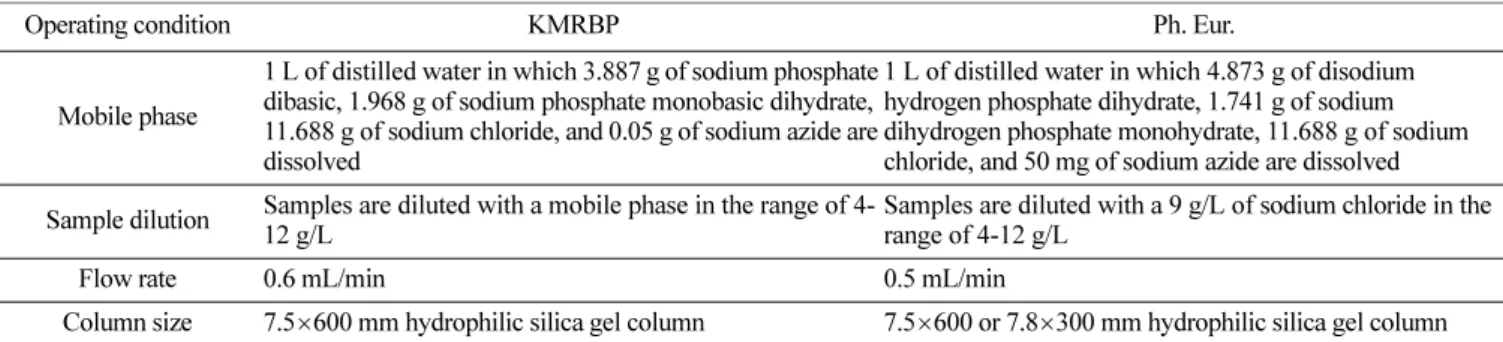
Size-exclusion high-performance liquid chromatography (SE- HPLC) analysis was performed using a hydrophilic silica gel column connected to an Alliance e2695 HPLC system, 2998 Photodiode Array detector, and Empower 3 data management software (Waters, MA, USA). The operating conditions were different between the KMRBP and Ph. Eur., as shown in Table 1.
Other conditions, including the room temperature for the column oven, 5
oC for autosampler, 20 µL for injection volume, 60 min for run time, and 280 nm for UV wavelength, were identical. The mobile phase and sample dilution buffer were filtered before use through a 0.22-µm filter (Corning, NY, USA).
Standardization of operating conditions
The effects of different operating conditions between KMRBP and Ph. Eur., including the mobile phase, sample dilution buffer, flow rate, and column size, on the results were investigated. For this purpose, human immunoglobulin (molecular size) BRP was analyzed with only one operating condition being different, based on the method used for KMRBP. In addition, Samples 4, 5, 6, and 7 were analyzed to compare the results of different column sizes. Taking these comparison results into consideration, we determined the operating conditions to be used, with an aim of improving the method.
Validation of the method
Specificity, injection repeatability, intermediate precision, robustness, and reproducibility were examined to validate the standardized method. Specificity was observed for the mobile phase and 9 g/L sodium chloride to determine whether other components were detected at the retention time of the human immunoglobulin
Table 1. Different operating conditions between the KMRBP and Ph. Eur
Operating condition KMRBP Ph. Eur.
Mobile phase
1 L of distilled water in which 3.887 g of sodium phosphate dibasic, 1.968 g of sodium phosphate monobasic dihydrate, 11.688 g of sodium chloride, and 0.05 g of sodium azide are dissolved
1 L of distilled water in which 4.873 g of disodium hydrogen phosphate dihydrate, 1.741 g of sodium dihydrogen phosphate monohydrate, 11.688 g of sodium chloride, and 50 mg of sodium azide are dissolved Sample dilution Samples are diluted with a mobile phase in the range of 4-
12 g/L
Samples are diluted with a 9 g/L of sodium chloride in the range of 4-12 g/L
Flow rate 0.6 mL/min 0.5 mL/min
Column size 7.5 ×600 mm hydrophilic silica gel column 7.5 ×600 or 7.8×300 mm hydrophilic silica gel column
component in the reference standard. Injection repeatability was measured using 10 repeated analyses of the reference standard.
Intermediate precision was evaluated for multiple days, analysts, and instruments to identify differences in each characteristic.
Robustness is a measure of the capability of the method to remain unaffected by small but deliberate variations in method parameters.
9)The method was validated for robustness using columns with different batch numbers. Reproducibility was assessed for an inter- laboratory study and should be considered in the case of standardization of a method for inclusion in an official compendium.
9)In the present study, the reference standard was analyzed in four laboratories using the standardized method to assess the differences in the results of samples tested among laboratories.
Application of the validated method for analysis of human immunoglobulin products
The validated method was used to analyze seven types of human immunoglobulin products approved and distributed in Korea in terms of the levels of monomer, dimer, and polymers/
aggregates in the final products. Human immunoglobulin (molecular size) BRP was used as a reference standard for the test. The results obtained by using the validated method were compared with those obtained using the current KMRBP method.
Statistical analysis
Data are representative of three injections, excluding the data for injection repeatability. The results were analyzed statistically and the mean, standard deviation (SD), and coefficient of variation (CV) were determined using Microsoft Excel 2016 (Microsoft, WA, USA). In the standardization of operating conditions and validation of the method, the acceptance level of CV was set as ≤1% for monomer/dimer and ≤10% for polymers/
aggregates.
Results
Standardization of operating conditions for molecular size distribution test
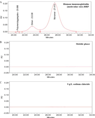
The SE-HPLC chromatogram of human immunoglobulin products shows a principal peak corresponding to the monomer and a peak corresponding to the dimer just to the left. Any peak with a retention time less than that of the dimer corresponds to polymers/aggregates (Fig. 1A). As shown in Table 2, a molecular size distribution test was conducted using different operating conditions stated in the KMRBP and Ph. Eur., including the mobile phase, sample dilution buffer, and flow rate. The CV values for the peak areas of the monomer/dimer and polymers/
aggregates were 0.01-0.06 and 3.57-9.32%, respectively. However, the peak areas of polymers/aggregates obtained using the 7.5×
600 mm column were higher than those obtained using the 7.8×
300 mm column, with a CV of 9.17-34.31% (Table 2). These results suggested that the column with dimensions of 7.5×600 mm was more sensitive in separating the polymers/aggregates than that having the dimensions of 7.8×300 mm. Based on these results, the following operating conditions were determined for the molecular size distribution test: mobile phase, 1 L of distilled water containing 4.873 g of disodium hydrogen phosphate dihydrate, 1.741 g of sodium dihydrogen phosphate monohydrate, 11.688 g of sodium chloride, and 50 mg of sodium azide; sample dilution buffer, 9 g/L of sodium chloride; flow rate, 0.5 mL/min; and column, 7.5×600 mm.
Validation of the standardized molecular size distribution method
Specificity
The chromatogram of the mobile phase and sample dilution buffer showed no peak in the retention time corresponding to the monomer, dimer, and polymers/aggregates for human immuno-
Fig. 1. SE-HPLC chromatogram of (A) human immunoglobulin
(molecular size) BRP, (B) mobile phase, and (C) 9 g/L sodium
chloride.
globulin (Fig. 1B and 1C). These results suggest that the method has an appropriate selectivity for the separation of human immunoglobulin molecules, without any interference from the mobile phase and sample dilution buffer.
Injection repeatability
Injection repeatability measured using multiple injections of a homogeneous sample indicates the performance of the HPLC instrument under the same chromatographic conditions and day tested.
10)The peak areas of monomer/dimer and polymers/
aggregates were 99.699±0.012 and 0.102±0.010%, respectively (Table 3). The CV for the peak areas of the monomer/dimer and polymers/aggregates were 0.01 and 9.34%, respectively.
Intermediate precision
The precision of a method indicates the closeness of agreement between a series of measurements obtained from multiple sampling of the same homogeneous sample under the stated conditions.
9)Intermediate precision data, including different days, analysts, and instruments, are shown in Table 3. The CV for the peak areas of monomer/dimer and polymers/aggregates were 0.00- 0.01 and 2.87-4.28%, respectively, indicating good intermediate precision within those characteristics.
Robustness
The standardized molecular size distribution method was further validated for robustness in terms of different column batch numbers (Table 3). The CV for the peak areas of monomer/dimer and polymers/aggregates were 0.00 and 3.41 %, respectively, suggesting that the method was not robust to changes in the column batch number.
Reproducibility
Four laboratories, including a national control laboratory and domestic manufacturers of human immunoglobulin products, participated in this reproducibility study. These laboratories were referred to as their randomly allocated code number, namely Laboratories 1 to 4 (Table 3). Reproducibility was very satisfactory with CV for the peak areas of monomer/dimer and polymers/aggregates of 0.27 and 7.02%, respectively.
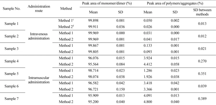
Comparison of molecular size distribution results between the validated and current KMRBP methods
The peak areas of monomer/dimer and polymers/aggregates were 99.805-99.969 and 0.026-0.133%, respectively, in the products intended for intravenous administration (Table 4). The peak areas of monomer/dimer and polymers/aggregates were Table 2. Comparison of the results according to the change of operating conditions
Operating condition Sample Method Peak area of monomer/dimer (%) Peak area of polymers/aggregates (%)
Mean SD CV Mean SD CV
Mobile phase
Human immunoglobulin
(molecular size) BRP
Method 1
a99.751 0.003
0.01 0.098 0.001
Method 2
b99.733 0.001 0.113 0.005 8.36
Sample dilution buffer
Method 1 99.761 0.062
0.06 0.104 0.001
Method 2 99.667 0.006 0.109 0.004 3.57
Flow rate Method 1 99.788 0.013
0.03 0.114 0.001
Method 2 99.833 0.010 0.097 0.005 9.32
Column size
Method 1 99.814 0.006
0.05 0.085 0.003
34.31
Method 2 99.731 0.009 0.044 0.001
Sample 4 Method 1 95.565 0.063
0.53 4.315 0.153
11.56
Method 2 96.419 0.305 3.581 0.305
Sample 5 Method 1 98.930 0.015
0.27 1.070 0.015
31.93
Method 2 99.401 0.091 0.595 0.087
Sample 6 Method 1 96.763 0.043
0.45 3.224 0.043
15.25
Method 2 97.561 0.048 2.439 0.048
Sample 7 Method 1 95.704 0.005
0.39 4.271 0.002
Method 2 96.388 0.029 3.612 0.029 9.17
a
Method 1: Method applying operating condition according to the current KMRBP.
b
Method 2: Method applying operating condition described in the Ph. Eur.
95.200-98.714 and 1.286-4.800%, respectively, in the products for intramuscular administration. The results of both methods showed that the level of polymers/aggregates in the products for intramuscular administration was higher than those in the products for intravenous administration. The SD of the peak areas of polymers/aggregates between the two methods was 0.012-0.021 and 0.039-0.389%, respectively, for the products intended for intravenous and intramuscular administration.
Discussion
Protein-based biopharmaceuticals are purified to obtain high purity during the manufacturing process, but monomeric proteins are aggregated and formed as polymers/aggregates especially during fermentation, refolding, purification, formulation, and storage.
11)Aggregation of proteins refers to the process by which individual protein molecules are composed of two or more proteins that aggregate into a stable complex, where the individual proteins are monomers.
12)Studies have shown that these polymers/
aggregates can increase the immune response to an active protein monomer, and the effect of the immune response on the active protein monomer can lower efficacy or even give rise to adverse reactions.
13)It may also induce immediate-type hypersensitivity, such as anaphylaxis.
14)In terms of immunogenicity, a key feature of protein aggregation is that it is not readily dissociated and maintains some of the folded secondary or tertiary structure due
to aggregation. Because of this feature, protein aggregates tend to induce even higher immunogenicity compared to monomers, although monomers may exhibit some immunogenicity.
12)In particular, polymers/aggregates of human immunoglobulin products have the potential to cause adverse reactions, such as rashes, headaches, fever, vomiting, and tachycardia, by activating complements.
15)However, it remains unclear whether all forms of protein aggregates exhibit immunogenicity and adverse reactions, and what additional factors are involved in the production of protein aggregates. For these reasons, NRAs worldwide have established and ascertained the levels of polymers/aggregates that can be present in human immunoglobulin products and have verified the level of polymers/aggregates present in the final product through national lot release test.
Various methods have been developed and used to evaluate the level of protein aggregation. Size-exclusion chromatography is the most widely used method for the separation and quantification of protein monomers, dimers, and polymers, and it separates biomolecules based on their hydrodynamic radius.
16)The stationary phase of the column used for size-exclusion chromatography is composed of spherical porous particles with well-controlled, fine pore size. When an aqueous buffer solution passes through it, the biomolecules diffuse. In other words, size-exclusion chromatography can be used for separating biomolecules based on the physical size of the molecule rather than their chemical nature. In addition, size-exclusion chromatography has the advantage of a weak Table 3. Validation results for injection repeatability, intermediate precision, robustness, and reproducibility
Validation parameter Peak area of monomer/dimer (%) Peak area of polymers/aggregates (%)
Mean SD CV Mean SD CV
Injection repeatability 99.699 0.012 0.01 0.102 0.010 9.34
Intermediate precision
Day 1 99.793 0.006
0.01
0.106 0.006
4.28
Day 2 99.788 0.006 0.110 0.003
Day 3 99.788 0.002 0.112 0.005
Analyst 1 99.761 0.008
0.01
0.134 0.001
3.48
Analyst 2 99.755 0.002 0.143 0.000
Analyst 3 99.756 0.012 0.143 0.001
Instrument 1 99.788 0.002
0.00 0.112 0.005
Instrument 2 99.790 0.005 0.112 0.002 2.87
Robustness Column 1 99.798 0.002
0.00 0.100 0.002
Column 2 99.797 0.002 0.097 0.004 3.41
Reproducibility
Laboratory 1 99.728 0.003
0.27
0.193 0.004
Laboratory 2 99.085 0.008 0.220 0.000 7.02
Laboratory 3 99.123 0.012 0.210 0.000
Laboratory 4 99.243 0.076 0.187 0.006
leaching condition that minimally affects the structural form and surrounding environment of a protein.
17)In this study, the operating conditions of the molecular size distribution test of KMRBP and Ph. Eur. were compared. Our results showed no difference in the results caused by changes in the mobile phase, sample dilution buffer, and flow rate. Therefore, it was reasonable to adopt the three operating conditions of Ph.
Eur. in the KMRBP. Currently, only a 7.5×600 mm column is used for the molecular size distribution test for national lot release in Korea. Although previous studies have shown that molecular size distribution data obtained with columns of different sizes cannot be compared without further consideration, the NIFDS uses only one column size to ensure consistency in lot release test.
18)In a comparison between two columns with dimensions of 7.5×600 and 7.8×300 mm, as proposed by the Ph. Eur., the peak area for polymers/aggregates obtained using the former was two times higher than that obtained using the latter. As human immunoglobulin (molecular size) BRP showed very low levels of polymers/aggregates, an additional study using four different types of products for intramuscular administration was conducted. The results showed that in all four samples, the peak areas of polymers/aggregates obtained using a 7.5×600 mm column were more than 1.2 times higher than those obtained using a 7.8×300 mm column. As polymers/aggregates play a critical role in the occurrence of adverse reactions, this finding also supports the
conclusion that the use of a 7.5×600 mm column, in which the distribution of polymers/aggregates was measured the highest among the two columns, is preferable for national lot release.
Validation of the method is crucial to ensure that a selected method will yield reproducible and reliable results that are adequate for the intended use; thus, it is important to adequately define both the operating conditions and purpose.
19)In this study, the method was validated for parameters including specificity, injection repeatability, intermediate precision, robustness, and reproducibility. In this method, only human immunoglobulin molecules were separated based on size, without any interference from the mobile phase or the sample dilution buffer. Injection repeatability, intermediate precision, robustness, and reproducibility studies revealed the CV for the peak areas of monomer/dimer and polymers/aggregates as 0.00-0.27 and 2.87-9.34%, respectively, for multiple injections, days, analysts, instruments, batch numbers of columns, and laboratories. The European Directorate for the Quality of Medicines & Healthcare has established the human immunoglobulin (molecular size) BRP, and it is required by the Ph. Eur. that the retention time for dimer peak with a relative to retention time for monomer peak should be determined to be approximately 0.85 for this reference standard.
2,5,20)In the validation study, the retention time of the dimer peak relative to the retention time of the monomer peak was identified as 0.850- 0.857.
Table 4. Molecular size distribution of human immunoglobulin products obtained using two methods
Sample No. Administration
route Method
Peak area of monomer/dimer (%) Peak area of polymers/aggregates (%)
Mean SD Mean SD SD between
methods Sample 1
Intravenous administration
Method 1
c99.898 0.001 0.050 0.002
0.013
Method 2
d99.911 0.036 0.026 0.000
Sample 2 Method 1 99.969 0.000 0.031 0.000
0.012
Method 2 99.969 0.001 0.041 0.017
Sample 3 Method 1 99.867 0.001 0.133 0.001
0.021
Method 2 99.805 0.001 0.093 0.001
Sample 4
Intramuscular administration
Method 1 96.076 0.015 3.924 0.015
0.270
Method 2 95.564 0.084 4.412 0.058
Sample 5 Method 1 98.714 0.023 1.286 0.023
0.351
Method 2 98.074 0.038 1.926 0.038
Sample 6 Method 1 96.582 0.042 3.418 0.042
0.039
Method 2 96.721 0.150 3.366 0.001
Sample 7 Method 1 95.909 0.013 4.091 0.013
0.389
Method 2 95.200 0.040 4.800 0.040
c
Method 1: Method standardized and validated in the present study.
d