• 검색 결과가 없습니다.

Dental Management in a Child Patient with Glucose-6-phosphate Dehydrogenase Deficiency : A Case Report

N/A
N/A
Protected

Academic year: 2021

Share "Dental Management in a Child Patient with Glucose-6-phosphate Dehydrogenase Deficiency : A Case Report"

Copied!
8
0
0

로드 중.... (전체 텍스트 보기)

전체 글

(1)

Department of Pediatric Dentistry, School of Dentistry, Chonnam National University

Glucose-6-phosphate dehydrogenase (G6PD) deficiency is an X-linked recessive disorder and is the most common enzyme deficiency worldwide. Although this disease is rare in Korea, its prevalence may increase due to an increase of multicultural families. Patients with this deficiency are prone to hemolytic anemia provoked by specific drugs commonly used or prescribed in pediatric dentistry. It is necessary for pediatric dentists to have accurate knowledge of a patient's medical history and to consult with a pediatric hematologist to avoid oxidative stress that can lead to hemolysis. The most effective treatment is prevention of exposure to factors that may trigger anemia. Appropriate education regarding risk factors is essential for patients diagnosed with this disease. This case report aimed to discuss the drugs and dental management methods that should be cautious in children with G6PD deficiency.

Key words : Glucose-6-phosphate dehydrogenase deficiency, Hemolytic anemia, Oxidative stress, Pediatric dentistry Abstract

Corresponding author : Seonmi Kim

Department of Pediatric Dentistry, School of Dentistry, Chonnam National University, 33 Yongbong-ro, Buk-gu, Gwangju, 61186, Korea Tel: +82-62-530-5660 / Fax: +82-62-530-5669 / E-mail: [email protected]

Received May 25, 2021 / Revised July 20, 2021 / Accepted July 1, 2021

Ⅰ. Introduction

Glucose-6-phosphate dehydrogenase (G6PD) is an enzyme that catalyzes the first reaction in the pentose phosphate pathway. G6PD converts glucose-6-phosphate into 6-phospho- gluconolactone, which converts glucose to pentose sugars for nucleic acid synthesis[1]. This step leads to the concomitant reduction of nicotinamide adenine dinucleotide phosphate (NADP) to its reduced form (NADPH), which helps preserve the reduced form of glutathione (GSH)[2,3]. GSH functions as an antioxidant and eliminates hydrogen peroxide (H₂O₂) from cells to protect against oxidative damage[3]. Therefore, defi- ciency of G6PD may result in the excessive accumulation of H₂O₂ and oxidative damage, which can lead to an abnormal

destruction of cell walls (Fig. 1). In most cells, this deficiency is inconsequential since other mitochondrial processes sustain a supply of natural antioxidants to eliminate damaging reactive oxidative species[4]. However, as red blood cells (RBCs) do not contain mitochondria, the pentose phosphate pathway is their only source of NADPH[1,5]. Therefore, defense against oxida- tive damage is strongly dependent on G6PD activity in RBCs.

G6PD deficiency is the most common human enzyme de- fect, and more than 400 million people worldwide have this deficiency. The prevalence of G6PD deficiency differs according to geographical location, with the highest prevalence reported in Africa, southeast Asia, and the Mediterranean region[1].

Since the first case was reported in 1990[6], a few reports

have been published in Korea, although none have been pub-

(2)

lished in the field of pediatric dentistry. This report presents a rare case of a child with G6PD deficiency and discuss the main considerations and precautions to take into account during dental treatment for pediatric patients.

Ⅱ. Case Report

A 4-year-old boy visited the Department of Pediatric Den- tistry at Chonnam National University Dental Hospital with a chief complaint of multiple caries. The patient had a history of hemolytic anemia of unknown cause, that persisted despite iron supplementation. After several hematologic tests, the child was diagnosed with G6PD deficiency at the age of 2 year and 9 months. No medication for G6PD deficiency was prescribed to the patient and the patient was advised not to take any drugs or consume food that could cause hemolysis.

On intraoral examination, multiple dental caries, poor oral hygiene with moderate plaque accumulation, and right pos- terior crossbite were detected (Fig. 2). The parents mentioned that the patient experienced mild dental pain and bleeding during toothbrushing. On radiographic examination, multiple

dental caries and root resorption of the maxillary primary cen- tral incisor due to severe caries were observed (Fig. 3).

The patient’s compliance was evaluated as 2nd grade by Frankl behavioral classification. The patient had never been treated under anesthesia or sedation. Given the patient’s lack of cooperation, sedative treatment was planned. The patient’s systemic condition and therapeutic approaches, such as pre- cautions related to using sedative medication, were discussed with a pediatric hematologist. Avoiding the use of midazolam as a sedative agent and lidocaine as a local anesthetic agent was recommended. Based on the information collected, treat- ment using chloral hydrate, hydroxyzine, and nitrous oxide (N₂O) inhalation sedation was decided. The patient’s parents fully understood the procedure and agreed to the planned treatment by signing a written informed consent. The patient was orally administered 1.5 g chloral hydrate (Pocral

®

, Hanlim, Korea) and 20 mg hydroxyzine (Dudrizine

®

, Sama, Korea), and the N₂O concentration was maintained at 40% during treat- ment. The treatment involved extractions, pulp therapy, and restorations was performed under local anesthesia using a to- tal 3.6 mL of bupivacaine. The procedure proceeded smoothly Fig. 1. Pentose phosphate pathway in red blood cells.

NADP+ = Nicotinamide adenine dinucleotide phosphate, NADPH = Reduced nicotinamide adenine dinucleotide phosphate,

GSSG = Oxidized glutathione, GSH = Reduced glutathione

(3)

Fig. 2. Initial intraoral photographs prior to treatment. (A) Crossbite on the right posterior teeth. (B, C) Multiple dental caries were observed.

Fig. 3. (A) Initial panoramic radiograph of the patient. (C) Root resorption of the maxillary primary central incisor. (B, D-H) Dental caries on all primary molars.

without any problems. In order to monitor any hemolytic at- tacks, the patient and his parents were advised to visit a pe- diatrician 3 days after the dental treatment.

Periodic follow-up was performed every 3 months (Fig. 4A, 4B). During the regular examination, an ectopic eruption of the

mandibular right first molar was observed, and a piston elastic

appliance was used for molar uprighting (Fig. 4C). Orthodontic

treatment for crossbite of the right posterior teeth is planned

to initiate around the time the patient turned 7 years old.

(4)

Table 1. The analysis of the patient’s enzyme activity in RBC

Enzyme activity in RBC Result Reference interval Unit

Glucose-6-phosphate dehydrogenase <0.10 1.74 - 2.71 mg/min/gHb

Pyruvate kinase 1.48 1.04 - 3.24 mg/min/gHb

Pyrimidine 5’-nucleotidase 10.7 3.45 - 47.1 ug/min/gHb

Hexose Kinase 74 225 - 515 ug/min/gHb

Triosephosphate isomerase 2000 409 - 835 ug/min/gHb

Adenosine deaminase 242.5 259.8 - 622.4 ug/min/gHb

Fig. 4. (A, B) Intraoral photographs obtained at 16 months follow-up. (C) Piston elastic appliance on right mandibular first molar.

Ⅲ. Discussion

G6PD deficiency is an X-linked, hereditary genetic defect that has a higher incidence in males than in females[1]. A son can acquire this disease if his mother is an asymptomatic car- rier. In the current case, the patient was born between a Ko- rean father and a Filipino mother, and it was presumed that he inherited a genetic factor from his mother. The patient's mother also had a history of anemia. Diagnosis of the patient was confirmed by using ultra performance liquid chromatog- raphy-tandem mass spectrometry, which is a method used to measure enzymatic activity in RBCs. The patient’s test result revealed a decrease in G6PD activity (Table 1).

Blackwell et al .[7] reported that the incidence of G6PD de- ficiency was estimated to be less than 0.9% in Korean males.

Saha[8] reported a high frequency of G6PD deficiency in Sin- gaporean Chinese, Thai, Filipino, and Taiwanese individuals, ranging from 6.7 - 13.0%, while Koreans had a low frequency of 3.5%. The recent studies failed to detect any known G6PD deficiency variants in the Korean population, due to the small study population[9,10]. It is difficult to determine the exact prevalence of G6PD deficiency in Korea based on the reported content so far.

According to a recent report from the Ministry of Justice's Yearbook of Immigration and Foreigners Policy[11], the num- ber of foreigners and immigrants residing in Korea has been increasing every year. The proportion of multicultural births was highest in Jeonbuk at 8.1%, followed by Jeju and Jeonnam at 7.8%[12]. Nationality of foreigners and naturalized immi- grants who gave multicultural birth is in the order of Vietnam, China, Philippines[12]. These countries have a high prevalence of G6PD deficiency, with Vietnam 0.4 - 9.1%, China 1.6 - 9.0%, and the Philippines 4.5 - 25.7%, respectively[13,14]. This can leads to ethnic diversity and an increase in the number of multicultural families. So the prevalence of G6PD deficiency in Korea may increase in the near future.

This disorder exhibits no overt or distinctive clinical

signs[15]. It is difficult to detect if the patient has not yet been

diagnosed with G6PD deficiency. Newborn babies with severe

neonatal jaundice, particularly those of Mediterranean or Afri-

can ancestry, are quite likely to have G6PD deficiency[1]. Data

from a series of studies suggest that about a third of all male

newborn babies with neonatal jaundice have G6PD deficien-

cy[16-18]. At the time of history taking, it is necessary to dif-

ferentially diagnose G6PD deficiency in patients with neonatal

jaundice or symptoms of anemia without a clear cause. There-

(5)

fore, thorough and careful medical and family history taking are particularly important.

The severity of the disease is determined by the magnitude of enzyme deficiency. As for the genotype-phenotype correla- tions, the World Health Organization has categorized G6PD deficiency into 5 classes (Table 2) based on residual enzymatic activity and clinical manifestations[19]. In this case, the patient was categorized with a class II disorder.

Fortunately, most G6PD-deficient patients usually have no clinical manifestations and remain asymptomatic. The disor- der generally manifests as acute hemolysis, which arises when RBCs undergo oxidative stress triggered by agents such as drugs, infection, or the ingestion of fava beans[1]. There is con- siderable debate among researchers as to which drugs should be avoided (Table 3) in patients with G6PD deficiency[20]. Pre- vious reports have recommended avoiding drugs that cause oxidative stress and induce methemoglobinemia[21,22]. Local anesthetic agents, such as prilocaine, articaine, and lidocaine, and topical anesthetics, such as benzocaine, have been re- ported to induce methemoglobinemia in patients with G6PD deficiency[23,24]. Altikat et al .[25] studied the effects of certain anesthetic agents on the enzymatic activity of G6PD in vitro.

It was found that isoflurane, sevoflurane, diazepam and mid- azolam had an inhibitory effects on G6PD activity, whereas halothane and ketamine had no effect on G6PD activity. In this case, based on previous reports and advice from a hematolo- gist, sedation was performed using chloral hydrate, hydroxy- zine, and N₂O instead of midazolam, and bupivacaine was selected as a local anesthetic instead of lidocaine.

Sklar[26] reported that acetaminophen overdose as a cause of hemolysis in patients with G6PD deficiency. There are sev- eral reports suggesting that acetaminophen can be adminis- tered safely if recommended doses are not exceeded[22,27].

Najafi et al .[28] conducted a study to evaluate the risk of he- molysis after short-term administration of analgesics, including

paracetamol, ibuprofen, tramadol, sufentanil and parecoxib, in therapeutic dosages in children with G6PD deficiency. They re- ported that short-term administration of these drugs in thera- peutic dosages did not increase the risk of hemolysis in the study population.

Whether a specific drug directly causes a hemolytic crisis in patients with G6PD deficiency is often difficult to establish. As Table 3. Drugs and chemicals associated with substantial hemoly- sis in patients with G6PD deficiency

Safe Controversially

discussed Unsafe

General anesthesia

Dexmedetomi- dine

Glycopyrrolate Ketamine Propofol Thiopental Halothane N

2

O

Midazolam Remifentanil Sevoflurane Isoflorane

Diazepam

Muscle

relaxant Rocuronium Succinylcholine Local

anesthesia Bupivacaine Prilocaine

Lidocaine Benzocaine Phenazopyridine

Narcotic analgesic

Pethidine Sufentanil Remifentanil Tramadol

Alfentanil Fentanyl

Antipyretic

or Analgesic Ibuprofen Parecoxib

Aspirin Metamizole Acetaminophen

Acetanilide Diclofenac

Antibiotic Amikacin

Penicillin Chloramphenicol Streptomycin Trimethoprim

Dapsone

Co-trimoxazole

Ciprofloxacin

Gentamicin

Sulfanilamide

(6)

pharmacokinetics can vary among individuals, an agent that appears to be safe in some individuals may not necessarily be safe for all. It is important to understand that a few case re- ports describing drug-disease interactions are not sufficient to establish causality[4].

According to Elyassi and Rowshan[22], infection is probably the most common factor that induces hemolytic anemia in patients with G6PD deficiency. While the mechanism underly- ing this reaction is not yet known, it is theorized that during phagocytosis, leukocytes release active oxygen species that cause oxidative stress in RBCs[1-3]. Quereshy et al .[29] re- ported cases of patients with G6PD deficiency who developed hemolytic anemia secondary to maxillofacial infection caused by severely decayed teeth. In order to prevent infection, the patient in this case and his parents were given instructions on how to brush effectively to maintain oral hygiene. It was moni- tored through regular check-ups, and these instructions were reiterated at every visit. Additionally, the patient was recom- mended to undergo fluoride application every 3 months after the initial treatment.

Clinical signs and symptoms of hemolysis typically occur within 24 - 72 hours after exposure to a triggering agent[22].

This makes it difficult for a dentist to identify a hemolytic cri- sis in patients who visit the outpatient clinic for a short time.

General anesthesia typically masks the immediate signs of hemolysis[30]. Dentists should inform high-risk patients and their parents to be aware of signs and symptoms of a hemo- lytic crisis[22]. The clinical manifestations of hemolytic anemia include a sudden increase in temperature, nausea, vomiting, cyanosis, headache, fatigue, dyspnea, and dark urine[30,31]. It is also important to maintain a close follow-up to check for the occurrence of hemolytic crisis. In this case, there were no physical symptoms indicative of hemolytic anemia during the 1 week follow-up.

In most cases of acute hemolysis, no specific treatment is necessary[22]. Treatment of hemolysis consists of eliminating the triggering factor and controlling the clinical symptoms[3].

Hemoglobin levels recover after 8 - 10 days[1,22]. The most effective management strategy for G6PD deficiency is to pre- vent hemolysis by avoiding oxidative stressors[1,3]. Clinicians and patients must stay alert and prepare to avoid any factors that may trigger severe clinical manifestations of the deficien- cy.

Ⅳ. Summary

Although G6PD deficiency is a very rare disease in Korea, its prevalence might increase due to the immigrant from different ethnic origins and geographical regions and the consequent increase of multicultural families. Pediatric dentists should be able to recognize children with suspected G6PD deficiency through thorough medical history and family history taking.

In cases of treatments that may lead to pain and anxiety in children, since the procedure itself can induce oxidative stress, stress relieving methods for patients should be considered.

Pediatric dentists should consider that drugs commonly used in dental treatment can cause hemolysis in patients with G6PD deficiency, and knowledge of this disease is essential for plan- ning and safe management of pediatric patients.

Authors’ Information

Jiyoon Jeon https://orcid.org/0000-0001-8543-3849 Sunmi Yang https://orcid.org/0000-0002-9802-0282 Namki Choi https://orcid.org/0000-0003-4830-8568 Seonmi Kim https://orcid.org/0000-0001-5103-767X

References

1. Cappellini MD, Fiorelli G : Glucose-6-phosphate dehydroge- nase deficiency. Lancet , 371:64-74, 2008.

2. Beutler E : G6PD deficiency. Blood , 84:3613-3636, 1994.

3. Frank JE : Diagnosis and management of G6PD deficiency.

Am Fam Physician , 72:1277-1282, 2005.

4. Bubp J, Jen M, Matuszewski K : Caring for Glucose-6-phos- phate Dehydrogenase (G6PD)-Deficient Patients: Implica- tions for Pharmacy. Pharmacol Ther, 40:572-574, 2015.

5. Goi T, Shionoya Y, Sunada K, Nakamura K : General Anes- thesia in a Glucose-6-Phosphate Dehydrogenase Deficiency Child: A Case Report. Anesth Prog , 66:94-96, 2019.

6. Lee MJ, Lee Se, Park SC, et al . : Glucose-6-phosphate dehy- drogenase deficiency. J Korean Pediatr Assoc , 33:212-219, 1990.

7. Blackwell RQ, Ro IH, Yen L : Low incidence of erythrocyte G6PD deficiency in Koreans. Vox Sang , 14:299-303, 1968.

8. Saha N : Distribution of Glucose-6-Phosphate dehydroge- nase phenotype sin five East Asian population groups. Ann Acad Med Singapore , 13:494-497, 1984.

9. Goo YK, Ji SY, Kim JY, et al . : First evaluation of glucose-

(7)

11. Ministry of Justice Korea Immigration Service : 2019 Yearbook of Immigration Foreigner Policy. Available from URL: https://www.immigration.go.kr/immigration/1511/

subview.do?enc=Zm5jdDF8QEB8JTJGYmJzJTJGaW1t- aWdyYXRpb24lMkYyMjYlMkY1MzA0MDklMkZhcnRjb- FZpZXcuZG8lM0ZwYXNzd29yZCUzRCUyNnJnc0Jnbm- RlU3RyJTNEJTI2YmJzQ2xTZXElM0QlMjZyZ3NFbmRkZ- VN0ciUzRCUyNmlzVmlld01pbmUlM0RmYWxzZSUyNn- BhZ2UlM0QxJTI2YmJzT3BlbldyZFNlcSUzRCUyNnNyY2hD- b2x1bW4lM0QlMjZzcmNoV3JkJTNEJTI2 (Accessed on May 3, 2021).

12. Statistics Korea : Vital Statistics of Immigrants in 2019.

Available from URL: http://kostat.go.kr/portal/eng/pressRe- leases/1/index.board?bmode=read&bSeq=&aSeq=387061

&pageNo=3&rowNum=10&navCount=10&currPg=&searc hInfo=srch&sTarget=title&sTxt=2019 (Accessed on June 18, 2021).

13. Nkhoma ET, Poole C, Beutler E, et al . : The global preva- lence of glucose-6-phosphate dehydrogenase deficiency: A systematic review and meta-analysis. Blood Cells Mol Dis , 42:267-278, 2009.

14. Padilla C, Nishiyama K, Shirakawa T, Matsuo M : Screening for glucose-6-phosphate dehydrogenase deficiency using a modified formazan method: a pilot study on Filipino male newborns. Pediatr Int , 45:10-15, 2003.

15. Greenwood M, Meechan JG : General medicine and sur- gery for dental practitioners. Part 2-metabolic disorders. Br Dent J , 208:389-392, 2010.

16. Matthay KK, Mentzer WC : Erythrocyte enzymopathies in the newborn. Clin Haematol , 10:31-55, 1981.

17. Kaplan M, Hammerman C, Beutler E, et al . : Acute hemo- lysis and severe neonatal hyperbilirubinemia in glucose- 6-phosphate dehydrogenase deficient heterozygotes. J Pediatr, 139:137-140, 2001.

18. Corchia C, Balata A, Meloni GF, Meloni T : Favism in a fe- male newborn infant whose mother ingested fava-beans before delivery. J Pediatr , 127:807-808, 1995.

19. WHO Working Group : Glucose-6-phosphate dehydro-

glucose-6-phosphate dehydrogenase deficiency. Can An- aesth Soc J , 20:390-392, 1973.

22. Elyassi AR, Rowshan HH : Perioperative management of the glucose-6-phosphate dehydrogenase deficient patient: A review of literature. Anesth Prog , 56:86-91, 2009.

23. Srikanth MS, Kahlstrom R, Oh KH, et al . : Topical benzo- caine (hurricaine) induced methemoglobinemia during en- doscopic procedures in gastric bypass patients. Obes Surg , 15:584-590, 2005.

24. Hegedus F, Herb K : Benzocaine-induced methemoglobin- emia. Anesth Prog , 52:136-139, 2005.

25. Altikat S, Ciftci M, Buyukokuroglu ME : In vitro effects of some anesthetic drugs on enzymatic activity of human red blood cell glucose-6-phosphate dehydrogenase. Polish J Pharmacol, 54:67-71, 2002.

26. Sklar GE : Hemolysis as a Potential Complication of Acet- aminophen Overdose in a Patient with Glucose-6-Phos- phate Dehydrogenase Deficiency. Pharmacotherapy , 22:

656-658, 2002.

27. Beutler E : Glucose-6-phosphate dehydrogenase deficiency.

N Engl J Med , 324:169-174, 1991.

28. Najafi N, Velde AV, Poelaert J : Potential risks of hemolysis after short-term administration of analgesics in children with glucose-6-phosphate dehydrogenase deficiency. J Pe- diatr, 159:1023-1028, 2011.

29. Quereshy FA, Gold ES, Powers MP : Hemolytic anemia in a glucose-6-phosphate dehydrogenase-deficient patient trig- gered by a maxillofacial infection. J Oral Maxillofac Surg , 58:805-807, 2000.

30. Wang X, Qing Y, Wang Q, et al . : Anesthetic Considerations of a Surgical Patient with Favism: A Case Report. J Blood Disord Transfus , 9:1-4, 2018.

31. Hernández-Pérez D, Butrón-Téllez Girón C, Pozos-Guillén

A, et al . : Dental Considerations in Children with Glucose-

6-phosphate Dehydrogenase Deficiency (Favism): A Review

of the Literature and Case Report. Case Rep Dent , 2015:1-

4, 2015.

(8)

국문초록

포도당-6-인산탈수소효소 결핍증 환아의 치과적 관리

전지윤ㆍ양선미ㆍ최남기ㆍ김선미 전남대학교 치의학전문대학원 소아치과학교실

포도당-6-인산탈수소효소 결핍증은 X 염색체 열성으로 유전되는 세계에서 가장 흔한 효소 결핍증이다. 이 질환은 우리나라에서는

드물게 나타나지만, 다문화 가정의 증가로 유병률이 높아질 가능성이 있다. 이 결핍증의 주된 문제는 소아치과에서 일반적으로 사용

되거나 처방되는 일부 약물에 의해 용혈성 빈혈이 유발될 수 있다는 점이다. 소아치과 의사는 환자의 병력에 대한 정확한 지식을 갖고

용혈을 유발할 수 있는 산화 스트레스를 피하기 위해 소아 혈액 전문의와 상담해야 한다. 가장 효과적인 치료는 빈혈을 유발할 수 있

는 인자에 노출되지 않도록 예방하는 것이다. 이 질환으로 진단받은 환자들에게 위험인자에 대한 적절한 교육은 필수적이다. 이 증례

보고는 G6PD 결핍증 환아에서 주의해야 할 약물과 치과적 관리 방법에 대해 논의하고자 하였다.

수치

Fig. 2. Initial intraoral photographs prior to treatment. (A) Crossbite on the right posterior teeth
Table 1. The analysis of the patient’s enzyme activity in RBC

참조

관련 문서

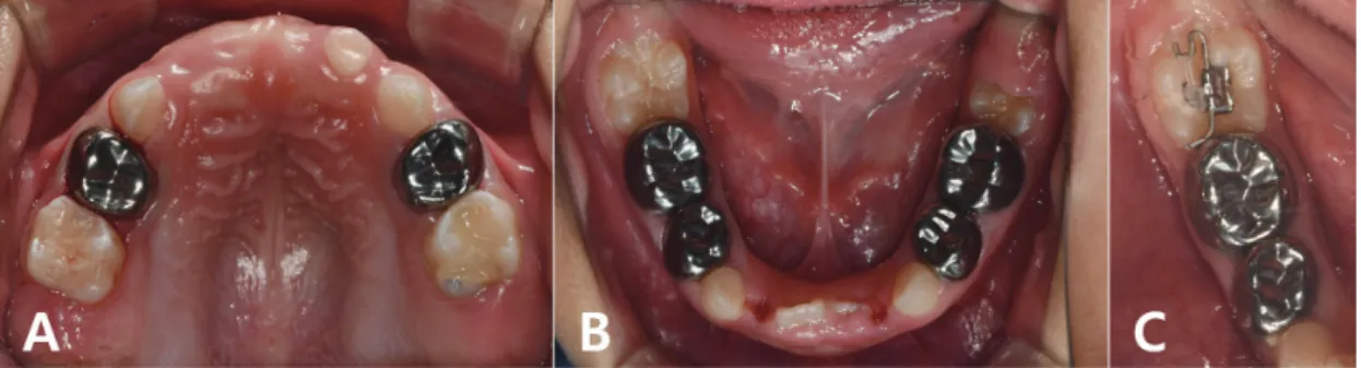
On the radio- graphic examination, interproximal dental caries of the right deciduous maxillary molars and the both right and left decidu- ous mandibular molars were

Endodontic treatment of both mandibular second premolars had been initiated 2 days previously in a local dental clinic, and the root canals were temporarily filled with a

Above all, the primary objective of dental management for patient with osteopetrosis is to maintain healthy oral condi- tions through periodic oral hygiene management to

We report a case of isolated celiac artery dissection with splenic infarction in a patient with PS deficiency that improved with conservative treatment.. The patient’s management did

Case report of cerebral creatine deficiency syndrome with novel mutation of SLC6A8 gene in a male child in Bangladesh.. Muhammad Mizanur Rahman

A 4-year-old boy with Swyer-James syndrome visited Seoul National University Dental Hospital Department of pediatric dentistry for caries treatment.. Clinical and

GENERALIZED HYPERCEMENTOSIS WITH ARRESTED DENTAL ERUPTION IN A CEREBRAL PALSY PATIENT : A CASE REPORT.. Byurira Kim, Yeji Sun, Je Seon Song,

DENTAL MANAGEMENT ASSOCIATED WITH ERUPTION DISORDERS IN A PATIENT WITH EHLERS-DANLOS SYNDROME : A CASE REPORT.. Dallae Jin, Chong-Chul Kim, Sang-Hoon Lee, Jung-Wook Kim, Young-Jae