Ⅰ. 서 론
가부키 증후군은 특징적인 얼굴 형태, 작은 골격적 이상, 지문 이상, 경증부터 중증까지의 지능 장애, 생후 성장 결핍 의 다섯 가지 징후를 보이는 드문 유전적 질환이다. 가부키
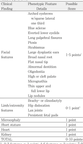
증후군의 진단의 점수 평가 방법은 다음과 같다(Table 1)1). 일본에서의 유병률은 대략 32,000명당 1명으로 보고되었 고, 전세계적인 유병률은 아직까지 알려지지 않았으나, 다 양한 인종에서 보고되었다. 한국의 경우 가부키 증후군은 드물게 보고되고 있으나, 이는 진단에 대한 인식과 진단도 구의 부족으로 인한 것으로 사료된다2). 가부키 증후군 환아 는 성장 결핍으로 인하여 약 1/3이 구순구개열을 보이고, 약 72%에서 높은 아치형의 구개를 보이므로 이에 따른 섭 식 장애, 발음의 어려움 등이 수반된다3).
가부키 증후군 환자의 치과적 소견으로는 구순구개열 외
- 104 -
◆ 증 례
가부키 증후군(Kabuki syndrome) 환자의 치과적 관리의 치험례
강힘찬1∙남옥형1∙김미선2∙최성철1∙김광철2∙이효설1*
1경희대학교 치의학전문대학원 소아치과학교실
2강동경희대학교 치과병원 소아치과
DENTAL MANAGEMENT OF A PATIENT WITH KABUKI SYNDROME : A CASE REPORT
Him Chan Kang
1, Ok Hyung Nam
1, Mi Sun Kim
2, Sung Chul Choi
1, Kwang Chul Kim
2, Hyo-Seol Lee
1*
1
Department of Pediatric Dentistry, School of Dentistry, Kyung Hee University
2
Department of Pediatric Dentistry, Kyung Hee University Hospital at Gangdong
Kabuki syndrome is characterized by typical facial features (elongated palpebral fissures with eversion of the lateral third of the lower eyelid; arched and broad eyebrows; short columella with depressed nasal tip;
large, prominent, or cupped ears), minor skeletal anomalies, persistence of fetal fingertip pads, mild to moderate intellectual disability, and postnatal growth deficiency.
A 6-year-old male with kabuki syndrome was referred from the local dental clinic for left facial swelling and dental caries on all primary molars. He was treated for acute periapical abscess with incision and drainage under physical restraint, and left maxillary second primary molar was extracted. Other caries treatment was performed under general anesthesia.
As the syndrome involves many different medical problems, special cares should be considered. Dental treatment should be carried out in comprehensive consultation system. [J Korean Dis Oral Health Vol.13,
No.2: 104-107, December 2017
]Key words :
Kabuki syndrome, General anesthesia, Dental managementAbstract
*Corresponding author : Hyo-Seol Lee
26, Kyungheedae-ro, Dongdaemun-gu, Seoul, 02447, Korea Department of Pediatric Dentistry, School of Dentistry, Kyung Hee University
Tel: +82-2-958-9440, Fax: +82-2-966-4572 E-mail: [email protected]
Received: 2017.06.16 / Revised: 2017.06.26 / Accepted: 2017.07.27
https://doi.org/10.12655/KADH.2017.13.2.104 J Korean Dis Oral Health 13(2) 2017
ISSN (print): 1738-8813 ISSN (online): 2287-7134
J Korean Dis Oral Health 13(2) 2017
- 105 -
에도 부분 무치증, 기형치, 소치증, 작은 악궁, 편측 후방부 교차교합 등이 있다4). 그러나 여러 의학적 문제 및 협조도의 부족으로 인해 치과 치료 시 여러 어려움을 겪게 된다.본 증례는 좌측 안면부의 종창과 다수의 치아우식증을 주 소로 내원한 가부키 증후군 환자를 물리적 속박, 전신마취 하에서 부작용 없이 양호한 치료 결과를 얻은 증례이다.
Ⅱ. 증 례
만 6세 남환이 왼쪽 얼굴이 부었다는 것을 주소로 내원하 였다. 임상 및 방사선 구강검사 결과 환아는 상악 좌측 제2
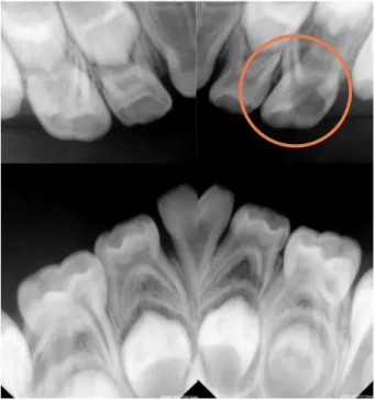
유구치의 심한 우식으로 인한 안면부 종창과 발적, 다발성 의 우식증을 보였다(Fig. 1). 환아의 종창이 악화되어, 배농 및 절개 후 지속적인 항생제를 투여하기로 결정하고 입원 및 의과적 진료의뢰를 하기로 결정하였다.
치료의 목표는 종창 완화 및 원인치와 전반적인 치아의 우식 치료로 하였다. 통증과 종창의 완화를 위해 첫 내원 당 일 상악 좌측 제2유구치 협측 부위에 절개 및 배농술을 시 행하였다. 술 후 경과 관찰 및 지속적인 항생제 투여를 위해 당일 입원하였고, 입원 5일째 경결감과 종창이 감소하여 퇴 원지시 하였다.
본 환아는 과거 급성 폐쇄성 후두염과 발작으로 인해 벤 조다이아제핀(benzodiazepine) 정맥 주사 투여 시 호흡 저 하를 보였던 병력이 있었다. 이러한 의과적 병력과 환아의 협조 상태를 고려하여 종창 및 통증이 재발될 수 있는 상악 좌측 제2유구치를 근관 치료가 아닌 물리적 속박 하에 외래 에서 발거하기로 결정하였고, 퇴원 후 3일째 발거 시행하였 다. 이후 환아의 발작 가능성과 벤조다이아제핀 투여 시 호 흡 저하 병력으로 인해 상악 제2유구치를 제외한 모든 유구 치 치아우식증의 치료는 전신마취 하에 시행하기로 결정하 였다(Fig. 2). 전신마취 전 혈액검사, 소변검사, 심전도 검 사, 방사선 흉부 촬영검사상 특이소견은 없었다.
좌측 하완에 정맥로 확보 후, 로큐로늄(Rocuronium)을 20mg을 정주하고, 흡입마취제인 세보플루레인(Sevoflurane) 을 이용하여 전신마취를 유도하였다. 근이완 상태를 확인 후 구강을 통해 기관내 삽관을 시행하였고, 레미펜타닐 (Remifentanyl)과 2% 세보플루레인을 이용하여 치료 중 마취를 유지하였다.
Table 1. Proposed Phenotypic Scoring System for Kabuki Syndrome
Clinical Phenotypic Feature Possible
Finding Details Score
Arched eyebrows w/sparse lateral one third Blue sclerae
Everted lower eyelids Long palpebral fissures Ptosis
Strabismus
Facial Large dysplastic ears
1-5 points
1features Broad nasal root
Flat nasal tip Abnormal dentition Oligodontia High or cleft palate Micrognathia Thin upper and
full lower lip Lip nodules
Brachy- or clinodactyly Limb/extremity Hip dislocation
0-1 point
2features Lax joints
Persistent fetal pads
Microcephaly 1 point
Short stature 1 point
Heart 1 point
Kidney 1 point
TOTAL 0-10 points
1. 0-3 features = 1 point; 4-6 features = 2 points; 7-9 features = 3 points; 10-12 features = 4 points; 13-15 features = 5 points
2. 0-1 feature = 0 points; 2-4 features = 1 point
Fig. 1. Extraoral frontal view of pre-operation; Left facial swelling.
J Korean Dis Oral Health 13(2) 2017
- 106 -
전신마취 하에서 상악 우측 제2유구치, 상악 좌측 제1유 구치, 하악 우측 제2유구치, 하악 좌측 제1유구치에 부분치 수절제술과 스테인리스 스틸 기성금속관 수복, 상악 우측 제1유구치, 하악 우측 제1유구치에 치면열구전색술, 하악 좌측 제2유구치에 레진 수복을 진행하였다. 그 후 전악 치 면 세마 후 전신마취를 종료하였고, 3개월 후 정기검진을 계획하였다(Fig. 3, 4).치료 중 생징후는 안정적이었으며 저산소증은 보이지 않 았다. 마취에서 회복 후 환아는 마취 회복실에서 30분 정도 회복 후 병동으로 이동하여 안정을 취하였다. 퇴원 시 특별 한 이상 징후 보이지 않았으며, 정기적인 검진을 진행하기 로 하였다.
Ⅲ. 총괄 및 고찰
가부키 증후군은 환자에 따라 질환의 증상, 심각도가 다 양하게 나타난다. 어떤 환자들은 부분적인 얼굴 기형을 보 이지만, 전신적으로 심각한 질환을 보이는 경우도 있다. 이 증후군의 일반적인 특징으로는 선천적인 심장질환, 지적 장 애, 비뇨 생식기 장애, 구순 구개열, 위장 장애, 안과 장애, 치과적 장애가 나타난다. 또한 감염과 자가면역질환에 대한 감수성 증가, 발작증세, 내분비적 장애, 섭식장애, 청력 손 실 등이 나타나기도 한다5-7).
가부키 증후군의 진단은 특정 병리적 기전보다는 임상적 인 특징에 기인하며, 그 양상이 다양하여 명확한 진단을 내 리는 것이 어렵다. 임상적으로는 주로 안모가 진단의 기준 에 포함되는데, 길게 연장된 눈꺼풀 틈새, 하안검 가측의 외 번, 아치형의 넓은 눈썹, 짧은 비주와 함께 처진 코의 끝, 크 고 도드라지거나 술잔 형태의 귀 등을 평가하게 된다6-9). 이 러한 안모 형태와 특징적인 선천적인 기형을 함께 보일 때 유전자 검사를 통해 최종 진단을 고려해 볼 수 있다.
임상적으로 진단된 가부키 증후군 환자의 약 52 - 76%
는 상염색체 상에 있는 KMT2D 유전자의 병적인 변이가 발견되었고, 약 10%정도에서는 X염색체 상에 있는 KDM6A 유전자의 병적인 변이를 보였다. 약 30%의 환자 에서는 유전적인 원인이 아직 밝혀지지 않았다10-14).
가부키 증후군 환자의 약 17%는 발작을 보이며 특히 자 극이 심하거나 스트레스를 받을 때 발작이 일어날 확률이 증가한다15). 이로 인해 치과 시술이 진행되는 어떤 시점에서 도 발작이 일어날 수 있다. 뿐만 아니라, 정신지체를 동반하 는 가부키 증후군 환자들은 대부분 치료에 대한 협조도가 불량하여 물리적 속박 등의 방법으로 치료를 강행하게 되는 데, 이는 환자의 발작을 더욱 악화시킬 수 있으므로 진정법 이나 전신마취를 통한 치과 치료가 고려되어야 한다.
본 증례 환아의 경우 가부키 증후군 환자가 보이는 치과 적 소견인 부분 무치증, 기형치, 소치증, 작은 악궁, 편측 후
Fig. 3. Intraoral view of pre-operation. Fig. 4. Intraoral view of post-operation.
Fig. 2. Periapical radiographic view of pre-operation; Severe
Dental caries of maxillary second primary molar.
J Korean Dis Oral Health 13(2) 2017
- 107 -
방부 교차교합 등은 보이지 않았다. 다만 벤조다이아제핀 사용 시 저산소증 병력과 다발성 치아우식증으로 인해 치료 할 치아가 다수였기 때문에 전신마취를 통한 치과 치료가 계획되었다.또한 상악 좌측 제2유구치의 심한 우식으로 인해 응급 치 료가 필요하였으나 발작 소견과 과거 병력, 이에 대한 단시 간 내 의과와의 협진이 불가한 상황이었기 때문에 근관 치 료를 보다는 발거를 시행하기로 결정하였다. 이에 따라 상 악 좌측 제2유구치 조기 발거에 의한 상악 좌측 제1대구치 의 근심 이동이 예상되었다. 제1대구치의 후방이동을 위한 장치로는 맹출 전에는 원심편자 간격유지장치(distal shoe space maintainer), 가철성 장치가 가능하며, 맹출 후에는 Finger spring, pendulum, distal Jet, Jones jig, Niti- coils, head-gear 등이 사용될 수 있으나 환아의 협조도를 고려하여 추후 고정성 장치인 pendulum을 사용하여 상악 좌측 제1대구치의 후방이동을 얻기로 하였다16). 그리고 환 아의 구강 위생 관리가 어려운 점을 고려하여, 정기적인 검 진 및 제1대구치 맹출 후 치면열구전색술을 고려해보기로 하였다.
Ⅳ. 요 약
본 증례는 가부키 증후군 환아의 상악 좌측 제2유구치의 심한 우식으로 인하여 좌측 협부 종창이 관찰되었고, 물리 적 속박 하에 절개 및 배농술을 시행 후 유구치는 발거되었 다. 이후 전신마취 하에 남은 우식치아의 치료가 진행되었 다. 가부키 증후군은 환자에 따라 다양한 의학적 소견을 보 이므로 전신마취 하 치과 치료가 계획 될 경우, 의과와의 협 진 및 철저한 치료계획이 필수적이다.
REFERENCES
1. Makrythanasis P, van Bon BW, Hoischen A, et al. : MLL2 mutation detected in 86 patients with Kabuki syndrome; a genotype-phenotype study.
Clin Genet, 84:539-545, 2013.
2. Cheon CK, Ko JM : Kabuki syndrome: clinical and molecular characteristics. Korean J Pediatr, 58:317-324, 2015.
3. Iida T, Park S, Kato K, Kitano I : Cleft palate in Kabuki syndrome: a report of six cases. Cleft Palate Craniofac J, 43:756-761, 2006.
4. Matsune K, Shimizu T, Maeda T, et al. : Craniofacial and dental characteristics of Kabuki syndrome. Am J Med Genet, 98:185-190, 2001.
5. Matsumoto N, Niikawa N : Kabuki make-up
syndrome: a review. Am J Med Genet C Semin Med Genet, 117C:57-65, 2003.
6. Armstrong L, Abd El Moneim A, Allanson JE, et al. : Further delineation of Kabuki syndrome in 48 well-defined new individuals. Am J Med Genet A, 132A:265-272, 2005.
7. Schrander-Stumpel CT, Spruyt L, Schrander JJ, et al. : Kabuki syndrome: Clinical data in 20 pa- tients, literature review, and further guidelines for preventive management. Am J Med Genet A, 132A:234-243, 2005.
8. Wilson GN : Thirteen cases of Niikawa-Kuroki syndrome: report and review with emphasis on medical complications and preventative manage- ment. Am J Med Genet, 79:112-120, 1998.
9. Hannibal MC, Buckingham KJ, Bamshad MJ, et al. : Spectrum of MLL2 (ALR) mutations in 110 cases of Kabuki syndrome. Am J Med Genet A, 155A:1511-1516, 2011.
10. Li Y, Bogershausen N, Wollnik B, et al. : A mu- tation screen in patients with Kabuki syndrome.
Hum Genet, 130:715-724, 2011.
11. Micale L, Augello B, Merla G, et al. : Mutation spectrum of MLL2 in a cohort of kabuki syn- drome patients. Orphanet J Rare Dis, 6:38, 2011.
12. Paulussen AD, Stegmann AP, Schrander- Stumpel CT, et al. : MLL2 mutation spectrum in 45 patients with Kabuki syndrome. Hum Mutat, 32:E2018-2025, 2011.
13. Banka S, Veeramachaneni R, Donnai D, et al. : How genetically heterogeneous is Kabuki syn- drome?: MLL2 testing in 116 patients, review, and analyses of mutation and phenotypic spec- trum. Eur J Hum Genet, 20:381-388, 2012.
14. Miyake N, Mizuno S, Matsumoto N, et al. : KDM6A point mutations cause Kabuki syn- drome. Hum Mutat, 34:108-110, 2013.
15. Niikawa N, Kuroki Y, Reynolds JF, et al. : Kabuki make-up (Niikawa-Kuroki) syndrome: A study of 62 patients. Am J Med Genet, 31:565- 589, 1988.
16. Kennedy DB, Turley PK : The clinical manage- ment of ectopically erupting first permanent mo- lars. Am J Orthod Dentofacial Orthop, 92:336- 345, 1987.