Vol. 21, No. 2, 2009 147
Received June 3, 2008, Accepted for publication August 5, 2008 Reprint request to: Beom Joon Kim, M.D., Department of Dermatology, Chung-Ang University Yong-San Hospital, 65-207, Hangangro 3-ga, Yongsan-gu, Seoul 140-757, Korea. Tel: 82-2-748-9898, Fax: 82-2- 6359-9573, E-mail: beomjoon@unitel.co.kr
Ann Dermatol Vol. 21, No. 2, 2009
CASE REPORT
A Case of Faun Tail Naevus Treated by Intense Pulsed Light
Hye In Lee, M.D., Yong Kwan Rho, M.D., Beom Joon Kim, M.D., Myeung Nam Kim, M.D.
Department of Dermatology, College of Medicine, Chung-Ang University, Seoul, Korea
A faun tail is abnormal lumbar hypertrichosis that is characterized by a wide, often triangular or lozenge-shaped patch of coarse hair, and this hair is usually several inches long. Faun tail is a rare entity. A 36-year-old male presented with a triangular shaped hair tuft with terminal hair on the lumbosacral area, and he’d had this unusual hair since birth.
There were no neurologic signs or abnormality on his spine X-ray. The MRI scan showed disc degeneration and loss of lodordosis. We report here on a rare case of faun tail, which was a form of localized hypertrichosis on the lumbosacral area, and this was successfully treated with intense pulsed (IPL) light. (Ann Dermatol 21(2) 147∼149, 2009)
-Keywords-
Faun tail, Hypertrichosis, Intense pulsed light
INTRODUCTION
Hypertrichosis is the excessive growth of hair on the non-androgen dependent areas of the body. Primary hypertrichosis has been classified based on the age of onset as to whether it is congenital or acquired, and the extent of distribution takes on either localized or ge- neralized forms. Primary localized hypertrichosis may occur as hypertrichosis cubiti involving the elbows, an- terior cervical hypertrichosis, posterior cervical hyper- trichosis or a faun tail deformity1. A faun tail is a lock of coarse, terminal hair situated on the lumbosacral area.
According to the Korean dermatologic articles2-5, only four cases with faun tail have been reported in association with
tethered cord syndrome or spina bifida occulta. We report herein on an interesting case of a bazaar faun tail naevus on a 36-year-old male.
CASE REPORT
A 36-year-old man was referred to our dermatology outpatient clinic for the treatment of a congenital focal hypertrichotic area on the lumbosacral region because it was cosmetically embarrassing. The dermatologic exami- nation revealed a localized, 3.0×5.0 cm sized, reverse triangular shaped hair tuft on the lumbosacral region.
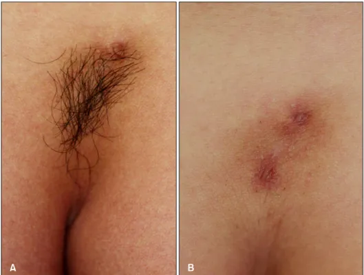
Coarse, dark, terminal hairs were observed and there was a single skin colored protruding papule within the hairs (Fig. 1A). The lesion had been present since his birth. The past medical history was noncontributory except for psoriasis vulgaris. No motor weakness or sensory changes were observed on the neurological examination. His height was estimated to be 156 cm. There was no history of a similar lesion in the family and relatives. The com- plete blood cell count, erythrocyte sedimentation rate and blood chemistry were all within the normal ranges. Simple lumbosacral radiography showed no specific finding. The magnetic resonance imaging scan showed no specific finding, except for a left subarticular protrusion of L4-5 and the loss of lordosis of the lumbar spine. The skin biopsy from the hypertrichotic patch demonstrated normal epidermis and a superficial perivascular lymphocytic infil- tration with mature terminal hair follicles in the dermis (Fig. 2). The other biopsy revealed a skin tag. A diagnosis of faun tail without any underlying neurologic disease was made. He was periodically treated with intense pulsed light (IPL) for hair removal and he was well satisfied with the result (Fig. 1B).
HI Lee, et al
148 Ann Dermatol
Fig. 1. (A) A localized reverse triangular shaped hair tuft with terminal hairs on the lumbosacral area. (B) The area after 3 treat- ment sessions with intense pulsed light (IPL) at 3 week intervals.
Fig. 2. The histologic findings showed a normal epidermis and a superficial perivascular lymphocytic infiltration in the terminal hair follicles in the dermis (H&E, ×100).
DISCUSSION
Hypertrichosis is a condition of excessive hair growth that must be distinguished from hirsutism, which is cha- racterized by an androgen-dependent hair pattern with an excessive body and facial terminal hair distribution in a male pattern1. Localized hypertrichosis can be categorized into the acquired and congenital forms. Congenital lo- calized hypertrichosis is present at birth or early in life without any inducing factors1. Congenital localized hyper-
trichosis is most commonly located in the sacral area and this is called “faun tail”.
The mechanisms for excess hair growth have not been well established. Clinical evidence exists that localized or site-specific factors may be important in determining the fate of a hair follicle. Conversion of vellus or fine caliber hairs to thicker terminal hairs occurs as a result of hormonal changes, but only at androgendependent sites such as the beard, chest, axillae and groin6. Congenital localized hypertrichosis in hamartomas or underlying neu- rologic abnormalities may represent an alternative path- way. Recent research has suggested the tremendous plasticity of the hair follicle, its mesenchyme and the surrounding connective tissue sheath, which allows for dramatic transformations of hair follicles Localized hyper- trichosis may therefore be a reflection of an abnormal signal for enlargement of the follicular papilla, a prolon- gation of the normal anagen hair growth cycle or both7. The clinical importance of a midline cutaneous posterior anomaly is that they are frequently associated with un- derlying defects such as diastematomyelia, meningocele, spina bifida, kyphoscoliosis or chest deformities. As both skin and nervous tissue are of an ectodermal origin, ano- malies of these tissues may occur simultaneously. When the congenital localized hypertrichosis is located away from the spine in apparently normal skin, it has been named as simple nevoid hypertrichosis, and it is usually a totally benign lesion8. The cutaneous lesions that should
A Case of Faun Tail Naevus Treated by Intense Pulsed Light
Vol. 21, No. 2, 2009 149 raise a higher degree of suspicion include hypertrichosis,
dimples, aplasia cutis, lipoma, hemangioma, dermoid cyst or sinus, acrochordons, true tail, pseudotail and conge- nital scarring9,10. Thus, localized hypertrichosis may be a clue for an underlying defect, some of which are surgi- cally correctable at an early age. Sacral hypertrichosis is also known as faun tail naevus, and this is the most common skin lesion that is evident at birth11, as was seen in our patient. Hairy patches are most frequently asso- ciated with tethered cord and diastematomyelia12. Al- though our case demonstrated no underlying neurologic problem associated with his localized hypertrichosis, accurate screening modalities such as MRI are necessary to detect any underlying dysraphic anomalies and to prevent the occurrence or progression of the neurologic deficit by timely intervention before neurological damage has occurred.
As for the management of hypertrichosis, long-term re- moval of unwanted hair is a challenge. The need for treatment depends on the degree of hypertrichosis and the psychosocial needs of the patient. The currently available treatment methods for removal of excessive hair include bleaching, trimming, waxing, physical and chemical de- pilatories, electrolysis, intense pulsed light therapy and laser hair removal13. A variety of laser devices and intense pulsed light therapy are now available. All are based on the principle of selective photothermolysis; the melanin pigment in the hair follicle provides the chromophore for selective targeting of hair follicles, while the surrounding dermis is spared. Therefore, at deeply penetrating wave- lengths in the 600∼1,100 nm range, melanin absorption may be used for selective photothermolysis of hair fol- licles14. Intense pulsed light source (590∼1200 nm) the- rapy for hair removal seems to be more effective for darker hair15. Because our patient’s lumbosacral hair was dark and thick like his axillary hair, we choose intense pulsed light therapy for hair removal. Occasionally, post- treatment erythema, edema, blisters and hyperpigmenta- tion can occur.
The aim of this paper is to draw attention to faun tail and to emphasize the responsibility of physicians and cos- meticians to the recognize early in patients’ lives lumbar paraspinal skin lesions so that appropriate early treatment can be administered for the possible underlying anomalies before the irreversible sequelae develop. Although other-
wise benign, these disorders may result in cosmetic dis- figurement and psychosocial trauma for the patients.
These patients should be adequately advised of the available treatment methods for both temporary and permanent hair removal.
REFERENCES
1. Vashi RA, Mancini AJ, Paller AS. Primary generalized and localized hypertrichosis in children. Arch Dermatol 2001;
137:877-884.
2. Choi JS, Lee WH, Lee JB. Circumscribed hypertrichosis and blue nevus with spina bifida occulta. Korean J Dermatol 1981;19:365-369.
3. Suhr GB, Lee JS, Lee JH, Park JK. A case of spina bifida occulta with faun-tail nevus. Korean J Dermatol 1988;26:
759-763.
4. Kim SW. Faun tail. Ann Dermatol 1993;5:47-50.
5. Bak H, Kim JY, Chung YL, Kim SC. A case of faun tail associated with tethered cord syndrome. Korean J Dermatol 2004;42:781-783.
6. Drolet B. Birthmarks to worry about. Cutaneous markers of dysraphism. Dermatol Clin 1998;16:447-453.
7. McAtee-Smith J, Hebert AA, Rapini RP, Goldberg NS. Skin lesions of the spinal axis and spinal dysraphism. Fifteen cases and a review of the literature. Arch Pediatr Adolesc Med 1994;148:740-748.
8. Tobin DJ, Gunin A, Magerl M, Handijski B, Paus R. Plasticity and cytokinetic dynamics of the hair follicle mesenchyme:
implications for hair growth control. J Invest Dermatol 2003;120:895-904.
9. Bertolino AP, Freedberg IM. Hair. In: Fitzpatrick TB, Eisen AZ, Wolff K, editors. Dermatology in general medicine.
New York: McGraw-Hill, 1987:627-666.
10. Ettl A, Marinkovic M, Koornneef L. Localized hypertrichosis associated with periorbital neurofibroma: clinical findings and differential diagnosis. Ophthalmology 1996;103:942- 948.
11. Kumar R, Singh SN. Spinal dysraphism: trends in northern India. Pediatr Neurosurg 2003;38:133-145.
12. Tavafoghi V, Ghandchi A, Hambrick GW Jr, Udverhelyi GB.
Cutaneous signs of spinal dysraphism. Report of a patient with a tail-like lipoma and review of 200 cases in the litera- ture. Arch Dermatol 1978;114:573-577.
13. Trueb RM. Causes and management of hypertrichosis. Am J Clin Dermatol 2002;3:617-627.
14. Liew SH. Laser hair removal: guidelines for management.
Am J Clin Dermatol 2002;3:107-115.
15. Olsen EA. Methods of hair removal. J Am Acad Dermatol 1999;40:143-155.