100
pISSN 2288-6575 • eISSN 2288-6796 http://dx.doi.org/10.4174/astr.2014.87.2.100 Annals of Surgical Treatment and Research
CASE REPORT
Extremely rare case of extrahepatic duct phytobezoar treated with intraoperative transenteral endoscopy
Jung Min Bae, Yong Kook Lee1
Department of Surgery,Yeungnam University College of Medicine, Daegu, 1Department of Internal Medicine, Dongguk University Gyeongju Hospital, Gyeongju, Korea
INTRODUCTION
Bezoar stones are concrete masses of indigestible material.
The phytobezoar is a common type of bezoar. It is composed of indigestible vegetable-like material. The incidence has been reported as 0.4% in the general population [1]. Phytobezoars are commonly reported in patients who have had previous gastric surgery [2,3]. Most commonly, bezoars produce mechanical obstructive symptoms but when they cause gastrointestinal bleeding or perforation, bezoars are associated with high mortality [4]. Therefore, it is very important to remove a bezoar to prevent mortality and morbidity. The treatment of bezoar is endoscopy or surgery.
We recently experienced a 79-year-old woman diagnosed with extrahepatic duct phytobezoar. Although biliary phytobezoar is extremely rare, a few cases of extrahepatic duct phytobezoar have been reported previously [5,6]. We successfully treated this case with intraoperative transenteral endoscopic removal.
This report concerns an extremely rare case of extrahepatic
duct phytobezoar and intraoperative transenteral endoscopic treatment including imaging and review of the literature.
CASE REPORT
A 79-year-old woman came to the Emergency Department due to severe cramping abdominal pain and vomit for 2 days.
She had a 40-year history of repeated hepatobiliary operation because of cholecystitis and postoperative complication. But, she and her family could not recall the precise operation name, type and its postoperative complication. She had a history of intestinal obstruction 6 months prior and was treated conservatively.
All vital signs, including body temperature, pulse, and blood pressure, were within normal limits. Laboratory findings in Emergency Department were unremarkable. Chest and abdomen radiographs were also unremarkable. The patient underwent and abdomino-pelvic CT scan to evaluate the cause of abdominal pain. The CT scan revealed dilatation
Received November 11, 2013, Reviewed January 8, 2014, Accepted January 14, 2014
Corresponding Author: Jung Min Bae
Department of Surgery, Yeungnam University College of Medicine, 317-1, Daemyung-dong, Nam-gu, Daegu 705-030, Korea
Tel: +82-53-620-3580, Fax: +82-53-624-1213 E-mail: [email protected]
Copyright ⓒ 2014, the Korean Surgical Society
cc Annals of Surgical Treatment and Research is an Open Access Journal. All articles are distributed under the terms of the Creative Commons Attribution Non- Commercial License (http://creativecommons.org/licenses/by-nc/3.0/) which permits unrestricted non-commercial use, distribution, and reproduction in any medium, provided the original work is properly cited.
Phytobezoar is a rare cause of gastro-intestinal tract obstruction. Common sites of phytobezoar are the stomach and small bowel. Naturally, extrahepatic duct phytobezoar is near impossible due to anatomical structure and location such as ampulla of vater, common bile duct and bifurcation of bile duct. Here, we present an extremely rare case of extrahepatic duct phytobezoar that resulted in abdominal pain. We successfully treated the case with extraoperative transenteral endoscopic removal of phytobezoar. For its great rarity and particular treatment approach, we report this case with review of literature.
[Ann Surg Treat Res 2014;87(2):100-103]
Key Words: Phytobezoar, Extrahepatic, Endoscopy
Annals of Surgical Treatment and Research 101 and a large stone of the extrahepatic duct with air-biliary
gram, parenchymal atrophy of the left hepatic lobe and no remarkable findings in the right hepatic lobe (Fig. 1). An endoscopic retrograde cholagio-pancreatography (ERCP) was then performed to evaluate the biliary tract. There was nothing found in the common bile duct on ERCP (Fig. 2).
Our results lead us to consider extrahepatic duct stone with ob struction and change of biliary drainage system due to the previous hepatobiliary operation. Surgical procedure was indi- cated and exploration was performed thereafter. There was severe fibrous adhesion between diaphragm, liver, stomach, small bowel, transverse colon and peritoneum, especially in the right upper quadrant area. There was jejuno-jejunostomy between the jejunal limb from the duodenum and jejunal limb from hepatico-jejunostomy 15 cm distally from the Treitz
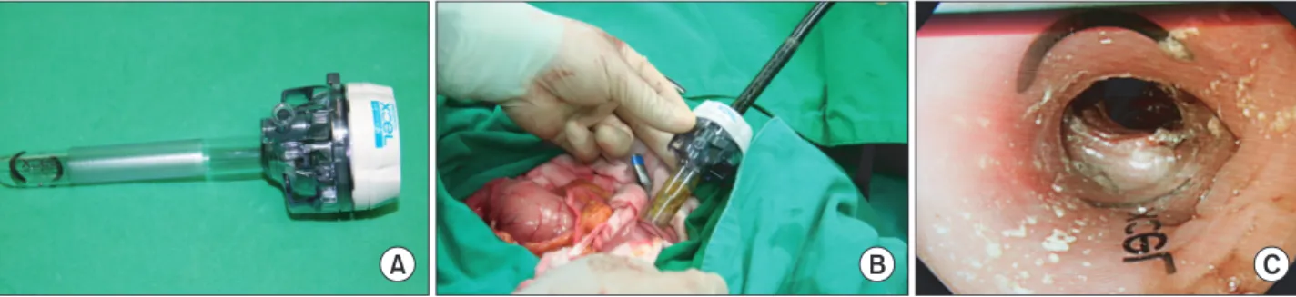
ligament (Fig. 3). There was a hepatico-jejunostomy site in the porta hepatis area. Small enterotomy was performed below the jejuno-jejunostomy site (Fig. 3). Intraoperative endoscopy was performed by transenterotomy using a 15-mm Xcel trocar (Ethicon Inc., Somerville, NJ, USA) (Fig. 4). There were hepatico- jejunostomy sites and concrete materials in the extrahepatic duct just above the stoma (Fig. 5). Endoscopic fragmentation, removal and saline irrigation were performed successfully. The removed mass-like material was not stone but phytobezoar composed of indigestible vegetable and food-like materials (Fig.
6). The patient’s postoperative course was uneventful and she is doing well following surgery.
Fig. 1. The CT scan revealed dilatation and large stone of intrahepatic duct with air-biliary gram, parenchymal atrophy of left hepatic lobe and unremarkable right hepatic lobe. (A) Transverse section view. (B) Coronal section view.
Fig. 2. Endoscopic retrograde cholangiopancreatography.
There was not founded common bile duct.
Fig. 3. Schematic outline of operation field appearance.
Jung Min Bae and Yong Kook Lee: Extrahepatic duct phytobezoar treated with intraoperative transenteral endoscopy
102
Annals of Surgical Treatment and Research 2014;87(2):100-103
DISCUSSION
Phytobezoar is a common type of bezoar. It composed of indigestible vegetable-like material. The incidence has been reported as 0.4% in the general population [1]. Common site of phytobezoar are the stomach and small bowel. Phytobezoars are commonly reported in patients who have had previous gastric surgery [2,3]. However, our patient had not had gastric surgery but cholecystectomy and hepatico-jejunostomy due to postcholecystectomy complication.
Phytobezoar in the biliary tract is very rare and few cases of
extrahepatic duct phytobezoar are reported in the literature [5,6]. That seems to be related to increased intestinal luminal pressure which allows reflux of indigestible vegetable and diverse food materials from the distal small bowel when especially, intestinal obstruction is developed [5].
Therefore, there is the possibility of formation of phytobezoar in the intra- or extrahepatic biliary tract in cases of new entry formation from the hollow viscus to the biliary tract such as choledoco-jejunostomy, choledoco-duodenostomy, hepatico- jejunostomy, cholecysto-duodenostomy, sphincteroplasty, etc.
Most commonly, the phytobezoar is a rare cause of gastro- intestinal tract obstruction. Maybe, the cause of pain was intrahepatic duct obstruction in our patient. In other clinical manifestations, when they cause gastrointestinal bleeding or perforation, bezoars are associated with a high mortality [4].
Therefore, it is very important to remove a bezoar to prevent mortality and morbidity.
Gastroscopy is a useful diagnostic tool of gastric phytobezoars and CT scan is the most useful diagnostic method of small bowel phytobezoar [7]. Differentiated diagnosis between stone and phytobezoar in the biliary tract is difficult because stones and concrete phytobezoars are radiolucent with similar shapes and the biliary tract is a very unusual site of phytobezoar. Natu- rally, extrahepatic duct phytobezoar is near impossible due to the anatomical structure and location such as the ampulla of vater, common bile duct and bifurcation of common hepatic Fig. 4. Intraoperative transenteral endoscopy. (A) 15-mm Xcel trocar (Ethicon Inc., Somerville, NJ, USA). (B) Transenteral endoscopy using Trocar. (C) Inner appearance of transenteral endoscopy.
Fig. 5. Endoscopic procedure appearance. (A) There is a phyto- bezoar in hepatico-jejunos tomy stoma. (B) Hepatico-jejuno- stomy stoma appearance after endoscopic removal of phyto- bezoar.
Fig. 6. The removed phytobezoar.
Annals of Surgical Treatment and Research 103 duct. So, extrahepatic duct phytobezoar is difficult to diagnose
preoperatively.
Surgical or endoscopic removal are treatments of choice for phytobezoars. Especially, surgical removal is the treatment of choice for phytobezoars that endoscopy cannot approach or fail.
We successfully treated a case of extrahepatic duct phy tobezoar with intraoperative transenteral endoscopic removal. Intra- operative transenteral endoscopy is an alternative treatment or diagnostic modality that can evaluate unknown small bowel bleeding or unknown small bowel intraluminal lesion during operation [8]. The benefit of using trocar during intraoperative endoscopy is its easy approach in the small bowel, aseptic from endoscopic equipment, and anchoring effect like the human mouth and neck [8].
In this patient, the gastroscopy and ERCP could not approach and evaluate the hepatico-jejunostomy site preoperatively because of Roux-en Y anastomosis between the jejunal limb from duodenum and the jejunal limb from hepatico-jejunostomy
(Fig. 3). On surgical exploration, the entire gastro intestinal tract must be explored to exclude other bezoar and prevent recurrent bowel obstruction caused by residual bezoar [9]. Other nonsurgical treatment options include sodium bicarbonate powder, a liquid diet, enzymatic digestion with various agents, endoscopic fragmentation and oral intake (or endoscopic injection ) of ‘Coca-Cola’ [10].
In conclusion, we have presented a case of extrahepatic duct phytobezoar successfully treated with intraoperative tran- senteral endoscopic removal. Although it is considered extre- mely rare, it can be one of the possible causes of extrahepatic duct obstruction. For its great rarity and particular treatment approach, we report this case with review of the literature.
CONFLICTS OF INTEREST
No potential conflict of interest relevant to this article was reported.
1. McKechnie JC. Gastroscopic removal of a phytobezoar. Gastro enterology 1972;62:
1047-51.
2. Angelelli G, Magliocca M, Zaccheo N, Vinci R, Rotondo A. Inte stinal obstruction caused by phytobezoar: computerized tomo graphy findings: report of 3 cases.
Radiol Med 1997;93:789-91.
3. Robles R, Parrilla P, Escamilla C, Lujan JA, Torralba JA, Liron R, et al. Gastrointestinal bezoars. Br J Surg 1994;81:1000-1.
4. Singh SK, Marupaka SK. Duodenal date seed bezoar: a very unu sual cause of partial gastric outlet obstruction. Austra- las Radiol 2007;51 Spec No.:B126-9.
5. Kim Y, Park BJ, Kim MJ, Sung DJ, Kim DS, Yu YD, et al. Biliary phytobezoar resulting in intestinal obstruction. World J Gastro- enterol 2013;19:133-6.
6. Lamotte M, Kockx M, Hautekeete M, Holvoet J, Hubens H. Biliary phytobezoar:
a medical curiosity. Am J Gastroenterol 1995;90:1346-8.
7. Delabrousse E, Brunelle S, Saguet O, Destrumelle N, Landecy G, Kastler B.
Small bowel obstruction secondary to phytobezoar CT findings. Clin Imaging 2001;25:44-6.
8. Bae JM, Lee HK, Bae JD, Choi EA, Jung KH,
Jung BW, et al. Trocar(R)(Ethicon) used intraoperative endoscopy in acute lower gastrointestinal bleeding. J Korean Surg Soc 2004;66:424-9.
9. Ezzat RF, Rashid SA, Rashid AT, Abdullah KM, Ahmed SM. Small intestinal obstruc- tion due to phytobezoar: a case report. J Med Case Rep 2009;3:9312.
10. Ha SS, Lee HS, Jung MK, Jeon SW, Cho CM, Kim SK, et al. Acute intestinal ob- struction caused by a persimmon phy- to bezoar after dissolution therapy with Coca-Cola. Korean J Intern Med 2007;
22:300-3.
REFERENCES
Jung Min Bae and Yong Kook Lee: Extrahepatic duct phytobezoar treated with intraoperative transenteral endoscopy