Journal of Bacteriology and Virology 2006. Vol. 36, No. 4 p.271 – 278
재조합 사람 파필로마바이러스 16형 L1 바이러스 유사입자의 제조과정 중의 잔류 숙주세포 DNA의 검출과 정량
식품의약품안전청 생물의약품평가부
1, 연세대학교 생명공학과
2, 국립독성연구원 독성연구부 유전독성팀
3손화경
1, 2·정혜성
1·김영림
1·김순남
1·신진호
1·민홍기
1·성백린
2·박순희
1,3*Detection and Quantification of Residual Cellular DNA in the Production of Recombinant HPV-16 L1 Virus-Like Particles
Hwa-Kyung Son1,2, Hye-Sung Jeong1, Young-Lim Kim1, Soon-Nam Kim1, Jin-Ho Shin1, Hong-Ki Min1, Baik-Lin Seong2 and Sue-Nie Park1,3*
1
Department of Biologics Evaluation, Korea Food and Drug Administration, 194 Tongil-ro Eunpyung-gu Seoul, 122-704, Korea,
2Department of Biotechnology, Yonsei University, 134 Sinchon-dong,
Seodaemun-gu, Seoul 120-749, Korea,
3Genetic Toxicology Division, National Institute of Toxicological Research, 5 Nokbun, Eunpyung-Gu, Seoul 122-704, Korea
Received : November 1, 2006 Accepted : November 25, 2006
A number of recombinant proteins isolated from cell sources are being produced for biopharmaceuticals. Although most biopharmaceuticals are highly purified, there is a safety concern that such recombinant products could be contaminated with impurities including adventitious virus, mycoplasma, endotoxin and oncogenic DNA. Residual DNA in recombinant biopharmaceuticals is a potential risk factor and must be evaluated and removed to meet the regulatory guidelines. Recombinant HPV type 16 L1 VLPs, recombinant protein produced in Spodoptera frugiperda (Sf ) 9 insect cells, is a HPV subunit vaccine candidate which has been studied as a preventive vaccine of cervical cancers. In this study, we performed detection and quantification of residual cellular DNA in the production of recombinant HPV type 16 L1 VLPs. HPV-16 L1 VLPs were purified by processes including detergent lysis, sonication treatment, sucrose cushion centrifugation, CsCl equilibrium density centrifugation, and DNase treatment which was added to inactivate residual cellular DNA after CsCl centrifugation step. We have developed a precise assay based on a dot-blot hybridization using digoxigenin random primed labeling DNA probes for the detection and quantification of residual cellular DNA during the purification process and final products. Detection limit of residual cellular DNA was 0.1 ng in this assay and the amount of residual cellular DNA in the final product was 0.5 ng~1 ng per 100 µg of protein. This study describes safer and more sensitive methods alternative to radioactive techniques employed for residual cellular DNA quantification of biopharmaceuticals produced by recombinant protein technology and presents method validation data demonstrating precision and reproducibility.
Key Words: HPV-16 L1 VLPs, Residual cellular DNA, Biopharmaceuticals
*교신저자: 박순희. 122-704, 서울특별시 은평구 녹번동 5번지, 국립독성연구원 유전독성팀 Phone: 02-380-1792, Fax: 02-380-1795, e-mail: suenie@kfda.go.kr
**본 연구는 식품의약품안전청 "WHO 인증 자궁경부암 원인바이러스 관련 기술확립 연구사업"의 지원에 의하여 이루어진 것임.
271
서 론
자궁경부암에서 발견되는 사람 파필로마바이러스 (Human papillomavirus, HPV)는 항문암, 구강암, 특히 편도선암에서 도 발견된다. 전 세계적으로 매우 빈번하게 발생되는 자궁경 부암과 관련이 깊은 사람 파필로마바이러스의 16형은 고위 험군 상피 내 종양의 형성과 관련이 깊으며, 방치되었을 경 우 자궁암으로 진전될 수 있다 (1,25). HPV 16형의 바이러스 유사입자 (Virus-like particles, VLPs)는 Spodoptera frugiperda (Sf) 9이라는 곤충세포나 yeast로부터 생산되는 백신후보물질 이고, 자궁경부암의 예방백신으로서 연구되고 있다 (11,17).
HPV의 L1 바이러스 캡시드 단백질은 비어있는 캡시드나 다 양한 발현시스템에서 생산된 VLPs에서 조립될 수 있다 (10, 12,16,29). VLPs는 실제 바이러스와 형태적으로 매우 유사하 나 바이러스의 유전체를 포함하지 않는 특징을 가지고 있으 며 (7), 주요한 캡시드 단백질 L1의 자가 조립으로 얻어진 VLPs에 의해 형성된 면역원성은 동물모델에서 파필로마바 이러스의 감염에 대한 방어 작용을 유도할 수 있고, 이것은 중화항체가 형태적으로 독립적인 항원결정기를 인식한다는 것을 나타낸다 (31). 이러한 VLPs를 이용한 백신 생산의 경 우 오염물질처럼 바람직하지 않는 면역반응이나 독성반응이 일어나지 않도록 적절히 정제되어야 한다 (6).
최근 세포와 재조합 DNA vector를 이용한 특정유전자의 발현에 대한 연구가 활발히 진행되고 있으며, 생물의약품의 생산을 위한 제조공정에 이용되고 있다. 하지만, 연속적인 세포주 (continuous cell lines)로부터 생산되는 생물의약품의 경우 제조공정 중에 외부로부터 유입되거나 초기 세포주에 서 유래하는 외래 바이러스, 발암성 DNA, 마이코플라즈마 (mycoplasma), 내독소 (endotoxin) 등과 같은 불순물에 오염 될 가능성을 가지고 있다 (26). 따라서 이들 생물의약품에 대 한 철저한 안전성 평가가 절실히 요구되고 있다 (14). 연속적 인 세포주는 또한 성장조절 유전자의 통제력상실에 의해 세 포가 무한정한 수명을 갖게 되기 때문에, 그러한 세포주의 DNA는 다른 세포들에게 세포의 성장불능이나 발암성과 같 은 능력을 전달할 수 있는 잠재성을 갖게 된다 (8). 지금까지 숙주세포의 잔류 DNA는 어떠한 위험요인으로서도 고려되어 오지 않았다. 그러므로 연속적인 세포주가 바이러스 백신의 제조에 사용되기 위해서는, 바이러스 입자에 발암성의 숙주 세포 DNA가 포함될 가능성이 주의 깊게 고려되어야 한다.
1986년 WHO study group은 연속적인 세포주로부터 생물 의약품 생산 시, 세포의 잔류 DNA가 one dose 당 100 pg이 나 그 이하 일 경우에는 위험성을 무시할 수 있다고 결정했 다. 이 한계에 대한 결정에서 주목해야 할 문제는 DNA 자체
에 있는 것이 아니라, 활성화된 발암유전자를 암호화하는 특 정 DNA 단편을 최소화해야 한다는 것에 있다 (32). 숙주세 포의 잔류 DNA와 관련된 생물의약품의 안전성은 정제과정 동안 오염 가능한 DNA량을 감소시키거나, DNA의 생물학 적 활성을 가능한 한 불활화시킴으로서 평가될 수 있다 (8).
생물의약품에서 DNA 불순물의 검출과 정량을 위해서 PCR (polymerase chain reaction) (9,19,30), total DNA threshold assay (2), 그리고 방사성동위원소를 이용하거나 또는 비-방 사성동위원소 실험법을 이용한, 혹은 바이오틴 처리를 하거 나 변형시킨 probe를 이용한 dot-blot hybridization 실험법과 같은 많은 실험법이 개발되어 왔다 (20,21). PCR 분석법이 가장 민감한 실험법이긴 하지만 각 핵산 단편을 위한 sequ- ence 특이적인 probe가 디자인 되어야 하기 때문에 DNA나 RNA의 검출법으로서는 일반적으로 적절하지 않다 (21). 이 중에서 가장 흔하게 사용되는 검출법은 핵산용액을 직접 적 용시키는 non-radioactive labeling probes를 이용하여 오염된 핵산을 고정시키는 dot-blot hybridization 실험법이다. 한번 고 정된 DNA나 RNA는 표적핵산의 sequence와 빠르게 반응 하게 되어 정량적, 정성적 결과를 모두 얻을 수 있다. 또한, non-radioactive labeling을 이용한 핵산 probes를 사용하면 일 반적인 hybridization 실험법이 갖고 있던 장애요소들을 없애 줄 수 있다. 생물의약품에서 잔류 DNA는 잠재적인 위험요 인이기 때문에 그것은 규제기관에서 권장하는 가이드라인에 맞는 제품을 생산하기 위해서 제거되어야 하고 평가되어야 한다.
본 연구에서는 곤충세포로부터 생산된 HPV 16형 L1 재 조합 단백질의 VLPs를 정제하는 과정에서 숙주세포의 잔류 DNA를 검출하고 정량하였다. HPV 백신후보물질의 정제과 정에 숙주세포의 잔류 DNA의 정량과 검출에 관한 연구에 대해서는 아직까지 보고된 바 없으며 재조합 HPV 16형 L1 단백질은 Sf9 숙주세포로부터 유래된 발암성 DNA 단편에 의해 오염 될 수 있는 이론적 가능성을 가지고 있다. HPV-16 L1 VLPs는 sucrose cushion 초원심분리와 CsCl 밀도구배 초 원심분리를 포함한 6단계에 의해 정제되었으며, 잔류 DNA 를 불활화 시키기 위해서 CsCl 밀도구배 초원심분리과정 후 에 DNase를 처리하였다. 각 단계의 시료와 최종 생산물에는 숙주세포 유래 DNA에 특이성을 갖는 probe를 이용하여 dot- blot hybridization을 시행하였다. 이 연구에 사용된 실험법은 잔류 DNA의 정량과 검출을 위해 사용된 radioactive techni- ques에 대한 새로운 대안이며, 검증 데이터는 정확성과 재현 성을 나타낸다.
재료 및 방법
1. 세포와 바이러스
Sf9 곤충세포 (Invitrogen, USA)는 Grace's insect medium (Gibco-BRL, USA)에 10% (v/v) fetal bovine serum과 1% (v/v) antibiotic-antimycotics를 첨가하여 27℃ incubator에서 배양하 였고, 0.5 ml spinner flask (Bellco, USA)에 1.0×10⁶ cells/ml을 첨가하여 3.0×10⁶ cells/ml까지 배양시킨 것을 바이러스 감염 에 이용하였다. HPV 16형 L1 VLPs의 정제를 위해서 HPV 16형 L1의 주된 캡시드 단백질을 발현하는 재조합 바이러 스는 국제 암센터 세포종양학 실험실 (Laboratory of Cellular Oncology, National Cancer Institute)의 John T Schiller박사로부 터 분양 받아 사용하였다.
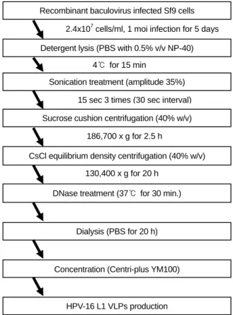
2. HPV-16 L1 VLPs의 정제와 Immunoblot 분석 Sf9 cells (200 ml; 2.4×10⁶ cells per ml)에 재조합 baculovirus (rHPV-16 L1)를 1 multiplicity of infection (MOI)로 감염시켰 고 감염 5일째에 세포를 수확하였다. 세포는 2,500 rpm에서 10분 동안 원심분리과정을 거쳐서 0.5% Nonidet P-40 (NP-40) 을 포함하는 PBS를 사용하여 15분 동안 ice에서 처리 후 30초 간격으로 15초 동안 3회 초음파 (Vibra cell, Sonics &
Materials inc., USA) 처리되었다. 세포 분쇄물은 4℃에서 Beckman SW41 rotor (186,700 x g for 2.5 h)를 사용하여 40%
(w/v) sucrose/PBS cushion 초원심분리를 거쳐, 40% CsCl/PBS 평형밀도구배 초원심분리 (130,400 x g for 20 h, SW41 rotor, USA)를 시행하였다. VLPs fractions의 존재를 확인하기 위 하여 Immunoblot 분석을 시행하였고, primary antibody로는 HPV-16 L1 monoclonal antibody인 CamVir-1 (Chemicon, USA) 을 blocking solution (1% Bovine Serum Albumin)에 1:500으 로 희석하여 사용하였고, secondary antibody로는 alkaline phosphatase가 결합된 anti-mouse IgG antibody를 1:1000으로 희석하여 사용하였다. VECTASTAIN ABC-Amp reagent (Vector laboratories, Burlingame CA, USA)로 약 10분 동안 반응시킨 후, NBT/BCIP (Vector laboratories, USA)로 검출 확인하였다.
VLPs를 포함하는 Immunoreactive fractions은 잔류 DNA의 활성을 불활화 시키기 위해서 37℃에서 30분 동안 DNase (Sigma, USA)를 처리한 후, PBS에서 20시간 동안 투석을 거쳐, 2,500 rpm에서 10분 동안 Centriplus YM-100 (Millipore, USA)를 사용하여 농축하였다 (Fig. 1). 각 정제단계로부터 얻 어진 시료의 단백질 농도는 Micro BCA kit (Pierce, Rockford, USA)을 사용하여 측정하였다.
3. HPV-16 L1 VLPs의 전자현미경 분석
정제된 HPV-16 L1 VLPs를 확인하기 위해서는 전자현미경 (JEOL. JP/JEM-1010, Japan)을 사용하였으며, CsCl 평형밀도 구배 초원심분리과정을 거친 시료와 DNase를 처리한 시료, 그리고 최종 시료를 carbon-coated grids에 2분 동안 흡착시 키고, 건조시킨 후에 2% uranyl acetate로 negative stain하여 50,000 또는 60,000배율로 관찰하였다.
4. Hybridization 실험법
1) Random primed labeling과 probe yields의 측정 숙주세포인 Sf9 cell로부터 Qiagen blood mini kit를 이용하 여 제조자의 사용설명서에 따라 DNA를 분리하였다. DNA 의 순도는 1.8~2.0 사이의 값을 가졌다. DNA 단편은 DIG High prime DNA Labeling and Detection kit (Roche Applied Science, Germany)를 이용한 random primer 반응에서 표지되 었고, probes 생산량은 제조자의 매뉴얼에 따라 성공적인 hy- bridization을 위한 probes의 적절한 양을 사용하기 위해 결정 되었다. Hybridization에 적절한 양의 probes를 첨가시키기 위 해 labeling 반응에서 생산된 DIG labeled probes의 양은 알
Recombinant baculovirus infected Sf9 cells
Detergent lysis (PBS with 0.5% v/v NP-40)
Sonication treatment (amplitude 35%)
Sucrose cushion centrifugation (40% w/v)
CsCl equilibrium density centrifugation (40% w/v)
DNase treatment (37℃ for 30 min.)
Dialysis (PBS for 20 h)
Concentration (Centri-plus YM100)
HPV-16 L1 VLPs production
2.4x10⁷cells/ml, 1 moi infection for 5 days
4℃ for 15 min
15 sec 3 times (30 sec interval)
186,700 x g for 2.5 h
130,400 x g for 20 h
Figure 1. Flowchart for the production of HPV-16 L1 VLPs.
Each step was individually assessed for the ability to remove residual cellular DNA.
려진 농도의 DIG-labeled positive control nucleic acid로 labeling 한 probes로 계단 희석하여 평가되었다.
2) 정제단계별 DNA의 추출
HPV-16 L1 VLPs의 정제단계별의 시료와 최종 생산물의 시료는 단백질 정량에 의해 각각 약 100 µg의 단백질로 균 등화한 다음 QIAmp DNA blood kit (Qiagen, USA) 실험방법 에 따라 DNA를 추출하였다. DNA는 TE buffer (10 mM Tris- HCl; 0.5 mM EDTA; pH 9.0) 100 µl에 녹여서 사용하기 전까 지 -20℃에 저장하였다.
3) DNA 고정을 위한 dot-blotting
DNA의 dot-blotting을 위하여 숙주세포 DNA stock 용액의 표준 DNA는 1000, 500, 250, 100, 50, 10, 5, 1, 0.5, and 0.1 ng의 농도로 희석하여 준비하였다. 표준 DNA stock 용액과 각 정 제단계에서 추출한 DNA 시료에 0.4 M NaOH와 10 mM EDTA를 첨가한 후, 100℃에서 10분 동안 heating 하였다. 각 시료는 6x SSC buffer에 미리 적셔진 nylon membrane (Roche Applied Science, Germany)에 spotting 되었다. 시료를 dot-blot 여과장치 (BIO-RAD, USA)를 사용하여 여과시킨 후, nylon membrane을 2x SSC buffer에서 수세하여 평형을 유지시키 고, vacuum dry oven (Heraeus Instruments, Germany)에서 2시 간 동안 80℃에서 baking 하였다.
4) Hybridization과 검출
DNA로 blotting 후 baking 된 membrane을 미리 온도를 맞 춘 hybridization buffer (DIG Easy Hyb, Roche Applied Science, Germany)를 가지고 42℃에서 30분 동안 pre-hybridization 시 켰다. Random primed labeling DNA probes를 끓는 수조에서 5 분 동안 heat denaturation 시킨 후, 42℃에서 미리 온도를 맞 춘 hybridization buffer를 첨가한 다음, pre-hybridization 된 membrane에 첨가하여 42℃에서 16시간 동안 반응시켰다.
Hybridization 반응 완료 후 membrane을 low stringency buffer (2x SSC containing 0.1% SDS)와 high stringency buffer (0.5x SSC containing 0.1% SDS)로 42℃에서 차례로 반응시켰다.
Washing buffer (0.1 M maleic acid, 0.15 M NaCl; pH 7.5; 0.3%
(v/v) Tween 20)에 membrane을 수세 후, blocking solution (Roche Applied Science, Germany)과 Anti-DIG-alkaline phosp- hatase를 포함하는 antibody solution을 차례로 반응 시키고, detection buffer (0.1 M Tris-HCl, 0.1 M NaCl, pH 9.5)에서 균일 화 시켰다. Membrane에 나타난 결과는 chromogenic alkaline phosphatase substrates (NBT/ BCIP)에 반응시켜 관찰하였다.
결 과
1. HPV-16 L1 VLPs 정제와 Immunoblot 분석
Sf9 곤충세포로부터 HPV-16 L1 VLPs 정제는 detergent
lysis와 sonication 처리 후 초원심분리 (Beckman, SW41 Ti, USA) 즉, 40% sucrose/PBS cushion 초원심분리와 40% CsCl 평형밀도구배 초원심분리에 의해 수행되었다. 초원심분리 에 의해 얻어진 각 fraction은 immunoblot을 수행하였으며, HPV-16 L1 monoclonal antibody인 CamVir-1 (Chemicon, Temecula CA, USA)과 alkaline phosphatase가 결합된 anti- mouse IgG antibody를 이용하여 VLPs를 포함하는 fractions 을 확인하였다. 11개의 fractions 중에서, HPV-16 L1 protein을 주로 포함하고 있었던 것은 No. 6, 7 fractions으로 확인되었 다 (Fig. 2).
2. 전자현미경에 의한 HPV-16 L1 VLPs의 검출 전자현미경에 의해 CsCl 평형밀도구배 초원심분리시료와 DNase 처리 시료, 그리고 최종 정제된 시료를 관찰한 결과, 각각의 시료에서 실제 바이러스 입자 크기와 거의 유사한 지 름 50~60 nm의 VLPs를 확인할 수 있었으며, DNase를 처리 한 시료는 처리하지 않은 시료와 비교했을 때 약 50%의 잔 류 DNA를 제거할 수 있었다. 또한 DNase의 처리에 의해 VLPs의 조립 (assembly)에 영향을 받지 않는 것을 확인하였 다 (Fig. 3).
3. 잔류 숙주세포 DNA의 검출
Random primed labeling 실험법을 사용한 dot-blot hybridi- zation assay의 검출한계는 DNA 0.1 ng 이었다 (Fig. 4). 검출 한계 0.1 ng의 유효성을 시험하기 위하여 반복성과 재현성 실험을 수행하였다. 각각 다른 3일에 걸쳐서 두 개의 다른 membranes에 각각 네 번씩 0.1 ng의 숙주세포 유래 DNA를 Figure 2. Immunoblot analysis of HPV-16 type L1 VLPs in each fraction after CsCl equilibrium density centrifugation. The fractions containing VLPs were identified by immunoblot analysis using HPV-16 L1 monoclonal antibody, CamVir-1 and anti-mouse IgG alkaline phosphatase.
blotting하여 반복성과 재현성을 확인하였다 (Fig. 5). 재조합 단백질의 정제과정에서 잔류하는 숙주세포 DNA의 검출과 정량을 위해서 실험방법에 묘사한 과정에 의해 재조합 bacu- lovirus에 감염된 Sf9 cell로부터 생산된 여섯 단계의 정제과 정 시료와 최종 생산물이 분석되었다. 각 정제단계에 존재하 는 숙주세포의 잔류 DNA의 양은, random primed labeling 실 험법을 사용하여 Sf9 cell DNA로부터 생산한 표준 DNA에 서 관찰된 명암 강도와 비교하여 결정되었다. 각 정제단계 시료와 최종 생산물로부터 추출한 DNA를 nylon membrane 에 blotting하고 거기에 Sf9 cell에 특이적인 random primed labeled probes를 적용시켰다. Hybridization assay는 stringency buffer를 사용하였고, blot의 신호는 실험과정에 묘사된 바와 같이, chromogenic alkaline phosphatase substrate (NBT/ BCIP) 를 이용하여 검출하였다. Dot-blot hybridization 실험법에 의 한 결과는 Fig. 6에 제시하였다. 잔류 DNA의 양을 감소시키 기 위해 CsCl 평형밀도구배 초원심분리단계를 거친 시료에 DNase를 처리하여 최종 생산물에 존재하는 숙주세포의 잔류
Day 1 Day 2 Day 3
1 2 1 2 1 2
100 nm
A B C
Figure 3. Identification of HPV-16 L1 VLPs by electron micrograph. To confirm VLPs assembled, each preparation was applied to carbon-coated grids with an equal volume of VLPs suspension. After drying, the carbon-coated grids containing VLP were negatively stained with 2% uranyl acetate, and were observed at x 50,000 or x 60,000 electron microscope (JEOL.JP/JEM-1010, Japan). (A) CsCl ultracentrifuged sample; (B) DNase treated sample; and (C) final product. Arrows indicate examples of assembled HPV VLPs. Assembled VLPs were shown in all three samples (A, B, C).
1
1000 500 250 100 50 10 5 1 0.5 0.1 DNA (ng)
2
Figure 4. Detection limit of residual cellular DNA. The limit of detection of the hybridization assay was evaluated at 0.1 ng with Random primed labeling kit. The figure shows 1000, 500, 100, 50, 10, 5, 1, 0.5, and 0.1 ng of targeted host cell derived-DNA (Sf9) spotted onto nylon membrane and assayed as described in materials and methods section. Detection limit of residual cellular DNA was 0.1 ng in this assay.
Figure 5. Reproducibility and repeatability studies of hybridi- zation assay. In order to validate the detection limit at 0.1 ng of random primed labeling DNA using the host-derived DNA, the reproducibility test was carried out on three different days. For the repeatability test, the DNA of 0.1 ng was blotted four times onto two separate membranes.
DNA의 양은 50% 이상 감소하였다. 그 결과 단백질 100 µg 당 숙주세포의 잔류 DNA는 약 0.5~1 ng 이었다.
고 찰
세계보건기구 (WHO)와 미국 식품의약국 (FDA)과 같은 규제기관은 생물의약품에 존재하는 잔류 DNA의 양을 dose 당 10~100 pg 이하로 낮추어야 한다고 권고하고 있다. 그러 나 이러한 요구는 표준 순도를 의미하는 것이 아니라 분석적 목적에서의 권고로 해석된다 (6). 현재, 재조합 단백질에 대 한 일반적인 지침서에서 오염 DNA의 적절하고 허용될만한 기준에 대한 정확한 기준은 제시되어 있지 않다 (5). 더욱이 최종 백신 생산물에 존재하는 소량의 잔류 DNA가 위험요인 이 된다는 주장도 명확하지 않지만, 이러한 주장은 논쟁의 여지가 있다 (13,18,23). 그럼에도 불구하고 생물의약품에 존 재하는 숙주세포의 잔류 DNA는 안전성을 위해서 고려되어 야 하고 감소되어야 한다.
앞서, 본 연구실에서는 재조합 HPV-16형 L1 VLPs의 제조 과정 중에서 Japanese encephalitis virus (JEV)와 Bovine viral diarrhea virus (BVDV) 등의 제거 검증을 보여준 바 있다 (15).
본 연구에서는, 생물의약품을 오염시킬 수 있는 불순물의 검 사를 위하여 세포배양과정과 정제과정, 최종 생산물의 시료 에 대하여 몇 가지 테스트를 수행하였다. 각 단계의 시료에 endotoxin test와 Mycoplasma test를 수행한 결과, 어떠한 오 염물질도 존재하지 않음을 확인하였다 (Data not shown). 또 한 자궁경부암의 예방백신으로서 연구되고 있는 HPV 백신 의 후보물질이며, Sf9 곤충세포에서 생산된 재조합 HPV-16 L1 VLPs의 제조과정 중에서, 잔류하는 숙주세포 DNA를 검 출하고 정량하였다.
바이러스 유사입자는 sucrose cushion 초원심분리와 CsCl 평형밀도구배 초원심분리를 포함한 과정에 의하여 정제되었 다. 정제과정 후 최종 생산물에서 숙주세포의 잔류 DNA가 검출되었기 때문에, 잔류 DNA의 양을 감소시키기 위해서, CsCl 평형밀도구배 초원심분리단계를 거친 시료에 DNase를 처리하여, 최종 생산물의 단백질 100 µg 당 잔류 DNA 1 ng 이하로 감소시킨 결과, DNase를 처리하지 않은 시료에 비해 50% 이상의 잔류 DNA를 감소시켰다. 전자현미경 관찰 결과 DNase는 바이러스 유사입자의 조립을 방해하지 않았지만 잔 류 DNA는 여전히 존재하였다. One dose의 단백질 투여량은 생산된 단백질의 안전성과 유효성에 따라 결정될 것이므로, 잔류 DNA의 정량 평가는 임상실험 결과 후 안전성과 유효 성을 갖는 생물의약품의 최소 투여량에 따라 결정되어야 할 것이다. 현재, HPV-16 VLPs를 이용하여 자궁경부암 백신후 보물질을 개발한 Merck사와 GSK사의 경우, Merck사에서는 유효성 있는 백신의 one dose로서 40 µg을, GSK사에서는 20 µg을 사용하였다 (3,17).
본 연구에서 생산한 백신후보물질의 유효성을 위에 언급 한 두 백신후보물질과 같다고 가정하고 같은 양의 one dose 를 사용했을 때, 잔류할 것으로 예상되는 DNA는 400 pg/40 µg, 200 pg/20 µg으로 10~100 pg/one dose이라는 규제기관의 권장량보다는 높은 수치를 나타낸다. 사실상, 시험과정 자체 가 많은 다양성과 불일치를 갖는 picograms 범위의 DNA를 위해 완전한 제거 검증을 실현하기란 매우 어렵다 (27). 이것 은 천연 소아마비 백신의 전체 DNA의 20~50%가 여전히 DNase에 저항하는 DNA를 만드는 단백질과 관련이 있는 것 에 미루어 강조된다 (24). 또한 picograms 범위로 잔류 DNA 의 검출감도를 높이기 위해서는 32P-labelled probes의 사용에 대해 자주 논의된다 (4). 그러나 높은 검출감도와 넓은 적용 범위에도 불구하고 radio-labeling 기술은 안전성 척도 요구, 표지된 probe의 불안정성, 폐기물 처리의 어려움, 긴 시험 시간 등과 같은 방사성 probes와 관련된 많은 문제점들을 갖 Sample No
No Steps of process ng of DNA/ 100 µg of protein 1 Detergent lysis < 10 2 Sonication treatment < 10 3 Sucrose cushion centrifugation < 10 4 CsCl equilibrium
density centrifugation < 50 5 DNase treatment < 1.0
6 Dialysis < 0.5
7 Concentration < 1.0 8 Vero 9013 cells < 0.1 9 BHK-21 cells < 0.1 10 Buffer control < 0.1 1000 500 250 100 50 10 5 1 0.5 0.1
1 2 3 4 5 6 7 8 9 10 Standard DNA (ng)
Figure 6. Dot-blot hybridization assay for in process samples and final product. Compared to the standard DNA, the amount of residual cellular DNA in each process sample during purification process and in final product was determined (the amount of resi- dual DNA in final product was 0.5 ng~1 ng per 100 µg of pro- tein). Vero 9013 cells and BHK-21 cells were used for controls of specificity test in the hybridization assay.
는다. 그러나 안정한 비-방사성 표지를 이용한 핵산 probes 의 사용은 radio-labeling 기술을 이용한 일반적인 hybridization 실험법의 적용을 막는 주요한 장애들을 제거할 수 있다. 비- 방사성표지 방법은 안전하고 비교적 쉽고, 표지된 probes는 적어도 1년 동안 저장할 수 있다 (28). 그래서 본 연구에서 는 방사성 실험법의 대안으로서 digoxigenin random primed labeling 실험법을 사용하였다. 본 연구에서 숙주세포의 잔류 DNA를 완전히 제거하기 위해서는, 현재의 잔류 DNA 불화 화 단계에 추가적으로 최적 조건이 확립된 DNase 처리단계 가 적용되어야 할 것으로 사료된다. 숙주세포의 잔류 DNA 를 불활화 시키기 위한 또 다른 시도로서, 다양한 촉매효소 에 의해 주형으로서 사용되는 잔류 DNA와 오염 DNA의 능력을 변경시키는 beta-propiolactone (BPL)을 사용할 수 있 다. 또한, 추가적으로 시행되는 정제단계에서는 바이러스 유 사입자가 조립되지 않거나 파손되지 않아야 한다 (22).
본 연구에서는 정제과정을 통하여 DNA 불순물을 완전히 제거하지는 못했으나, 재조합 사람 파필로마바이러스 16형 L1 바이러스 유사입자의 정제과정에서 잔류하는 숙주세포 DNA를 검출하고 정량한 첫 번째 시도라는 점에서 중요하고, 본 연구에서 보여준 실험법에 대한 반복성과 재현성은 잔류 DNA를 검출하기에 충분히 우수하며, 향후에 생물의약품으 로서 사람 파필로마바이러스 유사입자의 안전성 평가를 위 한 중요한 지침이 될 것으로 사료된다.
참 고 문 헌
1) Bosch FX, Manos MM, Munoz N, Sherman M, Jansen AM, Peto J, Schiffman MH, Moreno V, Kurman R, Shah KV: Prevalence of human papillomavirus in cervical cancer:
A worldwide prospective. J Natl Cancer Inst 87: 796-802, 1995.
2) Briggs J, Panfili PR: Quantitation of DNA and protein im- purities in biopharmaceuticals. Anal Chem 63 (9): 850-859, 1991.
3) Diane M Harper ELF, Cosette Wheeler, Daron G Ferris, David Jenkins, Anne Schuind, Toufik Zahaf, Bruce Innis, Paulo Naud, Newton S De Carvalho, et al: Efficacy of a bivalent L1 virus-like particle vaccine in prevention of infection with human papillomavirus types 16 and 18 in young women:
a randomised controlled trial. The Lancet 364(9447): 1757 -1765, 2004.
4) Eaton L: Quantification of residual Escherichia coli DNA in recombinant biopharmaceutical proteins by hybridization analysis. J Pharm Biomed Anal 7: 633-638, 1989.
5) Europe Co: Recombinant DNA technology. European Phar- macopoeia Strasbourg: 18-26, 2001.
6) Food and Drug Administration: Points to consider in the production and testing of new drugs and biologicals produced by recombinant DNA technology. Office of Biologics Research and Review, Center for Drugs and Biologics, April 10, 1985.
7) Francoise B, Pierre C: Human papillomavirus vaccines.
Seminars in Cancer Biology 9: 431-445, 1999.
8) Grachev V, Magrath D: WHO Requirements for the Use of Animal Cells as in vitro Substrates for the Production of Bio- logicals (Requirements for Biological Susbstances No. 50).
Biologicals 26 (3):175-193, 1998.
9) Gregory CA, Rigg GP, Illidge CM, Matthews RC: Quanti- fication of Escherichia coli Genomic DNA Contamination in Recombinant Protein Preparations by Polymerase Chain Reac- tion and Affinity-Based Collection. Analytical Biochemistry 296(1): 114-121, 2001.
10) Hagensee ME, Yaegashi N, Galloway DA: Self-assembly of human papillomavirus type 1 capsids by expression of the L1 protein alone or by coexpression of the L1 and L2 capsid proteins. J Virol 67: 315-322, 1993.
11) Harper DM, Franco EL, Wheeler C, Ferris DG, Jenkins D, Schuind A, Zahaf T, Innis B, Naud P, De Carvalho NS, Roteli-Martins CM, Teixeira J, Blatter MM, Korn AP, Quint W, Dubin G: Efficacy of a bivalent L1 virus-like particle vaccine in prevention of infection with human papil- lomavirus types 16 and 18 in young women: a randomised controlled trial. The Lancet 364(9447): 1757-1765, 2004.
12) Heino P, Dillner J, Schwartz S: Human Papillomavirus Type 16 Capsid Proteins Produced from Recombinant Semliki Forest Virus Assemble into Virus-like Particles. Virology 214(2):
349-359, 1995.
13) Horaud F: Viral Vaccines and Residual Cellular DNA. Biolo- gicals 23 (3): 225-228, 1995.
14) International Conference on Harmonisation: Viral safety evaluation of biotechnology products derived from cell lines of human or animal origin. ICH harmonised tripartite guide- line, 1997.
15) Jeong HS, Shin JH, Park YN, Choi JY, Kim YL, Kim BG, Ryu SR, Baek SY, Lee SH, Park SN: Development of real- time RT-PCR for evaluation of JEV clearance during purifi- cation of HPV type 16 L1 virus-like particles. Biologicals 31(3): 223-229, 2003.
16) Kirnbauer R, Taub J, Greenstone H, Roden R, Durst M,
Gissmann L, Lowy D, Schiller J: Efficient self-assembly of human papillomavirus type 16 L1 and L1-L2 into virus-like particles. J Virol 67: 6929-6936, 1993.
17) Koutsky LA AK, WHeeler CM, Brown DR, Barr E, Alvarez FB, Chiacchier ini LM, Jansen KU: A controlled trial of a human papillomavirus type 16 vaccine. N Engl J Med 347(21): 1703-1705, 2002 Nov 21.
18) Krause PR, Lewis J, Andrew M. Safety of Viral DNA in Biological Products. Biologicals 26 (4): 317-320, 1998.
19) Lahijani R, Duhon M, Lusby E, Betita H, Marquet M:
Quantitation of host cell DNA contaminate in pharmaceutical- grade plasmid DNA using competitive polymerase chain reaction and enzyme-linked immunosorbent assay. Hum Gene Ther 9: 1173-1180, 1998.
20) Lokteff M, Klinguer-Hamour C, Julien E, Picot D, Lannes L, Nguyen T, Bonnefoy JY, Beck A: Residual DNA Quanti- fication in Clinical Batches of BBG2Na, a Recombinant Subunit Vaccine Against Human Respiratory Syncytial Virus.
Biologicals 29(2): 123-132, 2001.
21) Pepin RA, Lucas DJ, Lang RB, Lee N, Liao MJ, Testa D:
Detection of picogram amounts of nucleic acid by dot blot hybridization. Biotechniques 8(6): 628-632, 1990.
22) Perrin P, Morgeaux S: Inactivation of DNA by [beta]-propio- lactone. Biologicals 23(3): 207-211, 1995.
23) Petricciani JC: Recombinant DNA vaccines and therapeutics.
Lancet 356: 1438, 2000.
24) Petricciani JC, Horaud FN: DNA, Dragones and Sanity.
Biologicals 23: 233-238, 1995.
25) Pisani P, Parkin DM, N.Munoz, Ferlay J: Cancer and In- fection. Cancer Epidemiol Biomark Prev 6: 387-400, 1997.
26) Riggin A, Luu VT, Lobdell JK, Wind MK: A non-isotopic probe-hybridization assay for residual DNA in biopharma- ceuticals. J Pharm Biomed Anal 16(4): 561-572, 1997.
27) Robertson JS, Heath AB: A Collaborative Study on DNA Quantitation in Biological Products. Biologicals 23(3): 199 -205, 1995.
28) Roche Applied Science: DIG Application Manual for Nonra- dioactive In situ Hybridization. Application Manual 3rd edition:
9-13,
29) Sasagawa T, Pushko P, Steers G, Gschmeissner S, Hajiba- gheri M, Finch J, Crawford L, Tommasino M: Synthesis and assembly of virus-like particles of human papillomaviruses type 6 and type 16 in fission yeast Schizosaccharomyces pombe. Virology 206: 126-135, 1995.
30) Smith GR, Helf M, Nesbet C, Betita H, Meek J, Ferre F:
Fast and accurate method for quantitating E. coli host-cell DNA contamination in plasmid DNA preparations. Biotechni- ques 26: 518-526, 1999.
31) Touze A, El Mehdaoui S, Sizaret PY, Mougin C, Munoz N, Coursaget P: The L1 major capsid protein of human papil- lomavirus type 16 variants affects yield of virus-like particles produced in an insect cell expression system. J Clin Microbiol 36(7): 2046-2051, 1998.
32) WHO Study Group: Acceptability of cell substrates for pro- duction of biologicals. WHO Technical Report Series No.747, 1987.