Spontaneous Aggressive Conversion of Venous Drainage Pattern in Dural Arteriovenous Fistula Treated with Onyx Embolization
Yeongu Chung1, Seok Keun Choi1, Sung Ho Lee1, Eui Jong Kim2
Departments of 1Neurosurgery and 2Radiology, Kyung Hee University Hospital, College of Medicine, Kyung Hee University, Seoul, Korea
We report a case of dural arteriovenous fistula (DAVF) that showed spontaneous conversion of venous drainage pattern from Borden type II to type III within a four month period of follow-up. Upon admission, the patient presented with aggravated neurologic status and newly developed seizure. After admission, endovascular embolization was performed through the middle meningeal artery with Onyx®. Complete obliteration of dural arteriovenous shunt was confirmed by angiography, and the patient's clin- ical symptoms improved. Although most cases of DAVF show benign clin- ical course and conversion pattern, close follow-up is required to detect potential aggravation.
J Cerebrovasc Endovasc Neurosurg.
2016 December;18(4):396-401 Received : 1 August 2016 Revised : 30 November 2016 Accepted : 15 December 2016 Correspondence to Eui Jong Kim
Department of Radiology, Kyung Hee Univer- sity Hospital, College of Medicine, Kyung Hee University, 23 Kyungheedae-ro, Dongdaemun-gu, Seoul 02447, Korea
Tel : 82-2-958-8611 Fax : 82-2-968-0787 E-mail : [email protected]
ORCID : http://orcid.org/0000-0003-2183-8657
This is an Open Access article distributed under the terms of the Creative Commons Attribution Non- Commercial License (http://creativecommons.org/li- censes/by-nc/3.0) which permits unrestricted non- commercial use, distribution, and reproduction in any medium, provided the original work is properly cited.
Keywords Conversion of venous drainage pattern, Cortical venous reflux, Dural arte- riovenous fistula, Onyx embolization
INTRODUCTION
Dural arteriovenous fistula (DAVF), also called dural arteriovenous shunt or malformation, consists of acquired lesions within the dura mater.11)19) The etiology and natural history of these lesions remain unknown.18)19)23) However, the clinical presentations and assessed risk of the lesions have been correlated with the associated patterns of venous drainage. Cortical venous reflux is considered to be one important predictor of clinical course.7)8)18) Some authors have reported that some patients with DAVF show conversion of angiographic venous drainage pattern.4)11)12)18)19) Of these studies, most cases have exhibited chronological progression with spontaneous occlusion of the arteriovenous shunt.
Only a small percentage of cases (about 1.7-4%) showed
conversion to an aggressive lesion with symptomatic aggravation.11)18)19) Therefore, in cases with worsening symptoms and neurological status, close clinical and radiological follow-up is particularly necessary.
We examined a case of spontaneous venous drain- age pattern conversion from Borden type II to type III in a patient with DAVF. The patient presented with worsening neurological deficit and newly developed seizure within a four month period. The lesion was suspected to be caused by sinus thrombosis and re- routing of cortical venous reflux following venous in- farction with mild hemorrhagic transformation.
CASE REPORT
A 64-year-old female patient was admitted to our
A B
Fig. 1. Initial DSA of the patient (A : AP view of the right ECA angiography, B : AP view of the left ECA angiography) show hy- pervascular lesion and revealing dural arteriovenous fistula. (A) Right middle meningeal artery and superficial temporal artery supplied blood flow into superior saggital sinus via arteriovenous shunt. (B) Left middle meningeal artery supplied blood flow into superior saggital sinus via arteriovenous shunt. And there is stenosis at the proximal portion of superior saggital sinus. Cortical venous reflux is shown from superior saggital sinus to left cerebral hemisphere. DSA = digital subtraction angiography; AP = anteroposterior; ECA
= external carotid artery.
institute with mild right hemiparesis, numbness on the right upper extremity, nausea, vomiting, and dizziness. Neurologic examination revealed grade IV+
right-side motor weakness. A magnetic resonance im- age (MRI) scan of the brain showed multiple small, round and tubular low-signal intensity regions on the left frontal lobe, a finding suggestive of vascular malformation. Initial digital subtraction angiography (DSA) identified a hypervascular lesion and arterio- venous shunting lesion on both external carotid artery (ECA) angiograms (Fig. 1). Both lesions suggestive of DAVF showed abnormal retrograde flow from the middle meningeal artery (MMA) and the superficial temporal artery into the superior sagittal sinus, in- cluding cortical venous reflux via the arteriovenous shunt (Borden classification II). We planned to treat the lesions with an endovascular approach because they were correlated with the patient's symptoms and showed cortical venous reflux. However, the patient
and her family refused further treatment and re- quested hospital discharge.
At 4 months after hospital discharge, the patient was readmitted with repeated seizure that had been occurring for 3 weeks. The most recent seizure was a generalized tonic-clonic seizure with head turning and eyeball deviation to the right side; postictal con- fusion was also shown for 10 minutes. Brain MRI re- vealed subacute focal cerebral infarction with minimal hemorrhagic transformation on the left frontal lobe (Fig. 2). Antiepileptic drugs were initially loaded, and follow-up DSA was performed after 5 days when the patient's seizures were controlled by medication. DSA revealed a change of venous drainage pattern of the original lesions (Fig. 3). Abnormal venous reflux into the superior sagittal sinus was not present in the right ECA angiogram. Unexpectedly, only venous drainage directly into the subarachnoid cortical vein was ob- served, without contrast filling in the venous sinus in
A B C
Fig. 2. Brain MRI performed when second admission after four months later from initial radiologic examination, show subacute focal infarction with minimal hemorrhagic transformation. (A) T2-weighted flair image show high signal intensity of left frontal lobe sug- gesting subacute cerebral infarction. (B) GRE image show multiple small low signal lesions suggesting minimal hemorrhagic transformation. (C) Mild gyral enhancement at the left frontal lobe is observed in gadolinium enhancement image. MRI = magnetic resonance image; GRE = gradient echo.
A B C
Fig. 3. Follow-up DSA after five months later from initial DSA revealing the change of venous drainage pattern. (A) Disappearance of arteriovenous shunting lesion in right ECA (external carotid artery) angiography. (B) Retrograde cortical venous reflux is shown without enhancement of superior saggital sinus in AP view of left ECA angiography. (C) There is stenotic lesion between the re- fluxed cortical vein and superior saggital sinus in lateral view of left ECA angiography. DSA = digital subtraction angiography; ECA = external carotid artery; AP = anteroposterior.
the left ECA angiogram (Borden classification III).
Mild stenosis between the cortical vein and superior saggital sinus remained. We hypothesized that the hemiparesis and seizure arose from venous infarction with congestion of the left frontal lobe due to pro- gression and rerouting of the cortical venous reflux after venous sinus thrombosis. We planned to treat the lesions with trans-arterial endovascular emboliza- tion with Onyx® (a nonadhesive liquid embolic agent;
eV3 Neurovascular, Irvine, CA, USA).
Onyx® embolization was performed with successful obliteration of the arteriovenous shunt through the superselected left MMA in a single session (Fig. 4).
Abnormal vascular structures, including cortical ve- nous reflux, did not appear on any follow-up magnetic resonance angiography scans. The Glasgow outcome scale score was 2 when the patient was discharged, an improvement over the hemiparesis from her status at admission. Over a clinical follow-up period of 3 years, the patient did not experience any major
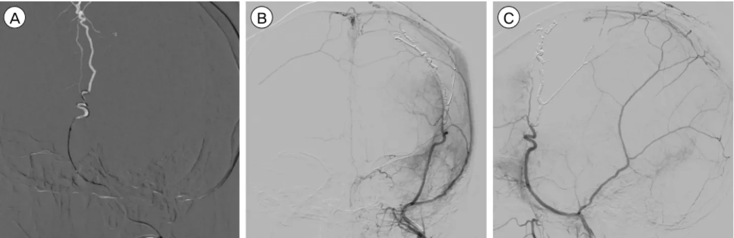
A B C
Fig. 4. After six days from second admission, Onyx embolization was performed. (A) Superselected left MMA with microcatheter and double micro-guidewire. AP view (B) and lateral view (C) after Onyx embolization show disappearance both of arteriovenous shunt- ing lesion and cortical venous reflux. MMA = middle meningeal artery; AP = anteroposterior.
problems.
DISCUSSION
Intracranial DAVF constitutes 10-15% of all intracranial vascular malformations, yet the natural course and pathogenesis of the lesions remain controversial.14)18)19)23)
It is generally accepted that the presentation of DAVF is dictated by the location and pattern of venous drainage. Moreover, the cortical venous reflux pattern is an important predictor of clinical course.7)8)17) DAVFs with an aggressive course have characteristic angio- graphic features such as cortical or leptomeningeal venous drainage and an associated varix.17) In a meta-analysis, Awad et al.2) reported a hemorrhage rate of 88% and a severe neurologic deficit in 12% of 100 aggressive cases among 377 patients with DAVF.
A previous study of Borden types II and III DAVFs reported an annual hemorrhage rate of 8.1% and an annual mortality rate of 10%.22) Another recent study reported an annual incidence of hemorrhage of 7.4%
for patients presenting with an intracranial hemor- rhage and 1.5% for those not presenting with hemorrhage.19) These discrepancies between the stud- ies are likely due to demographic differences, different patient inclusion criteria, and publication bias. Knowledge about the natural disease course and classification based on the venous drainage pattern can inform de-
cisions regarding the most appropriate treatment mo- dality for these patients.
However, DAVF is a dynamic disease that can un- dergo spontaneous angiographic venous drainage pat- tern conversion. Kim et al.11) reported pattern con- version as assessed by angiography in 18 of 112 cases (16.1%). Among these cases, the conversion rate to an aggressive lesion was 4%; all of these cases were asso- ciated with occlusion of the ipsilateral draining vein.
Moreover, several reports have described spontaneous closure of DAVF without any aggressive treatment.1)5)12) On the other hand, a few cases of conversion to ag- gressive lesions have been reported, and one study reported that benign AVFs have a 2% potential for angiographically verified conversion.18) Several theo- ries have been suggested to explain the conversion of venous drainage pattern, including sinus thrombosis, change of sinus wall structure, and flow dynamics.2)12)16) Cerebral sinus thrombosis can cause spontaneous re- gression with symptom resolution and can also force retrograde venous reflux through the cortical veins, predisposing the patient to an aggressive clinical course.11)18)21) Thus, cerebral sinus thrombosis can pro- mote the occlusion of venous outflow and result in dangerous rerouting. A histological study of DAVF suggested that microscopic thrombosis is always pres- ent, and that localizing thrombosis at the compartment or accessory mural channel of the drainage sinus may
cause shunt occlusions.15) Stenosis and thrombosis of the venous outlets have been reported to forecast later worsening of DAVFs. Our case also showed stenosis of the venous outlets and ultimately showed sponta- neous closure of the shunt into the superior saggital sinus and aggravated cortical venous reflux from Borden type II to type III.
Endovascular embolization has become the first line of treatment in patients with high-risk DAVFs, and surgery is considered only if endovascular treatments fail or are unfeasible. Trans-arterial approaches with embolic materials such as glue (NBCA: N-butyl cya- noacrylate), particles (PVA: polyvinyl alcohol), or Onyx® are performed to enable the embolic material to be pushed through the shunt into the proximal ve- nous outlet. In recent studies, Onyx® has demon- strated multiple advantages over other materials, with reported occlusion rates of 62.5-80%.6)13)20) However, in cases of potentially dangerous occult ECA-internal carotid artery or vertebral artery anastomosis, devel- opment of ischemic cranial nerve palsies, or in fistulas with multiple feeders, a trans-venous approach is recommended.17) Trans-venous approaches can be per- formed with detachable coils and particles in the most proximal venous outlet and have been the treatment of choice, especially for carotid cavernous DAVFs.
The shunt locations and cortical venous reflux pat- terns may predict the clinical manifestations and drainage patterns of DAVFs. Superior saggital sinus DAVFs, as in our case, represent about 8-13% of all intracranial fistulae.10) These DAVFs are often accom- panied by hemorrhage or progressive neurological deficits.24) These fistulae are characterized by bilateral supply from the MMA, ophthalmic artery, or posteri- or meningeal artery with critical venous drainage and require aggressive treatment.10) Despite the variety of treatment modalities available, trans-arterial emboliza- tion is recommended in superior saggital sinus DAVFs with adjuvant surgery or radiosurgery following failed embolization.3)9) In our case, we performed Onyx® embolization through the MMA, and successful ob- literation was achieved in a single session. Due to the
properties and increased predictability of Onyx®, more prolonged injections through single feeders like the MMA can be performed compared with other em- bolic materials.14) Moreover, the excellent flow control enables greater injection control, even when the vessel diameter is very small. With the newer and eas- ier-to-navigate micro-catheters and the advent of Onyx®, trans-arterial embolization has become the ac- cepted initial treatment for DAVF.
CONCLUSION
Although most cases of venous drainage pattern conversion in DAVF show benign clinical courses and do not develop symptoms, aggressive conversion sometimes occurs. Therefore, close clinical and radio- logical follow-up is needed to detect these lesions.
Angiographic features should be the most important factor in deciding treatment modality. Our results in- dicate that endovascular embolization with Onyx® is a reliable treatment with acceptable safety and efficacy.
Disclosure
The authors report no conflict of interest concerning the materials or methods used in this study or the findings specified in this paper.
REFERENCES
1. Al-Afif S, Nakamura M, Gotz F, Krauss JK. Spontaneous closure of a dural arteriovenous fistula. BMJ Case Rep.
2014 Jul;21;1-6.
2. Awad IA, Little JR, Akarawi WP, Ahl J. Intracranial du- ral arteriovenous malformations: factors predisposing to an aggressive neurological course. J Neurosurg. 1990 Jun;72(6):839-50.
3. Bertalanffy A, Dietrich W, Kitz K, Bavinzski G.
Treatment of dural arteriovenous fistulae (dAVF's) at the superior sagittal sinus (SSS) using embolisation com- bined with micro- or radiosurgery. Minim Invasive Neurosurg. 2001 Dec;44(4):205-10.
4. Chaudhary MY, Sachdev VP, Cho SH, Weitzner I Jr, Puljic S, Huang YP. Dural arteriovenous malformation of the major venous sinuses: an acquired lesion. AJNR Am J Neuroradiol. 1982 Jan-Feb;3(1):13-9.
5. Clarencon F, Biondi A, Sourour NA, Di Maria F, Iosif C, Nouet A, et al. Spontaneous closure of intracranial
dural arteriovenous fistulas: a report of 3 cases. Clin Neurol Neurosurg. 2013 Jul;115(7):971-5.
6. Cognard C, Januel AC, Silva NA Jr, Tall P. Endovascular treatment of intracranial dural arteriovenous fistulas with cortical venous drainage: new management using Onyx.
AJNR Am J Neuroradiol. 2008 Feb;29(2):235-41.
7. Davies MA, TerBrugge K, Willinsky R, Coyne T, Saleh J, Wallace MC. The validity of classification for the clin- ical presentation of intracranial dural arteriovenous fistulas.
J Neurosurg. 1996 Nov;85(5):830-7.
8. Fermand M, Reizine D, Melki JP, Riche MC, Merland JJ.
Long term follow-up of 43 pure dural arteriovenous fis- tulae (AVF) of the lateral sinus. Neuroradiology. 1987;29(4):
348-53.
9. Ghobrial GM, Marchan E, Nair AK, Dumont AS, Tjoumakaris SI, Gonzalez LF, et al. Dural arteriovenous fistulas: a review of the literature and a presentation of a single institution's experience. World Neurosurg. 2013 Jul-Aug;80(1-2):94-102.
10. Halbach VV, Higashida RT, Hieshima GB, Rosenblum M, Cahan L. Treatment of dural arteriovenous malfor- mations involving the superior sagittal sinus. AJNR Am J Neuroradiol. 1988 Mar-Apr;9(2):337-43.
11. Kim DJ, terBrugge K, Krings T, Willinsky R, Wallace C.
Spontaneous angiographic conversion of intracranial du- ral arteriovenous shunt: long-term follow-up in non- treated patients. Stroke. 2010 Jul;41(7):1489-94.
12. Luciani A, Houdart E, Mounayer C, Saint Maurice JP, Merland JJ. Spontaneous closure of dural arteriovenous fistulas: report of three cases and review of the literature.
AJNR Am J Neuroradiol. 2001 May;22(5):992-6.
13. Lv X, Jiang C, Zhang J, Li Y, Wu Z. Complications re- lated to percutaneous transarterial embolization of intra- cranial dural arteriovenous fistulas in 40 patients. AJNR Am J Neuroradiol. 2009 Mar;30(3):462-8.
14. Natarajan SK, Ghodke B, Kim LJ, Hallam DK, Britz GW, Sekhar LN. Multimodality treatment of intracranial dural arteriovenous fistulas in the Onyx era: a single cen- ter experience. World Neurosurg. 2010 Apr;73(4):365-79.
15. Piske RL, Campos CM, Chaves JB, Abicalaf R, Dabus G, Batista LL, et al. Dural sinus compartment in dural arte- riovenous shunts: a new angioarchitectural feature allowing superselective transvenous dural sinus occlusion treatment.
AJNR Am J Neuroradiol. 2005 Aug;26(7):1715-22.
16. Saito A, Furuno Y, Nishimura S, Kamiyama H, Nishijima M. Spontaneous closure of transverse sinus dural arte- riovenous fistula: case report. Neurol Med Chir (Tokyo).
2008 Dec;48(12):564-8.
17. Santillan A, Nanaszko M, Burkhardt JK, Patsalides A, Gobin YP, Riina HA. Endovascular management of in- tracranial dural arteriovenous fistulas: a review. Clin Neurol Neurosurg. 2013 Mar;115(3):241-51.
18. Satomi J, van Dijk JM, Terbrugge KG, Willinsky RA, Wallace MC. Benign cranial dural arteriovenous fistulas:
outcome of conservative management based on the natural history of the lesion. J Neurosurg. 2002 Oct;97(4):767-70.
19. Soderman M, Pavic L, Edner G, Holmin S, Andersson T. Natural history of dural arteriovenous shunts. Stroke.
2008 Jun;39(6):1735-9.
20. Stiefel MF, Albuquerque FC, Park MS, Dashti SR, McDougall CG. Endovascular treatment of intracranial dural arteriovenous fistulae using Onyx: a case series.
Neurosurgery. 2009 Dec;65(6 Suppl):132-9; discussion 139-40.
21. Tsai LK, Jeng JS, Liu HM, Wang HJ, Yip PK.
Intracranial dural arteriovenous fistulas with or without cerebral sinus thrombosis: analysis of 69 patients. J Neurol Neurosurg Psychiatry. 2004 Nov;75(11):1639-41.
22. van Dijk JM, terBrugge KG, Willinsky RA, Wallace MC.
Clinical course of cranial dural arteriovenous fistulas with long-term persistent cortical venous reflux. Stroke.
2002 May;33(5):1233-6.
23. Webb S, Hopkins LN. Intracranial dural arteriovenous fistulas: a treatment paradigm in flux. World Neurosurg.
2013 Jul-Aug;80(1-2):47-9.
24. Woo HH, Masaryk TJ, Rasmussen PA. Treatment of du- ral arteriovenous malformations and fistulae. Neurosurg Clin N Am. 2005 Apr;16(2):381-93.