Outcomes of Abdominal Total Gastrectomy for Type II and III Gastroesophageal Junction Tumors: Single Center’s
Experience in Korea
Kyoung Tai Kim, Oh Jeong, Mi Ran Jung, Seong Yeop Ryu, and Young-Kyu Park
Division of Gastroenterologic Surgery, Department of Surgery, Chonnam National University Medical School, Gwangju, Korea
Purpose: The aim of this study was to evaluate the surgical outcomes of abdominal total gastrectomy, without mediastinal lymph node dissection for type II and III gastroesophageal junction (GEJ) cancers.
Materials and Methods: We retrospectively reviewed surgical outcomes in 67 consecutive patients with type II and III GEJ cancers that were treated by the surgical resection between 2004 and 2008.
Results: Thirty (45%) patients had type II and 37 (55%) had type III tumor. Among the 65 (97%) patients with curative surgery, 21 (31%) patients underwent the extended total gastrectomy with trans-hiatal distal esophageal resection, and in 44 (66%) patients, abdominal total gastrectomy alone was done. Palliative gastrectomy was performed in two patients due to the accompanying perito- neal metastasis. The postoperative morbidity and mortality rates were 21.4% and 1.5%, respectively. After a median follow up of 36 months, the overall 3-years was 68%, without any differences between the Siewert types or the operative approaches (transhiatal ap- proach vs. abdominal approach alone). On the univariate analysis, the T stage, N stage and R0 resection were found to be associated with the survival, and multivariate analysis revealed that the N stage was a poor independent prognostic factor for survival.
Conclusions: Type II and III GEJ cancers may successfully be treated with the abdominal total gastrectomy, without mediastinal lymph node dissection in the Korean population.
Key Words: Surgery; Esophagogastric junction; Stomach neoplasms; Korea
Correspondence to: Oh Jeong
Division of Gastroenterologic Surgery, Department of Surgery, Chonnam National University Medical School, 160, Ilsim-ri, Hwasun- eup, Hwasun-gun 519-809, Korea
Tel: +82-61-379-7646, Fax: +82-61-379-7661 E-mail: [email protected]
Received February 23, 2011 Revised March 7, 2012 Accepted March 7, 2012
Copyrights © 2012 by The Korean Gastric Cancer Association www.jgc-online.org
This is an open-access article distributed under the terms of the Creative Commons Attribution Non-Commercial License (http://creativecommons.org/
licenses/by-nc/3.0) which permits unrestricted noncommercial use, distribution, and reproduction in any medium, provided the original work is properly cited.
Introduction
Despite the decreasing global incidence of gastric cancer, the gastroesophageal junction (GEJ) cancers have greatly increased in the Western countries.(1) However, distal gastric cancer is still a predominant type among gastric cancers in Korea and Japan(2,3) and patients are often diagnosed at an early stage with the use of
endoscopy for mass screening.(4-6) Besides, the epidemiology of GEJ cancers is also quite different from that of the West, in which type I GEJ cancer is extremely rare, while type II and III account for the types of the GEJ cancers in Korea.
Although achieving complete tumor resection (R0 resection) is the mainstay of surgical treatment for GEJ cancers, the operative approaches, especially regarding the need for thoracotomy, still remain controversial.(7) Because of the frequent tumor infiltration, at least 6 cm of grossly normal esophageal resection has been ad- vocated by several groups to achieve negative resection margins in the West.(8-10) Some investigators have even insisted that as long as 10 cm of esophageal resection is required to minimize the risk of positive proximal margins.(11,12) To secure these long proximal resection margins, thoracotomy often should be performed along with abdominal total gastrectomy. Possible metastasis to the medi-
astinal lymph nodes is another concern necessitating thoracotomy in GEJ cancer. However, the relationship between the operative approaches and survival still remains to be investigated.
Although GEJ cancer are surgically treated as another entity of esophageal cancer in the West, the surgical approach to GEJ cancers in Korea is just similar to that for gastric cancer, in which abdominal total gastrectomy without mediastinal lymph node dis- section is commonly performed. To date, there were few case and study about the evaluation of the outcome of abdominal total gastrectomy in GEJ cancer in asia. In this study, we evaluated the surgical outcomes of abdominal total gastrectomy with the selective transhiatal approach that was perform for type II and III GEJ can- cers to establish the optimal operative approach for these tumors and to define the prognostic factors. The aim of this study was to evaluate the short term and the long term results of the surgical procedure that was removal of complete tumor margin with the selective transhiatal approach or not.
Materials and Methods
1. Patients
From 2004 to 2008, 1775 patients underwent surgery for gastric carcinoma at our institution. By reviewing the operation record, pathological reports, and surgical specimen pictures, 67 (3.8%) pa- tients with type II and III GEJ cancers and who were treated by surgical resection were included in this study. GEJ cancers were defined as tumors whose center was within 5 cm proximal and distal of the anatomical cardia, and were classified according to the Siewert’s classification of GEJ cancer.(13)
All patients were preoperatively evaluated with endoscopy along with biopsy, abdominal computed tomography (CT) scanning, and a barium swallow study. Endoscopic ultrasound was performed in selected cases. None of the patients in this study received chemo- therapy or radiation therapy before the operation. After operation, the patients were followed up every six months for the first three years, and then every 12 months after that. Disease recurrence was detected by physical examination, tumor markers, endoscopy and abdominal CT scanning.
The data regarding the patient demographics, the operative procedures, the pathologic results and the short-term surgical out- comes was obtained from a retrospective review of the medical records. The histologic differentiation and staging were principally based on the 6th edition of the International Union Against Can- cer TNM classification for gastric carcinoma.(14) The information
regarding disease recurrence and survival were obtained during outpatient clinical visits and by telephone interviews.
2. Operative procedures
The choice of surgical approach was based on the goal of achieving complete macroscopic and microscopic tumor resection.
In general, abdominal total gastrectomy was performed with resec- tion of the distal esophagus with at least 3 cm of a gross surgical margin from the tumor. The transhiatal approach was performed in selected cases where complete resection was less likely to be achieved with the abdominal approach alone, based on the preop- erative examinations or the intraoperative findings. Intraoperative frozen section analysis was performed in every operation, and the adequacy of the extent of esophageal resection was confirmed.
For most patients, D2 lymph node dissection was performed as a standard procedure as is outlined by the principles of the Japanese Classification of Gastric Carcinoma.(15) And mediastinal lymph node dissection was not undertaken in this study.
The mode of reconstruction was mechanical esophagojejunos- tomy or jejunal interposition, and the mode was selected according to the surgeon’s preference.
3. Statistics
Statistical analysis was performed using the software SPSS ver- sion 12.0 statistical package for windows (SPSS Inc., Chicago, IL, USA). Chi-square tests or Fisher’s exact tests and the student’s t- test were used for comparisons as appropriate. Patient survival was calculated using the Kaplan-Meier method and the patient survival between groups was compared using the log rank test. Univariate and multivariate analysis were performed using the Cox regression models to identify the prognostic factors. A P-value <0.05 was considered statistically significant throughout the study.
Results
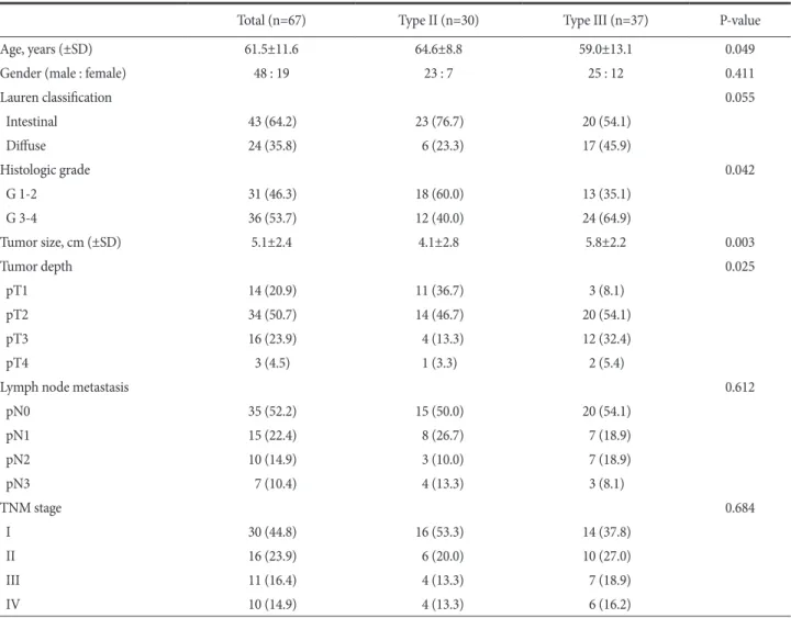
The patients consisted of 48 men and 19 women with a mean age of 61.5±11.6 years. The tumor location described in the final pathologic report was based on Siewert classification, and this re- vealed 30 type II and 37 type III tumors. No significant differences were observed between the type II and III tumors with respect to gender, Lauren’s classification, the N stage, and the presence of Barrett’s esophagus (Table 1). However, there were significant dif- ferences in age, the histologic grade, the tumor size, and the T stage between the two groups: an older age, more histologic grade G1/2
tumors, a smaller tumor size, and more pT1/2 stage tumors were the specific features of the type II tumors.
With respect to lymph node metastasis, both tumor types showed similar patterns of lymphatic spread. For both types of tu- mor, lymph nodes number 1, 2 and 3 were the most commonly in- volved lymph nodes, with spreading toward the celiac lymph nodes (Fig. 1).
1. Surgical outcomes
Of the 67 patients, 65 (97.0%) patients underwent surgery with a curative intent, and two patients underwent palliative gastrectomy due to disseminated peritoneal metastasis. Twenty-two (33%) patients required the transhiatal approach for securing esophageal resection margin, while 45 (67%) patients required the abdominal approach only (Table 2). As a result, the transhiatal approach was Table 1. The clinicopathological features of the GEJ cancers
Total (n=67) Type II (n=30) Type III (n=37) P-value
Age, years (±SD) 61.5±11.6 64.6±8.8 59.0±13.1 0.049
Gender (male : female) 48 : 19 23 : 7 25 : 12 0.411
Lauren classification 0.055
Intestinal 43 (64.2) 23 (76.7) 20 (54.1)
Diffuse 24 (35.8) 6 (23.3) 17 (45.9)
Histologic grade 0.042
G 1-2 31 (46.3) 18 (60.0) 13 (35.1)
G 3-4 36 (53.7) 12 (40.0) 24 (64.9)
Tumor size, cm (±SD) 5.1±2.4 4.1±2.8 5.8±2.2 0.003
Tumor depth 0.025
pT1 14 (20.9) 11 (36.7) 3 (8.1)
pT2 34 (50.7) 14 (46.7) 20 (54.1)
pT3 16 (23.9) 4 (13.3) 12 (32.4)
pT4 3 (4.5) 1 (3.3) 2 (5.4)
Lymph node metastasis 0.612
pN0 35 (52.2) 15 (50.0) 20 (54.1)
pN1 15 (22.4) 8 (26.7) 7 (18.9)
pN2 10 (14.9) 3 (10.0) 7 (18.9)
pN3 7 (10.4) 4 (13.3) 3 (8.1)
TNM stage 0.684
I 30 (44.8) 16 (53.3) 14 (37.8)
II 16 (23.9) 6 (20.0) 10 (27.0)
III 11 (16.4) 4 (13.3) 7 (18.9)
IV 10 (14.9) 4 (13.3) 6 (16.2)
The numbers in parenthesis are percentages. TNM stage is based on the sixth edition of UICC/AJCC TNM classification. GEJ = gastroesophageal junction; SD = standard deviation; UICC = International Union Against Cancer; AJCC = American Joint Committee on Cancer.
Fig. 1. Lymph node metastasis at each regional station in the patients with type II and III tumors. These two tumor types had similar patterns of lymphatic spread, with most commonly involving the lymph node station 1, 2, and 3 nodes and the spread was directed toward the celiac nodes.
performed for 19 (63%) of the 30 type II tumors and for 3 (8%) of the 37 type III tumors. The mean proximal margin length (cm) was 2.26±0.74 (range: 1.0~4.0 cm), and none of the patients revealed residual tumor cells at the esophageal resection margin. Combined spleen or pancreas resection was performed in 24 (35.8%) patients.
Sixty-four (95.5%) patients underwent lymphadenectomy of more than D2, and the mean number of harvested lymph nodes was 45±9.
A total of 17 (25.4%) patients developed postoperative compli- cations with one in-hospital death. The most common complica- tion was pancreatic fistula (n=5) and this was followed by abdomi- nal infection (n=4), and surgical wound infection (n=2). The mean hospital stay (in days) was 16±3.
2. Survival and the prognostic factors
With a median follow-up of 36 months (range: 13 to 58 months), the overall survival rate at 3 years was 68%: it was 70%
for patients with type II tumors versus 67% for the patients with type III tumors (P=0.546), and 72% for the transhiatal approach vs. 66% for the abdominal approach alone (P=0.811). A total of 14
patients developed recurrence during the follow up: there were 11 cases of peritoneal seeding, 3 cases of distant lymph node metasta- ses, 2 cases of hematogenous metastases and 4 cases of locoregional recurrences. No patients had recurrence at the mediastinal lymph nodes during the follow-up. Meanwhile, no significant difference of the recurrence pattern was observed according to the Siewert types or the surgical approaches.
On the univariate analysis, the T stage (T1-2 vs. T3-4), N stage (N0 vs. N+) and R0 resection were significantly associated with survival, while the Siewert type or the transhiatal approach was not.
Multivariate analysis of the significant variables on the univariate analysis showed that lymph node metastasis (hazard ratio=11.97, 95% confidence interval=2.657~63.98) was an independent factor for a poor prognosis (Table 3).
Discussion
Adenocarcinomas of the GEJ are biologically aggressive and they are typically diagnosed at an advanced stage of disease pro- Table 2. The surgical outcomes
Total (n=67) Type II (n=30) Type III (n=37) P-value
Curability 0.724
R0 resection 65 (97.0) 29 (96.7) 36 (97.3)
Non-R0 resection 2 (3.0) 1 (3.3) 1 (2.7)
Surgical approach <0.001
Total gastrectomy only 45 (67.2) 11 (36.7) 33 (91.7)
Combined trans-hiatal approach 22 (32.8) 19 (63.3) 3 (8.3)
Lymph node dissection 0.583
D2 or more 64 (95.5) 28 (93.3) 36 (97.3)
Less than D2 3 (4.5) 2 (6.7) 1 (2.7)
Reconstruction 1.000
Jejunal interposition 11 (16.4) 4 (13.3) 6 (16.2)
Esophagojejunostomy 56 (83.6) 26 (86.7) 31 (83.8)
Combined resection 24 (35.8) 7 (23.3) 17 (45.9) 0.096
Spleen 12 4 8
Distal pancreas 11 2 9
Others 1 1 0
Proximal margin (cm, ±SD) 2.26±0.74 2.10±0.64 2.38±0.80 0.120
Retrieved lymph nodes (±SD) 45±9 44±14 46±22 0.637
Hospital stay (d, ±SD) 16±3 15±4 16±5 0.438
Morbidity 17 (25.4) 10 (33.3) 7 (18.9) 0.178
Mortality 1 (1.5) 1 (3.3) 0 0.448
The numbers in parenthesis are percentages. SD = standard deviation.
gression. As a result, they are difficult to treat and the patient prog- nosis is poor even after curative surgical resection. The five-year survival rate after surgical resection has been reported to range only 16% to 32.3%.(11,16,17) Although such reports are rare, the GEJ cancers in East Asia show significant differences of their clinico- pathological features as compared with those observed in the West.
First, its incidence is very low despite of the high prevalence of gastric cancer. Second, distal esophageal adenocarcinomas related to Barrett’s mucosa (type I GEJ cancer) are rarely seen, and most GEJ cancers arise in the gastric cardia or subcardial region. Third, patients in these areas are often diagnosed at an early stage of disease because of the widespread use of endoscopy screening.(4-6) Thus, considering these distinctive features, abdominal total gastrectomy is frequently performed for GEJ cancers in Korea, rather than tho- raco-abdominal approach in the West, but only a few studies have reported surgical outcomes of this procedure. This study shows that abdominal total gastrectomy with selective trans-hiatal approach is appropriate in terms of securing safe proximal resection margin and acceptable postoperative morbidity. Besides, long term out- comes were also similar to the historical controls.
Achieving complete tumor resection is the mainstay of the sur- gical treatment for GEJ cancers. The surgical approaches to achieve R0 resection for these tumors vary widely from esophagectomy through the transthoracic or transmediastinal approach to extended total gastrectomy with transhiatal resection.(18) Because of the frequent tumor infiltration at the esophageal resection margin, ex- tended esophageal resection has been advocated by several research groups. Ito et al.(9) recommended that the surgical approach should
be directed based on the goal of obtaining at least 6 cm of a gross proximal resection margin to avoid residual cancer cells at the re- section margin, and this was generally consistent with that reported by Barbour et al.,(19) Bezzetti et al.,(8) and Mattioli et al.(10) How- ever, most series have not demonstrated a survival benefit for one operative approach over another,(20,21) and proximal resection margin infiltration does not universally translate to poor survival when performing curative resection.(22)
In general, it has been well accepted that the selection of the surgical approach to GEJ cancers should be tailored based upon achieving macroscopic and microscopic tumor resection.(23) In this study, with performing grossly normal esophageal resection of only 3 cm, complete (R0) tumor resection was performed in as many as 97% of the patients. The rate of R0 resection of this study was considerably higher than those studies in the West that reported a 55% to 75% rate of complete resection.(9,19,24) Unlike the strong correlation of a positive resection margin with the ex- tent of the surgical resection as observed by Mattioli et al.,(10) our study found no difference in R0 resection between the abdominal approach and the transhiatal approach. Complete tumor resection was accomplished via the abdominal approach alone in 43 (64%) patients, and the transhiatal approach was required only in 22 (33%) patients. Microscopic mural extension of cancer cells was very rare in our experience, as reflected by the fact that no positive resection margin was observed for all patients who underwent surgery with a curative intent. Therefore, our study suggests that the required extent of esophageal resection for GEJ cancers in Korea may differ from those insisted in the West, and the surgical approach could Table 3. The prognostic factors on the univariate and multivariate analysis
Variables Univariate Multivariate
HR (95% CI) P-value HR (95% CI) P-value
Age (≤65 yr vs. >65 yr) 0.593 (0.233~1.511) 0.274
Gender (male vs. female) 1.243 (0.472~3.273) 0.660
Siewert type (type 2 vs. 3) 1.325 (0.631~3.305) 0.547
T stage (T1-2 vs. 3-4) 3.853 (1.549~9.584) 0.004 2.377 (0.895~6.317) 0.082
N stage (N- vs. N+) 14.731 (3.368~64.434) < 0.001 11.97 (2.657~53.98) 0.001
Curability (R0 vs. R1-2) 6.058 (1.726~21.264) 0.005 1.776 (0.476~6.621) 0.393
Lauren classification (intestinal vs. non-intestinal) 1.137 (0.447~2.889) 0.788 Surgical approach (TG vs. TG+THA) 1.072 (0.834~1.329) 0.543 Histologic grade (G1-2 vs. G3-4) 1.546 (0.620~3.855) 0.349
Tumor size 1.190 (0.993~1.426) 0.060
HR = hazard ration; CI = confidence interval; TG = total gastrectomy; THA = transhiatal approach.
be tailored using a less extensive esophageal resection, such as the selective use of the transhiatal approach based on abdominal total gastrectomy.
The application of less extensive esophageal resection and the resulting high R0 resection rate in this study may be explained that the distinctive surgical epidemiology of GEJ cancers in Korea is different from those observed in the West. It is well known that the degree of mural extension is strongly correlated with the T stage.(12,25) In Western countries, most patients with GEJ cancers are typically diagnosed at an advanced stage as having T3 or T4 tumors, and lymph node metastasis.(18,19) On the contrary, our study found over 70% of the patients had tumors limited within the serosal layer (T1 or T2), and nearly 50% of the patients had no lymph node metastasis. Controversy continues concerning the opti- mal lymph node dissection when we perform GEJ cancer surgery.
de Manzoni et al.(16)’s study suggested to perform total Gastrec- tomy with D2 lymphadenectomy with advanced cardia cancer type II or III. Pedrazzani et al.(26)’s study said that chest nodal involve- ment rate was 46.2% in type I, 29.5% in type II, 9.3% for type III.
Siewert et al.(18)’s study suggested that lower mediastinal nodal involvement rate was 12% in type II, 5% in type III. So, our study performed D2 lymphadenectomy without mediastinal node dissec- tion. Moreover, most GEJ cancers in our experience were limited in the cardia or subcardial region, which means that the vast ma- jority of these tumors must be considered as gastric in origin and they should be treated as such. These characteristics of GEJ cancer observed in our study are consistent with those of other domes- tic reports from Korea.(4,6) However, further studies with a large number of patients are needed to ascertain the surgical epidemiol- ogy of GEJ cancers in Korea, and the optimal surgical approach should be determined based on these result.
In conclusion, our experience has shown that complete tumor resection can be achieved with less extensive esophageal resec- tion as compared to those performed in the West. To achieve R0 resection, abdominal total gastrectomy with the selective use of the transhiatal approach was appropriate for treating GEJ cancer in Korea. This may be because the biologic and clinical features of GEJ cancer in Korea are different from those of the West, yet further studies with a large number of patients will be needed to establish the proper surgical approach to GEJ cancer in Korea.
References
1. Parkin DM, Bray F, Ferlay J, Pisani P. Global cancer statistics,
2002. CA Cancer J Clin 2005;55:74-108.
2. Blaser MJ, Saito D. Trends in reported adenocarcinomas of the oesophagus and gastric cardia in Japan. Eur J Gastroenterol Hepatol 2002;14:107-113.
3. Lee JY, Kim HY, Kim KH, Jang HJ, Kim JB, Lee JH, et al. No changing trends in incidence of gastric cardia cancer in Korea.
J Korean Med Sci 2003;18:53-57.
4. Kim HJ, Kwon SJ. Analysis of clinocopathologic difference between type II and type III cancers in siewert classification for adenocarcinomas of the cardia. J Korean Gastric Cancer Assoc 2004;4:143-148.
5. Kodera Y, Yamamura Y, Shimizu Y, Torii A, Hirai T, Yasui K, et al. Adenocarcinoma of the gastroesophageal junction in Japan:
relevance of Siewert's classification applied to 177 cases resect- ed at a single institution. J Am Coll Surg 1999;189:594-601.
6. Park CH, Kang WK, Song KY, Bae JS, Kim JJ, Park SM, et al.
Adenocarcinoma of the gastro-esophageal junction: Applica- tion of Siewert's classification to the Eastern experience. J Ko- rean Gastric Cancer Assoc 2004;4:36-43.
7. von Rahden BH, Stein HJ, Siewert JR. Surgical management of esophagogastric junction tumors. World J Gastroenterol 2006;12:6608-6613.
8. Bozzetti F, Bonfanti G, Bufalino R, Menotti V, Persano S, An- dreola S, et al. Adequacy of margins of resection in gastrectomy for cancer. Ann Surg 1982;196:685-690.
9. Ito H, Clancy TE, Osteen RT, Swanson RS, Bueno R, Sugar- baker DJ, et al. Adenocarcinoma of the gastric cardia: what is the optimal surgical approach? J Am Coll Surg 2004;199:880- 886.
10. Mattioli S, Di Simone MP, Ferruzzi L, D'Ovidio F, Pilotti V, Carella R, et al. Surgical therapy for adenocarcinoma of the cardia: modalities of recurrence and extension of resection. Dis Esophagus 2001;14:104-109.
11. Mariette C, Castel B, Toursel H, Fabre S, Balon JM, Triboulet JP. Surgical management of and long-term survival after ad- enocarcinoma of the cardia. Br J Surg 2002;89:1156-1163.
12. Tsujitani S, Okuyama T, Orita H, Kakeji Y, Maehara Y, Sugima- chi K, et al. Margins of resection of the esophagus for gastric cancer with esophageal invasion. Hepatogastroenterology 1995;42:873-877.
13. Siewert JR, Stein HJ. Classification of adenocarcinoma of the oesophagogastric junction. Br J Surg 1998;85:1457-1459.
14. Sobin LH, Wittekind C, International Union against Cancer, eds. TNM Classification of Malignant Tumours. 6th ed. New
York: Willey-Liss, 2002.
15. Japanese Gastric Cancer Association. Japanese Classification of Gastric Carcinoma - 2nd English Edition -. Gastric Cancer 1998;1:10-24.
16. de Manzoni G, Pedrazzani C, Pasini F, Di Leo A, Durante E, Castaldini G, et al. Results of surgical treatment of adenocar- cinoma of the gastric cardia. Ann Thorac Surg 2002;73:1035- 1040.
17. Rüdiger Siewert J, Feith M, Werner M, Stein HJ. Adenocar- cinoma of the esophagogastric junction: results of surgical therapy based on anatomical/topographic classification in 1,002 consecutive patients. Ann Surg 2000;232:353-361.
18. Siewert JR, Feith M, Stein HJ. Biologic and clinical variations of adenocarcinoma at the esophago-gastric junction: relevance of a topographic-anatomic subclassification. J Surg Oncol 2005;90:139-146.
19. Barbour AP, Rizk NP, Gonen M, Tang L, Bains MS, Rusch VW, et al. Adenocarcinoma of the gastroesophageal junction: influ- ence of esophageal resection margin and operative approach on outcome. Ann Surg 2007;246:1-8.
20. Hulscher JB, van Sandick JW, de Boer AG, Wijnhoven BP, Tijs- sen JG, Fockens P, et al. Extended transthoracic resection com- pared with limited transhiatal resection for adenocarcinoma of
the esophagus. N Engl J Med 2002;347:1662-1669.
21. Sasako M, Sano T, Yamamoto S, Sairenji M, Arai K, Kinoshita T, et al; Japan Clinical Oncology Group (JCOG9502). Left thora- coabdominal approach versus abdominal-transhiatal approach for gastric cancer of the cardia or subcardia: a randomised con- trolled trial. Lancet Oncol 2006;7:644-651.
22. Kim SH, Karpeh MS, Klimstra DS, Leung D, Brennan MF.
Effect of microscopic resection line disease on gastric cancer survival. J Gastrointest Surg 1999;3:24-33.
23. DeMeester SR. Adenocarcinoma of the esophagus and cardia:
a review of the disease and its treatment. Ann Surg Oncol 2006;13:12-30.
24. Carboni F, Lorusso R, Santoro R, Lepiane P, Mancini P, Sper- duti I, et al. Adenocarcinoma of the esophagogastric junction:
the role of abdominal-transhiatal resection. Ann Surg Oncol 2009;16:304-310.
25. Casson AG, Darnton SJ, Subramanian S, Hiller L. What is the optimal distal resection margin for esophageal carcinoma?
Ann Thorac Surg 2000;69:205-209.
26. Pedrazzani C, de Manzoni G, Marrelli D, Giacopuzzi S, Corso G, Minicozzi AM, et al. Lymph node involvement in advanced gastroesophageal junction adenocarcinoma. J Thorac Cardio- vasc Surg 2007;134:378-385.