하지 심부 정맥 혈전증의 중재적 방사선적 치료
Interventional Radiologic Treatment of
Deep Venous Thrombosis in Lower Extremity
원 제 환 | 아주의대 영상의학과 | Je Hwan Won, MD
Department of Diagnostic Radiology, Ajou University School of Medicine E-mail : [email protected]
J Korean Med Assoc 2007; 50(1): 80 - 87
D
eep vein thrombosis (DVT) is a common disease; however, it can result in significant disabilities from pulmonary embolism and postphlebitic syndrome, especially when the iliofemoral vein is involved. Although anticoagulation can prevent thrombus propagation and recurrent venous thrombosis, it cannot dissolve the occluding thrombus or reduce venous outflow obstruction, leaving the patients suffering from postphlebitic syndrome.Catheter-directed thrombolysis, with direct delivery of a concentrated lytic agent into the clot, has been proposed as an alternative therapy to anticoagulation. Recent studies have demonstrated that early clot lysis through this technique rapidly restores venous patency, more effectively preserves valvular function, and reduces a risk of postphlebitic syndrome. To decrease clot burden, duration of treatment, and bleeding complications, mechanical thrombectomy may work synergistically with catheter-directed thrombolysis. After clot removal through those therapies, balloon angioplasty and stent placement are needed in patients with venous stricture (eg, iliac vein compression syndrome). Short- and long-term outcomes of stenting in iliofemoral DVT appear to be favorable.
When used in conjunction with each other and anticoagulation, these minimally invasive endovascular techniques allow a better resolution of venous clot burden and have the potential to lead to improved long-term outcomes in patients with DVT. This review introduces endovascular treatments of acute DVT in the lower extremities performed in the interventional radiology section.
Keywords : Deep vein thrombosis; thrombolysis; thrombectomy; percutaneous intervention
핵 심 용 어 : 심부정맥혈전증; 혈전용해술; 혈전제거술; 경피적 중재치료Abstract
만명 이상이 폐혈전색전증으로 사망한다(2). 폐혈전색전 증과 같은 급성 합병증 외에도 심부정맥혈전증은 만성 정맥 부전에 의한 postphlebitic syndrome을 일으킬 수 있다. Postphlebitic syndrome은 하지의 부종, 통증, 궤 양 및 정맥성 절뚝거림(venous claudication) 등의 증상 을 보이는 증후군으로 피부에 과다색소침착이 나타날 수 도 있다. 엉덩넙다리 심부정맥혈전증에 대한 기왕력이 있는 환자의 90%에서 이와 같은 증상이 나타날 수 있고 10
~
15%에서는 급성기의 항응고제 치료에도 불구하고 궤양이 생길 수 있다(3, 4). 따라서 심부정맥혈전증의 치 료의 목적은 하지의 증상 정도 및 기간을 감소시키는 것, 폐혈전색전증의 예방, 정맥혈전증의 재발 방지, 정맥 혈 류 복원 및 밸브기능 유지, postphlebitic syndrome의 예방으로 요약될 수 있다(5, 6).헤파린을 이용한 항응고요법은 심부정맥혈전증의 고 전적 치료방법으로 알려져 있으나 엉덩넙다리 심부정맥 혈전증의 환자의 44%에서는 전형적인 항응고요법에도 불구하고 절뚝거림의 증상이 나타나는 것으로 알려져 있 다(7). 항응고요법이 혈전으로 막힌 혈관을 재개통하는 것은 오직 환자 자신의 섬유소용해능력에 의존할 수 밖 에 없으므로 엉덩넙다리정맥과 같이 다량의 정맥혈전이 있는 경우, 환자의 통증과 부종이 치유되는 데 많은 시간 이 걸릴 뿐 아니라 적절한 헤파린 치료에도 불구하고 환 자의 6%에서만이 혈전이 완전히 용해되며 40%에서는 오히려 혈전이 더 진행된다(8, 9). 따라서 엉덩넙다리정
맥과 같이 혈전의 양이 많은 경우에 보다 적극적으로 정 맥을 개통해주려는 치료방법이 모색되어 왔다. 본 의학 강좌에서 가장 대표적으로 시행되고 있는 대안적 치료방 법인 혈관내치료(endovascular treatment)에 대해 소개 하고자 한다.
Catheter - directed Thrombolysis
과거의 혈전용해술은 주로 스트렙토키나제나 유로키 나제를 표재정맥을 통해 주입하는 전신주사요법이었다.
전신 혈전용해술(systemic thrombolysis)은 헤파린 단 독 투여에 비하여 혈전을 녹이는데 우수할 뿐 아니라 치 료 후 postphlebitic syndrome 빈도도 줄일 수 있다고 보고되었다(8, 10). 그러나 이와 같은 방법은 많은 양의 혈전용해제가 전신으로 투여되기 때문에 출혈과 같은 합 병증의 발생빈도가 높아진다. 반면 catheter-directed thrombolysis는 혈전이 있는 심부 정맥에 다수의 측공이 있는 카테터를 삽입하고 고농도의 혈전용해제를 투여하 기 때문에 혈전용해제의 효과를 높일 수 있고, 필요한 혈 전용해제의 용량 및 투여 시간도 줄일 수 있다. 따라서 전
Figure 1.Multi-side hole infusion catheter. Thrombolytic drug are infused through multiple side holes of infusion catheter
신적인 섬유소 용해 효과가 적어지게 되어 출혈과 같은 합병증을 줄일 수 있는 장점이 있다(11, 12). 또한 정맥의 밸브 주변의 혈전도 용해시키므로 시술 후에도 밸브 기 능을 유지시켜 줄 수 있다.
Catheter-directed thrombolysis의 주요 적응증은 하대정맥 및 오금정맥 침범 여부와 관계 없이 엉덩넙다 리정맥의 급성 심부정맥혈전증이며 넙다리정맥에만 국 한된 경우에도 적응증이 된다. 그러나 오금정맥 하방의 장딴지정맥에만 국한되어 있는 경우는 적응증이 아니다.
항응고제, 혈관조영제, 혈전용해제 등의 사용이 금기가 되는 환자들도 대상에서 제외된다. 혈전 용해제 사용이 금기가 되는 경우는 급성 내부 출혈, 최근 10일 이내의 위 장관 출혈, 최근의 뇌혈관 사고 병력, 최근의 주요수술 경 력, 혈전용해제에 대한 알레르기가 있는 경우 심장내 혈 전, 심한 고혈압, 임신의 경우 등이다(11, 13). 또한 골반 부의 암종증에 의한 심부 정맥 혈전증 및 생존기간이 얼
마 남지 않은 말기암 환자도 대상군에서 제외된다.
1. 시술방법
시술 전에 엉덩정맥이나 하대정맥에 움직이는 혈전 (free floating thrombus)가 있거나 재발성 폐동맥 색전 증이 있는 환자 및 시술 도중 혈전의 이동의 위험성이 높 은 환자에서는 하대정맥 필터를 설치한다.
시술은 우선 환자가 엎드린 자세에서 초음파 유도 하 에 오금정맥을 천자 후 6F sheath를 삽입한다. Sheath를 통하여 5F multi-side-hole catheter(Figure 1)를 혈전 부위에 위치시킨 후에 infusion pump를 통하여 유로키 나제를 투여한다. 유로키나제 농도는 유로키나제 50만 IU 을 생리식염수 250mL에 섞는 것이 효과적이라고 알려져 있다. 주입 속도는 문헌마다 약간씩 차이가 있으며 초기 논문에서는 시간당 15
~
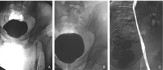
20만 IU을 주었으나 중대 합병 증의 발생을 줄이기 위해 최근에는 7~15만 IU으로 줄여Figure 2. A 59-year-old man with acute deep vein thrombosis
(A) Ascending venography shows extensive thrombus in left iliofemoral vein
(B) Multi- side- hole infusion catheter is embedded within thrombosed segment and urokinase is infused for 24 hours (C) After catheter directed thrombolysis, patency of the iliofemoral vein is restored
A B C
진행한다. 이 때 혈전의 완전용해는 남아있는 혈전이 5%
미만일 때로 정의한다(11, 14).
혈전의 완전용해를 보이고 정맥의 협착이 없는 경우에 는 더 이상의 인터벤션이 필요없다(Figure 2).
혈전의 완전 용해를 보이나 엉덩정맥의 협착이나 폐쇄 가 있는 경우에는 혈관확장술과 스텐트 삽입술을 시행 한다.
혈전의 완전 용해를 보이나 넙다리정맥의 협착이 있는 경우에는 혈관확장술만 시행한다.
부분적인 혈전 용해를 보이는 경우 fibrinogen 값이 100mg/dL 이상이고 출혈 합병증이 없다면 혈전 용해 제 주입을 계속한다.
엉덩정맥 및 넙다리정맥에 혈전 용해가 없는 경우에는 기술적 실패로 간주하고 더 이상의 인터벤션은 시행하 지 않는다.
혈전용해술이 끝난 후에도 정맥을 통한 헤파린의 투 여는 2~3일간 계속하며 퇴원 전에 lower molecular weight heparin의 피하 투여, warfarin의 경구투여 순으 로 항응고요법을 바꾼다. 보통 warfarin의 경구투여는 6개월 동안 계속되는데, 이 때 INR은 2
~
3 사이가 유지되 도록 한다.2. 결 과
초기의 보고들은 catheter-directed thrombolysis이 혈전을 빠르게 제거해 주는 데 효과적이라는 것을 보여
법 단독 치료와의 비교 연구에서는 catheter-directed thrombolysis를 받은 환자군에서 좀 더 나은 기능 및 복 지 지수(functional and well-being score)를 보이고 있다(17). 혈전용해술의 성공률은 혈전의 나이와 가장 관 계가 깊어서 10일 이내의 급성혈전에서 가장 높은 성공 률을 보이고 4주 이상의 만성혈전증에서는 성공률이 떨 어진다. 그 밖에도 악성종양 여부, 심부정맥혈전증의 기 왕력, 혈전의 용해 정도도 영향을 미친다. 악성종양의 경 우 응고 항진(hypercoagulable state) 때문에 정맥의 개 통률이 떨어지고 심부정맥혈전증의 기왕력이 있는 경우 도 치료성공률이 떨어진다.
3. 합 병 증
가장 흔한 합병증은 출혈이며 대부분 sheath 삽입 부 위 출혈이다. 이 외에도 혈전용해제의 전신적인 효과에 의한 원격 출혈이 발생할 수 있다. 수혈이 필요할 정도의 주요 출혈(major bleeding)의 빈도는 보고자마다 다양 하나 주로 혈전용해제의 종류, 용량 및 주입시간, 함께 주는 항응고제의 용량에 따라 달라지는 것으로 알려져 있다.
가장 많은 환자를 대상으로 한 National Venous Thrombolysis Registry에서의 주요 출혈의 빈도는 11%
였으나 주로 정맥 천자부위에서의 출혈이었다. 이 밖에 도 두개내 출혈은 0.4%, 증상이 있는 폐색전증은 1%의 빈도를 보였다(13).
기계적 혈전제거술 (Mechanical Thrombectomy)
Catheter-directed thrombolysis가 앞서 소개한 대 로 혈전이 있는 정맥의 빠른 개통 뿐 아니라 밸브의 기능 을 그대로 유지시키고 postphlebitic syndrome을 예방 한다는 점에서 매력적인 시술이 아닐 수 없다. 그러나 출 혈의 위험이 있다는 점, 혈전용해제 투여 동안에는 중환 자실 및 그에 준하는 집중적 감시가 필요하다는 점, 입원 기간이 길다는 점, 혈전 용해제의 높은 가격, 아직 무작위 전향적 임상연구가 없다는 점 등이 그 제한점으로 지적 되고 있다. 이를 극복하기 위하여 다양한 percutaneous mechanical thrombectomy device들이 개발되었다.
이들의 잠재적 장점은 보다 빠르게 정맥을 재개통 시키 고, 혈전용해제의 사용을 줄여 출혈 합병증의 위험을 줄 이며 입원기간을 단축시키는 등 전체적인 비용의 절감을 가져올 수 있다는 것이다(18).
현재 우리나라에서 가장 널리 쓰이는 기계적 혈전 제
거방법은 7~10F guiding catheter 혹은 introducer를 이용한 흡입술이다. 이 방법은 카테터에 음압을 주어 혈 전을 빨아내는 방법으로 매우 경제적이며, 급성혈전은 대부분 이러한 방법으로 제거할 수 있어 유로키나제 사 용을 피할 수 있거나 용량을 줄일 수 있다. 이 밖에도 현 재 국내에서 사용 가능한 기구는 Arrow Precutaneous Thrombectomy Device(Arrow international, Reading, USA)와 Oasis thrombectomy catheter(Boston scien- tific, Watertown, USA)가 있다. 전자는 나이티놀 철사 로 바스켓 모양을 만들어 고속으로 회전시켜 혈전을 분 쇄하는 것으로 회전시에 기구가 혈관벽에 닫기 때문에 혈관벽 및 밸브에 잠재적 손상을 줄 수 있다. 후자는 카테 터 끝에서 생리식염수를 빠르게 통과시키면 음압이 발생 되어 주변의 혈전이 microfragmentation되고 이것을 빨 아들이는 것이다. 그러나 이 기구는 직경이 큰 정맥에서 는 그 효과가 미미하다. 이 외에도 여러가지 기구들이 개 발되었으나 아직 임상적 경험은 많지 않다.
최근 연구에 의하면 catheter-directed thrombolysis
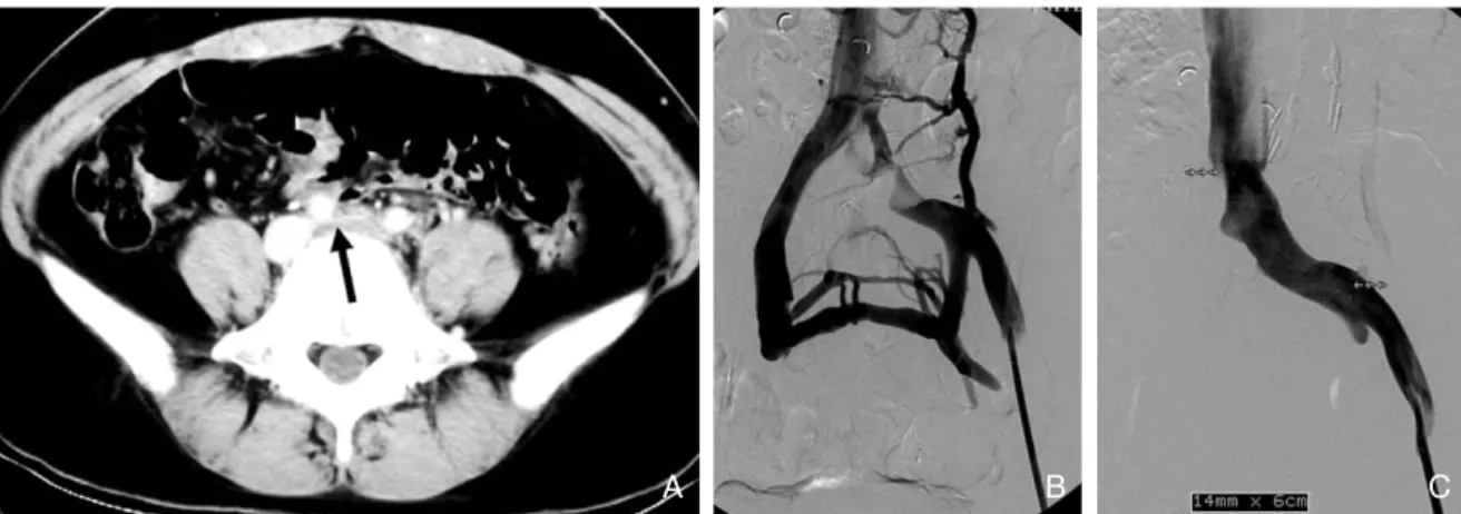
Figure 3.A 44-year-old woman with iliac compression syndrome
(A) CT scan shows compressed left iliac vein (arrow) between right common iliac artery and vertebra body
(B) Venography shows extrinsic compression of proximal left common iliac vein and collaterals to contralateral iliac vein (C) After placement of self expandable stent in left common iliac vein, venography shows widely patent left common
iliac vein
A B C
심부정맥혈전증의 원인이 되는 엉덩넙다리정맥의 폐 쇄는 엉덩정맥 압박증후군(iliac compression syn- drome), 골반부 방사선 치료, 골반암의 정맥 침범, 중심 정맥 카테터 시술 경력 등으로부터 발생할 수 있다(22).
이 중 가장 대표적인 엉덩정맥 압박증후군은 May- Thurner syndrome이라고도 불리는데, 왼쪽 엉덩넙다 리 정맥혈전증환자의 50
~
80%가 이 질환에 기인한다고 하며 하지의 심부정맥혈전증이 오른쪽보다 왼쪽에 2~3 배가 많이 발생하는 것도 이 질환 때문인 것으로 알려져 있다(23). 이 질환은 오른엉덩동맥과 척추뼈몸통 사이에 왼엉덩정맥이 눌려서 생긴다(Figure 3A). 이렇게 눌린 왼엉덩정맥은 지속적이고 만성적인 동맥의 박동에 의한 자극으로 인하여 정맥벽의 내막증식 및 돌기(spur)를 만 들고 결국엔 정맥의 폐쇄를 유발하게 된다(23, 24). 엉덩 정맥 압박증후군은 주로 30~40대의 여성에서 발생하며
심부정맥혈전증과 관련된 갑작스런 좌측 다리의 부종과 통증을 호소하거나 혈전증 없이 만성적인 다리의 부종 및 울혈 궤양(stasis ulceration)을 보일 수도 있다. 정맥 조영술 소견은 동맥의 압박에 의하여 엉덩정맥의 근위부 에 협착이나 폐쇄가 관찰되고 많은 측부 순환이 보인다 (Figure 3B).엉덩정맥 압박증후군의 혈관내 치료는 우선 동반된 심부정맥의 혈전을 용해술 및 기계적 혈전제거술 등으로 혈전을 제거한 후에 풍선확장술과 스텐트 설치술을 하게
아래쪽 정맥에 설치한 스텐트의 1년 개통률이 18%에 불 과하므로 서혜부 아래 정맥에는 풍선확장술 외에 스텐트 를 삽입하는 것은 피하는 것이 좋다(25).
결 론
하지의 급성 심부정맥혈전증 환자의 혈관내 치료는 빠 르게 정맥을 재개통 시키고 밸브의 기능을 유지시켜 주 며 postphlebitic syndrome과 같은 후유증을 줄여 줄 수 있다. 치료에 따른 합병증을 줄이고 치료기간을 단축 시키기 위해서는 catheter-directed thrombolysis와 기 계적 혈전제거술을 함께 시행하는 병합치료가 바람직하 며 정맥폐쇄가 있는 경우에는 풍선혈관성형술 및 스텐트 설치술로 정맥의 내경을 확보할 수 있다. 이와 같이 중재 적 방사선적 치료는 심부정맥혈전증의 진단에서부터 혈 전제거, 동반된 정맥폐쇄의 내경 확보 등 치료의 전 과정 을 효과적이고 안전하게 해결할 수 있다는 장점이 있다.
그러나 일차적 치료로 널리 이용되기 위해서는 무작위 전향적 임상연구 결과가 뒷받침 되어야 할 것이다.
참 고 문 헌
1. Anderson FA, Wheeler HB, Goldberg RJ, Hosmer DW, Pa- twardhan NA, Jovanovic B, Forcier A, Dalen JE. A population- based perspective of the hospital incidence and case-fatality
rates of deep vein thrombosis and pulmonary embolism. The Worcester DVT Study. Arch Intern Med 1991; 151: 933 - 938.
2. Haines ST. Venous thromboembolism: pathophysiology and clinical presentation. Am J Health Syst Pharm 2003; 60 (S 7):
S3 - 5.
3. O’Donnell TF Jr, Browse NL, Burnand KG, Thomas ML. The socioeconomic effects of an iliofemoral venous thrombosis. J Surg Res 1977; 22: 483 - 488.
4. Akesson H, Brudin L, Dahlstrom JA, Eklof B, Ohlin P, Plate G.
Venous function assessed during a 5 year period after acute ilio-femoral venous thrombosis treated with anticoagulation.
Eur J Vasc Surg 1990; 4: 43 - 48.
5. Augustinos P, Ouriel K. Invasive approaches to treatment of ve- nous thromboembolism. Circulation 2004; 110(9 S 1): I27 - 134.
6. Blum A, Roche E. Endovascular management of acute deep vein thrombosis. Am J Med 2005; 118 (S 8A): 31 - 36.
7. Delis KT, Bountouroglou D, Mansfield AO. Venous claudication in iliofemoral thrombosis: long-term effects on venous he- modynamics, clinical status, and quality of life. Ann Surg 2004;
239: 118 - 126.
8. Sherry S. Thrombolytic therapy for deep venous thrombosis.
Semin Intervent Radiol 1985; 4: 331 - 337.
9. Krupski WC, Bass A, Dilley RB, Bernstein EF, Otis SM. Pro- pagation of deep venous thrombosis identified by duplex ultrasonography. J Vasc Surg 1990; 12: 467 - 474 discussion 474 - 475.
10. Arnesen H, Hoiseth A, Ly B. Streptokinase of heparin in the treatment of deep vein thrombosis. Follow-up results of a prospective study. Acta Med Scand 1982; 211: 65 - 68.
11. Semba CP, Razavi MK, Kee ST, Sze DY, Dake MD. Throm- bolysis for lower extremity deep venous thrombosis. Tech Vasc Interv Radiol 2004; 7: 68 - 78.
12. Hood DB, Alexander JQ. Endovascular management of ilio-
femoral venous occlusive disease. Surg Clin North Am 2004;
84: 1381 - 1396, viii.
13. Mewissen MW, Seabrook GR, Meissner MH, Cynamon J, Labropoulos N, Haughton SH. Catheter-directed thrombolysis for lower extremity deep venous thrombosis: report of a na- tional multicenter registry. Radiology 1999; 211: 39 - 49.
14. Semba CP, Dake MD. Iliofemoral deep venous thrombosis:
aggressive therapy with catheter-directed thrombolysis.
Radiology 1994; 191: 487 - 494.
15. Bjarnason H, Kruse JR, Asinger DA, Nazarian GK, Dietz CA Jr, Caldwell MD, key NS, Hirsch AT, Hunter DW. Iliofemoral deep venous thrombosis: safety and efficacy outcome during 5 years of catheter-directed thrombolytic therapy. J Vasc Interv Radiol 1997; 8: 405 - 418.
16. Tarry WC, Makhoul RG, Tisnado J, Posner MP, Sobel M, Lee HM. Catheter-directed thrombolysis following vena cava filtration for severe deep venous thrombosis. Ann Vasc Surg 1994; 8: 583 - 590.
17. Comerota AJ, Throm RC, Mathias SD, Haughton S, Mewi- ssen M. Catheter-directed thrombolysis for iliofemoral deep venous thrombosis improves health-related quality of life.
J Vasc Surg 2000; 32: 130 - 137.
18. Murphy KD. Mechanical thrombectomy for DVT. Tech Vasc Interv Radiol 2004; 7: 79 - 85.
19. Kim HS, Patra A, Paxton BE, Khan J, Streiff MB. Adjunctive percutaneous mechanical thrombectomy for lower-extremity deep vein thrombosis: clinical and economic outcomes. J Vasc Interv Radiol 2006; 17: 1099 - 1104.
20. Lee KH, Han H, Lee KJ, Yoon CS, Kim SH, Won JY, Lee do Y.
Mechanical thrombectomy of acute iliofemoral deep vein thrombosis with use of an Arrow-Trerotola percutaneous thrombectomy device. J Vasc Interv Radiol 2006; 17: 487 - 495.
Plassmann L. Left iliac venous thrombosis caused by venous spur: treatment with thrombectomy and stent implantation.
11: 823 - 836.
Peer Reviewer Commentary
본 논문은 최근에 시작된 정맥 혈전증의 적극적인 치료방법에 대한 기본 이해와 치료방법에 대하여 기술하고 있다. 정 맥 혈전색전증은 발생빈도가 매우 높으나 정확하게 진단되는 빈도는 매우 낮아서 적절한 치료를 시행하지 못하여 심각 한 급성 및 만성 합병증을 초래할 수 있는 매우 위험한 질환으로, 최근에 세포 생물학, 분자 생물학의 발전으로 위험인 자와 병태생리가 밝혀지면서 다양한 치료방법이 시도되고 있다. 필자가 밝힌 대로 기본적인 치료방법으로 인식된 항응 고요법만의 문제점을 극복하기 위한 중재적 방사선적 치료의 초기 결과는 매우 고무적이나 장기적인 치료 결과가 아직 없고, 다양한 양상의 정맥 혈전색전증에 대한 기본적인 치료 원칙을 정립하여야 할 필요가 있음을 고려하여야 하겠다.
노 병 석 (원광의대 진단방사선과)