Lipopolysaccharide-injected Rats
I. Introduction
Acupuncture has long been renowned for its effective therapeutic benefits, such as relief of pain, immunomodulation and treat- ment of internal organic disorders in
Oriental medicine16,20,30). According to the theories of Oriental medicine, acupuncture acts on human body to sustain the Yin and Yang balance by regulating Qi, and finally displays its efficacy on various diseases1).
Particularly, many researchers have reported studies on the immunomodulatory effect of acupuncture3,32). For example, modulatory effect of acupuncture on immune
침자가 LPS를 주입한 흰쥐 시상하부 방실핵의 신경활성에 미치는 영향
손양선1⋅박히준1⋅김승태1⋅임사비나1
1경희대학교 한의과대학 경혈학교실
Effects of Acupuncture on the Neuronal Activation of Paraventricular Nucleus of Hypothalamus in
Lipopolysaccharide-injected Rats
Yang-Sun Son1, Hi-Joon Park1, Seung-Tae Kim1, Sabina Lim1
1Dept. of Meridian & Acupoint, College of Oriental Medicine, Kyung Hee University
국문초록
본 연구에서는 침의 면역조절작용을 통한 염증반응 억제효과를 연구하기 위하여 내독소를 주입한 흰쥐의 시상하부에서 염증반응의 중추인 방실핵의 신경활성에 미치는 영향을 관찰하였다. 흰쥐의 미정맥에 LPS와 생리식염수를 각각 주입하고 군에 따라 양측 소부(HT8)나 족삼리(ST36)에 1분간 침치료를 각각 시행하였다.
C-Fos는 신경활성을 자극하는 초기단계에 발현되는 유전자로서 신경계의 특정부위의 활성도를 측정하는 지 표로 널리 사용되고 있다. 본 연구자는 침자극이 LPS로 인한 염증반응에 미치는 영향과 그 기전을 알아보 기 위해 면역조직화학염색의 방법을 이용하여 대뇌 시상하부의 방실핵에서 c-Fos 면역활성을 측정하였다.
LPS를 주입한 군의 방실핵에서 생리식염수를 주입한 군에 비해 c-Fos 면역활성이 유의하게 증가한 반면 소부에 자침했을 때 LPS에 의해 증가된 c-Fos 면역활성이 유의하게 감소하였다. 족삼리에 자침한 군에서는 유의한 변화가 나타나지 않았다.
결론적으로 소부 침치료는 LPS로 인해 증가된 방실핵의 신경활성을 효과적으로 감소시켰고 이는 침의 면역조절 및 탁월한 염증억제 효과를 보여주는 결과일 뿐 아니라 침의 인체 항상성 유지를 통한 치료기전 에 대한 향후 연구의 중요한 실마리를 제공해주고 있다고 사료된다.
Keywords : Acupuncture, Lipopolysaccharide, Paraventricular nucleus, Neuronal activation, Immunomo- dulation.
■교신저자 : 임사비나, 서울특별시 동대문구 회기동 1 경희대학교 한의 과대학 경혈학교실, Tel. 02-961-0324, Fax. 02-961-7831, E-mail : [email protected]
The Journal of Korean Meridian & Acupoint
function has been reported in the traumatized rats5,7,8). In addition, other researchers observed that several immune- related indexes such as the natural killer cell activity, interferon-γ and interleukin-2 incre- ased in normal animals after acupuncture treatment27,39,40).
Lipopolysaccharide (LPS) is an endo- toxin derived from gram-negative bacterial cell wall. It has been used to induce the bacterial infection-like symptom in animals by central or peripheral injection. This infectious condition, including fever, is known to be activated by pro-inflammatory cytokines - e.g. interleukin-1β, tumor necro- sis factor-α- that stimulate the central immune regulatory center, the hypothalamus10,15). Stimulating hypothalamus via cytokine-induced pathway could be followed by the activation of hypothalamic-pituitary- adrenal (HPA) axis, which induces systemic symptoms such as decrease in feeding, drinking, social interactions and alterations in brain neurochemistry15). In fact, these cytokines activate the HPA axis mainly by stimulating the neurons in the paraventricular nucleus (PVN)10,18).
PVN has been known to represent a center for homeostasis. It contains specific neuronal projections to endocrine and auton- omic control sites33) and involves in the acute phase reaction of inflammatory response18,36). As a main integrative part of hypothalamus, the PVN is known to be activated in response to immune stress, e.g. via the systemic injection of endotoxin, and to act on the anterior pituitary gland by secreting corticotropin releasing factor (CRF) to stimulate the release of adrenocorticotropin hormone (ACTH)2,17). It is well known that
peripheral (i.v. or i.p.) injection of LPS stimulates the neurons in the PVN10,25), and in turn, increases the plasma CRF and ACTH through the activation of HPA axis12,13,26,34).
C-Fos, which is an immediate early gene expressed after different kinds of stimulation, has been used as neuronal marker14,29).
Integration of above known facts suggests a hypothesis that the anti-inflam- matory effects of acupuncture could be mediated by the activation of the PVN neurons. Therefore, I have carried out this study to investigate the effects of acupunc- ture on the neuronal activation of PVN in the LPS-injected rats using c-Fos immunohistoc- hemistry.
II. Materials and methods
1. Animals
Male Sprague-Dawley rats (n = 30) weighing 300-350 g were used in this experiment. All animals were supplied from Korean Experimental Animal Center and treated according to NIH guideline.
2. Materials
Lipopolysaccharide (LPS; E. coli, 055:B5, Sigma, USA) was used for animal infection model. For acupuncture treatment, sterilized needles (stainless steel, 0.25 mm o.d., Dongbang acupuncture INC, Korea) were used.
3. LPS injection
LPS (400 ㎍/kg, i.v.) diluted in normal
Lipopolysaccharide-injected Rats saline was injected to mimic the bacterial
infection of rats. Normal saline was injected intravenously for saline control groups.
4. Acupuncture treatment
Two acupuncture points were selected for the purpose of treatment and control. The acupuncture point, Sobu (HT8) is located between fourth and fifth metacarpal bones in the palm of forepaw. Control point, Joksamli (ST36) is located 5 mm laterally and distally from the anterior tubercle of the tibia.
HT8 has been used for the treatment of fever-related disorders because of its im- munomodulatory effects in Oriental medicine1,31). ST36 was selected for control acupoint in this experiment though it was reported to have various other effects16,19,20).
Acupuncture needles were bilaterally inserted into each acupoints to a depth of 5 mm and twisted at 2 Hz for 1 min. All treatments were performed 2 hrs after LPS- or saline- injection was treated.
5. Immunohistochemistry
Animals were anesthetized by sodium pentobarbital 4 hrs after injection for the preparation of perfusion. After checking for complete lack of response, the rats were transcardially perfused with 0.05 M phosphate-buffered saline (PBS) and then with 4% paraformaldehyde in 0.1 M phosphate buffer (PB) at pH 7.4. Brains were dissected, post-fixed in the same fixative overnight, and transferred into 30% sucrose for cryoprotection.
Serial frontal sections up to 40 μm thickness were made using a freezing microtome (Shandon, UK). Twelve sections
were collected from each brain for immuno- histochemistry, and steeped in storing solution with 30% glycerin and ethylene glycol in 0.2 M PB. For immunohistochemical procedure, free-floating tissue sections were washed three times for 15 min in 0.05 M PBS, and then incubated in 1% H2O2 in 0.05 M PBS for 15 min. After washing it three times with the PBS for the removal of bubbles, sections were incubated in the blocking solution with 1% BSA and 10% goat serum for 1 hr, and subsequently incubated overnight with c-Fos primary antibody (1:20000, Oncogene, MA) at room temperature. After washing it three times with PBS, sections were incubated for 1 hr with biotinylated anti-rabbit antibody (1:100, Vector laboratories, Burlingame, CA). Bound secondary antibody was then amplified with the Vector Elite ABC (Avidin biotin complex) kit for 1 h before washing it three times with PBS. For visualization, the antibody -avidin-biotin -peroxidase complexes were stained using 0.02% 3,3′- diaminobenzidine containing nickel chloride (40 mg/ml) (Nickel-DAB) in 0.05 M Tris buffer (pH 7.6) with 0.03% H2O2 for 3 to 5 min. After sufficient visualization has been achieved, reacted sections were mounted on gelatin-coated slide, and dried overnight.
The intensities of c-Fos-specific stain- ing were assessed in a quantitative fashion according to a microdensitometrical method based on optical density (mean gray scale) using an image analyzer (OPTIMAS version 6.5, Media Cybernetics, MD). Before starting the image analysis, the light source was adjusted to the brightness, which generates the best possible contrast between positive- and negative- staining cells. C-Fos immun- oreactivity in the PVN was analyzed
bilaterally, but in the results, it was expressed unilaterally, on condition that there were no significant differences between right and left part of PVN.
6. Data analysis
To compare the number of c-Fos-posi- tive cells in the PVN of each groups, results were expressed as a mean±SEM and analyz- ed by analysis of variance (ANOVA) followed by LSD test. Significant differences were considered for P < 0.05 values.
III. Results
In animals injected with saline solu- tion, a few numbers of c-Fos-positive cells
were observed in the PVN of hypothalamus 4 hrs after injection (Fig. 1A). The LPS-injected group showed a strong increase in the number of c-Fos-positive cells compared to the saline-injected group at the same concordance points (Fig. 1B).
However, this increase of c-Fos-positive cells in the PVN was counteracted by acupuncture on HT8 in LPS-injected rats (Fig. 1C). Acupuncture on ST36 did not alter the increased number of c-Fos-positive cells in the PVN in LPS-injected rats (Fig.
1D). The acupuncture treated- group (HT8) following saline-injection did not change the number of c-Fos-positive cells in the PVN (Fig. 1E).
The number of c-Fos-positive cells in the right side of PVN was noted because there was no significant difference between right and left part of PVN. The number of c-Fos-positive cells in the PVN per section was 29.00±5.85 (mean±SEM) in the saline- injected group. The number of PVN c-Fos- positive cells in the LPS-injected group was 203.00±41.24, which was significantly higher than that of the saline-injected group. The number of c-Fos-positive cells in the PVN of LPS-injected animals with acupuncture on HT8 was 52.83±11.95, which was significan- tly decreased compared to the LPS-injected group. The number of PVN c-Fos-positive cells, in LPS-injection group with acupunc- ture on ST36 and saline-injection group with acupuncture on HT8, were reported to values of 151.50±35.43, 38.17±8.93 respectively (Table 1).
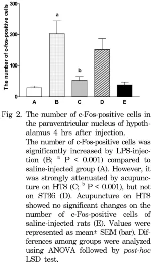
The number of c-Fos-positive cells in the PVN was significantly increased by LPS-injection and decreased after acupunc- ture on HT8, but not on ST36 (Fig. 2). These results indicate that acupuncture on HT8 Fig 1. C-Fos immunoreactivity in the same
sections of the PVN of hypothalamus 4 hrs after injection in rats.
A: Group with saline-injection B: Group with LPS-injection
C: Group with acupuncture on HT8 after LPS-injection
D: Group with acupuncture on ST36 after LPS-injection
E: Group with acupuncture on HT8 after saline-injection
Scale bar = 100 μm.
Lipopolysaccharide-injected Rats
inhibits the LPS-induced neuronal activation in the PVN of rats.
IV. Discussion
In this study, LPS injection (400 ㎍/kg, i.v.) showed a similar time-course distribu- tion of neuronal activation in the PVN to that described previously9,21,37). It is suggested that neurons in the PVN, stimulated after LPS-injection, may activate the HPA axis to cause the systemic symptoms of animals in this experiment.
According to the results, neurons in the PVN were activated 4 hrs after LPS-injec- tion, while these activation were significantly attenuated by acupuncture on HT8. In saline-injected rats, however, acupuncture on HT8 did not change the neuronal activation of PVN. These results suggest that acupuncture on HT8 may suppress the activation of HPA axis and consequently reduce the infection-like symptoms, e.g. fever in LPS-injected rats. In Oriental medicine, the acupoint HT8 is well known to express the Hyung Points (滎穴) among the Five Su Points (五輸穴), and has been believed to be effective in treating fever-related disorders from immune dysfunction1,31). This effect of HT8 was clarified in the LPS-induced
infection model via the method of c-Fos immunohistochemistry in this study.
Acupuncture on ST36 slightly suppre- ssed an increase of neuronal activation in the PVN of LPS-injected rats, but not to a significant value. Although ST36 has been used for the treatment of various diseases, e.g. pain, gastric disorder and stroke16,19,20), it is not effective for infectious condition. It may also demonstrate the fact that each acupoints shows a unique and diverse effect for the purpose of treating diseases.
The expression of the proto-oncogene c-Fos has been known as a useful marker for neuronal activation, and its expression in the Table 1. The number of c-Fos positive
neurons in the PVN of hypothalamus (mean
±SEM)
Group Animal The number of c-Fos-positive cells
Saline 6 29.00±5.85
LPS 6 203.00±41.24
LPS+HT8 6 52.83±11.95
LPS+ST36 6 151.50±35.43
Saline+HT8 6 38.17±8.93
Fig 2. The number of c-Fos-positive cells in the paraventricular nucleus of hypoth- alamus 4 hrs after injection.
The number of c-Fos-positive cells was significantly increased by LPS-injec- tion (B; a P < 0.001) compared to saline-injected group (A). However, it was strongly attenuated by acupunc- ture on HT8 (C; b P < 0.001), but not on ST36 (D). Acupuncture on HT8 showed no significant changes on the number of c-Fos-positive cells of saline-injected rats (E). Values were represented as mean± SEM (bar). Dif- ferences among groups were analyzed using ANOVA followed by post-hoc LSD test.
A B C D E
0 100 200 300
a
b
The number of c-fos-positive cells
various brain regions containing PVN after mild and acute physical stress has been reported14,29). C-Fos expression in the PVN after saline-injection might be caused by the stress from light immobilization and injection during the handling process. Recent report suggests that restraint and LPS-injection showed similar effect on c-Fos expression in the PVN with regard to the intensity and their distribution pattern38). Thus, significant difference in c-Fos immunoreactivity in the PVN between saline- and LPS-injected groups indicates the obvious induction of c-Fos expression by LPS.
Many c-Fos studies have been conducted for the investigation of analgesia effect of electroacupuncture (EA). These studies reported that brain stem, hypo- thalamus and spinal cord showed regional increases of c-Fos immunoreactivity after EA-induced analgesia22,24). C-Fos immuno- histochemistry has been also used to investigate the therapeutic effects of acupuncture. C-Fos immunoreactive cells were increased in the spinal cord after noxious stimulation and decreased by acu- puncture4). Acupuncture suppressed the c-Fos expression evoked by tooth pulp stimulation in the trigeminal subnucleus pars caudalis and the periaqueductal gray of rats28).
In the present study, c-Fos immunoreactivity was used for the evaluation of neuronal activation to examine the acupuncture-induced modulation on the process of inflammatory response in the PVN.
Previous reports showed the effects of acupuncture on peripheral immune system in normal or pathological conditions6,11,23, 25). Recently, it was reported that LPS-induced
cytokine mRNAs in the hypothalamus were reduced by acupuncture treatment with the attenuation of bacterial fever in rats30). The present immunohistochemical study was conducted in the same way with previous studies to prove the potent actions of acupuncture on the immune system, but we paid focus on specific area of rat brain, the PVN.
In conclusion, this study provided an evidence that stimulation on specific acu- point such as HT8 may exert anti-inflam- matory effect by down-regulating the expres- sion of immediate early genes of cytokine in PVN hypothalamus of LPS inflammation rat model.
Acknowledgement: This research was supported by a grant for Pain and Neuroscience Research from Vision 2000 project of Kyung Hee University. We thank Mrs. Mi-Sook Hong and Mrs. Kyung-Hee Lee for technical assistance.
References
1. Ahn KS. The Essence of Oriental Medicine.
Seoul : Sonamoo. 1999 : 19, 294-5.
2. Berkenbosch F, VanOers J, Del Ray, Tilders F, Besedovsky H. Cortico- tropin-releasing factor-producing neurons in the rat activated by interleukin-1.
Science. 1987 ; 238 : 524-6.
3. Bossy J. Immune systems, defense me- chanisms and acupuncture, fundamental and practical aspects. Am. J. Acupunct.
1990 ; 18 : 219-32.
4. Chang CJ, Huang ST, Hsu K, Lin A, Stoller ML, Lue TF. Electroacupuncture decreases c-Fos expression in the spinal cord induced by noxious stimulation of the rat bladder.
Lipopolysaccharide-injected Rats J. Urol. 1998 ; 160 : 2274-9.
5. Cheng XD, Wu GC, He QZ, Cao XD. Effect of continued electroacupuncture on induction of interleukin-2 production of spleen lymphocytes from the injured rats.
Acupunct. Electrother. Res. 1997 ; 22 : 1-8.
6. Du L, Jiang J, Cao X. Time course of the effect of elecroacupuncture on immunomodulation of normal rat. Chen Tzu Yen Chiu. 1995 ; 20 : 36-39.
7. Du LN, Jiang JW, Wu GC, Cao XD.
Naloxone and electroacupuncture improve the immune function of traumatized rats.
Sheng Li Xue Bao. 1998 ; 50 : 636-42.
8. Du LN, Wu GC, Cao XD. Modulation of orphanin FQ or electroacupuncture on immune function of traumatic rats.
Acupunct. Electrother. Res. 1998 ; 23 : 1-8.
9. Elmquist JK, Scammell TE, Jacobson CD, Saper CB. Distribution of Fos-like immunoreactivity in the rat brain following intravenous Lipopolysaccharide. J. Comp.
Neurol. 1996 ; 371 : 85-103.
10. Elmquist JK, Saper CB. Activation of neurons projecting to the paraventricular hypothalamic nucleus by intravenous lipopolysaccharide. J. Comp. Neurol. 1996
; 374 : 315-31.
11. Fujiwara R, Tong ZG, Matsuoka H, Shibata H, Iwamoto M, Yokoyama MM. Effects of acupuncture on immune response in mice.
Int. J. Neurosci. 1991 ; 57 : 141-150.
12. Harbuz MS, Jessop DS. Dissociation between c-fos mRNA in the paraventri- cular nucleus and corticosterone secretion in rats with adjuvant-induced arthritis. J.
Endocrinol. 1999 ; 163 : 107-13.
13. Hare AS, Clarke G, Tolchard S. Bacterial lipopolysaccharide-induced changes in FOS protein expression in the rat brain:
correlation with thermoregulatory
changes and plasma corticosterone. J.
Neuroendocrinol. 1995 ; 7 : 791-9.
14. Imaki T, Shibasaki T, Hotta M. Demura H. Early induction of c-fos precedes increased expression of corticotropin -releasing factor messenger ribonucleic acid in the paraventricular nucleus after immobilization stress. Endocrinol.1992 ; 131 : 240-6.
15. Jianping W, Dunn AJ. The role of interleukin-6 in the activation of the hypothalamo-pituitary-adrenocortical axis and brain indoleamines by endotoxin and interleukin-1β. Brain Res. 1999 ; 815 : 337-48.
16. Jin HO, Zhou L, Lee KY, Chang TM, Chey WY. Inhibition of acid secretion by electrical acupuncture is mediated via beta-endorphin and somatostatin. Am. J.
Physiol. 1996 ; 271 : 524-30.
17. Kakucska I, Qi Y, Clark BD, Lechan RM.
Endotoxin-induced cortico- tropin-releasing hormone gene expression in the hypothalamic paraventricular nucleus is mediated centrally by interleu- kin-1. Endocrinology. 1993 ; 133 : 815-21.
18. Kandel ER, Schwartz JH, Jessell TM.
Principles of Neural Science. New York : McGraw-Hill. 2000 : 960-81.
19. Kim EH, Kim YJ, Lee HJ, Huh Y, Chung JH, Seo JC, Kang JE, Lee HJ, Yim SV, Kim CJ. Acupuncture increases cell proliferation in dentate gyrus after transient global ischemia in gerbils.
Neurosci. Lett. 2001 ; 297 : 21-4.
20. Kim JH, Min BI, Schmidt D, Lee HJ, Park DS. The difference between electroacupuncture only and electroacu- puncture with manipulation on analgesia in rats. Neurosci. Lett. 2000 ; 279 : 149-52.
21. Konsman JP, Kelley K, Dantzer R.
Temporal and spatial relationships between Lipopolysaccharide-induced ex- pression of fos, interleukin-1beta and inducible nitric oxide synthase in rat brain.
Neuroscience. 1999 ; 89 : 535-48.
22. Lee JH, Beitz, AJ. The distribution of brain-stem and spinal cord nuclei associated with different frequencies of electroacupuncture analgesia. Pain. 1993
; 52 : 11-28.
23. Lundeberg T, Eriksson SV, Theodorsson E. Neuroimmunomodulatory effects of acupuncture in mice. Neurosci. Lett. 1991
; 128 : 161-4.
24. Pan B, Castro-Lopes JM, Coimbra A. C-fos expression in the hypothalamus-pituitary system induced by electroacupucnture or noxious stimulation. Neuroreport. 1994 ; 5 : 1649-52.
25. Petti F, Bangrazi A, Liguori A, Reale G, Ippoliti F. Effects of acupuncture on immune response related to opioid-like peptides. J. Tradit. Chin. Med. 1998 ; 18 : 55-63.
26. Rivest S, Laflamme N. Neuronal activity and neuropeptide gene transcription in the brains of immune-challenged rats J.
Neuroendocrinol. 1995 ; 7 : 501-25.
27. Sato T, Yu Y, Guo SY, Kasahara T, Hisamitsu T. Acupuncture stimulation enhances splenic natural killer cell cytotoxicity in rats. Jpn. J. Physiol. 1996
; 46 : 131-6.
28. Sheng LL, Nishiyama K, Honda T, Sugiura M, Yaginuma H, Sugiura Y. Suppressive effects of Neiting acupuncture on toothache: an experimental analysis on Fos expression evoked by tooth pulp stimulation in the trigeminal subnucleus pars caudalis and the periaqueductal gray of rats. Neurosci. Res. 2000 ; 38 : 331-9.
29. Smith MA, Banerjee S, Gold PW, Glowa J. Induction of c-fos mRNA in rat brain by conditioned and unconditioned stressors. Brain Res. 1992 ; 578 : 135-41.
30. Son YS, Park HJ, Kwon OB, Jung SC, Shin HC, Lim S. Antipyretic effects of acupuncture on the lipopoly- saccharide-induced fever and expression of interleukin-6 and interleukin-1β mRNAs in the hypothalamus of rats.
Neurosci. Lett. 2002 ; 319 : 45-8.
31. Stux G, Pomeranz B. Acupuncture. Berlin : Springer-Verlag. 1987 : 63-4, 114-9.
32. Sun T, Du LN, Wu GC, Cao XD. Effect of intrathecal morphine and electro-acupuncture on cellular immune function of rats and increasement of mu-opioid receptor mRNA expression in PAG following intrathecal morphine.
Acupunct. Electrother. Res. 2000 ; 25 : 1-8.
33. Swanson LW, Sawchenko PE.
Hypothalamic integration: organization of the paraventricular and supraoptic nuclei, Ann. Rev. Neurosci. 1983 ; 6 : 269-324.
34. Takemura T, Makino S, Takao T, Asaba K, Suemaru S, Hashimoto K.
Hypothalamic-pituitary-adrenocortical responses to single vs. repeated endotoxin Lipopolysaccharide administration in the rat. Brain Res. 1997 ; 767 : 181-91.
35. Wan W, Janz L Vriend CY, Sorensen CM, Greenberg AH, Nance DM. Differential induction of c-Fos immunoreactivity in hypothalamus and brain stem nuclei following central and peripheral administration of endotoxin. Brain Res.
Bull. 1993 ; 32 : 581-7.
36. Watkins LR, Maier SF, Goehler, LE.
Cytokine-to-brain communication: a review and analysis of alternative mechanisms. Life Sci. 1995 ; 57 : 1011-26.
Lipopolysaccharide-injected Rats 37. Yang WW, Krukoff TL. Nitric oxide
regulates body temperature, neuronal activation and interleukin-1beta gene expression in the hypothalamic paraven- tricular nucleus in response to immune stress. Neuropharmacology. 2000 ; 39 : 2075-89.
38. Yokoyama C, Sasaki K. Regional expressions of Fos-like immunoreactivity in rat cerebral cortex after stress; restraint and intraperitoneal Lipopolysaccharide.
Brain Res. 1999 ; 816 : 267-75.
39. Yu Y, Kasahara T, Sato T, Guo S, Liu Y, Asano K, Hisamitsu T. Enhancement of splenic interferon-γ, interleukin-2 and NK cytotoxicity by S36 acupoint acupuncture in F344 rats. Jpn. J. Physiol. 1997 ; 47 : 173-8.
40. Yu Y, Kasahara T, Sato T, Asano K, Yu GD, Fang JQ, Guo SY, Sahara M, Hisamitsu T. Role of endogenous interferon-γ on the enhancement of splenic NK cell activity by electroacupuncture stimulation in mice. J. Neuroimmunol.
1998 ; 90 : 176-86.