Adjuvant therapy using ex vivo-expanded allogenic
natural killer cells in hepatectomy patients with hepatitis B virus related solitary hepatocellular carcinoma: MG4101 study
Jong Man Kim1,*, Sung Yoo Cho2,*, Jinsoo Rhu1, Miyoung Jung2, Jung Hyun Her2, Okjae Lim2, Gyu-Seong Choi1, Eui-Cheol Shin3, Yu-Kyeong Hwang2, and Jae-Won Joh1
1Department of Surgery, Samsung Medical Center, Sungkyunkwan University School of Medicine, Seoul,
2Cell Therapy Research Center, GC LabCell, Yongin,
3Laboratory of Immunology and Infectious Diseases, Graduate School of Medical Science and Engineering, Korea Advanced Institute of Science and Technology, Daejeon, Korea
Backgrounds/Aims: Fewer reports have been published regarding hepatectomy patients with solitary hepatocellular car- cinoma (HCC) who received immunotherapeutic agents as adjuvant therapy. We evaluated the safety and efficacy of ex vivo-expanded allogenic natural killer (NK) cells in those patients with modified International Union Against Cancer (UICC) stage T3. Methods: From August 2014 to October 2015, five patients who underwent hepatic resection received ex vivo-expanded allogenic NK cells. Patients received five rounds of NK cells (2-3×10⁷ cells/kg) at postoperative 4, 6, 8, 12, and 16 weeks. This study is registered with ClinicalTrials.gov, number NCT02008929. Results: The median age of the five patients (three men and two women) was 44.8 years (range, 36-54 years). All had hepatitis B virus-re- lated HCC, and the median tumor size was 2.2 cm (range, 2.1-8.2 cm). None of the patients had any adverse events.
HCC recurrence developed in two patients at one year after hepatic resection, but four patients were alive at 3 years.
The two recurrence-free patients showed a higher ratio of CD8+ T lymphocyte populations before and after admin- istration of ex vivo-expanded allogenic NK cells compared with the three patients who experienced recurrence.
Conclusions: Immunotherapy using ex vivo-expanded allogenic NK cells in hepatectomy patients can be used safely.
Further studies should be investigated for efficacy. (Ann Hepatobiliary Pancreat Surg 2021;25:206-214) Key Words: Hepatocellular carcinoma; Natural killer cells; Immunotherapy; Safety; Efficacy
Received: September 11, 2020; Revised: October 10, 2020; Accepted: October 11, 2020 Co-Corresponding author: Jae-Won Joh
Department of Surgery, Samsung Medical Center, Sungkyunkwan University School of Medicine, 81 Irwon-ro, Gangnam-gu, Seoul 06351, Korea Tel: +82-2-3410-3466, Fax: +82-2-3410-0040, E-mail: jw.joh@samsung.com
Co-Corresponding author: Yu-Kyeong Hwang
Cell Therapy Research Center, GC LabCell, 107 Ihyeon-ro 30beon-gil, Giheung-gu, Yongin 16924, Korea Tel: +82-31-260-9853, Fax: +82-31-270-1450, E-mail: ykhwang@greencross.com
*Jong Man Kim and Sung Yoo Cho contributed equally to this work as co-first authors.
Copyright Ⓒ 2021 by The Korean Association of Hepato-Biliary-Pancreatic Surgery
This is an Open Access article distributed under the terms of the Creative Commons Attribution Non-Commercial License (http://creativecommons.org/
licenses/by-nc/4.0) which permits unrestricted non-commercial use, distribution, and reproduction in any medium, provided the original work is properly cited.
Annals of Hepato-Biliary-Pancreatic Surgery ∙ pISSN: 2508-5778ㆍeISSN: 2508-5859
INTRODUCTION
Liver resection (LR) is a curative treatment method for solitary hepatocellular carcinoma (HCC).1 However, the recurrence of HCC after LR is a major surgical limitation because the tumor recurrence rate exceeds 50% at 5 years after resection.2,3 In addition, cases with early HCC re- currence showed poor survival compared to those with late recurrence.4,5
The high incidence of HCC has led to efforts to devel- op adjuvant therapies to reduce recurrence. A number of
studies have explored adjuvant strategies.6 A recent multi- center trial that investigated the effect of sorafenib after surgical resection/radiofrequency ablation (RFA) failed to demonstrate any adjuvant effect of sorafenib on survival.7 This underlines the need for novel and effective adjuvant therapies to treat patients with HCC and to prevent re- currence after curative liver resection. However, the bene- fit of any form of adjuvant therapy remains unclear, and current scientific guidelines do not recommend adjuvant therapy in patients treated with resection.8-10
Immunotherapy is emerging as a new treatment strategy
the infusion of autologous or allogenic cells that are acti- vated and expanded ex vivo. Previous studies using im- munotherapy reported high response rates.11-13 Most of these studies are being carried out using modified T-cells, but we are increasingly interested in a similar adaptation of the immune system’s “natural killer”, or NK cells, which are able to attack cancer cells directly and quickly.
Recently, the development of allogenic NK cells has gained much attention because interaction of the relevant self-major histocompatibility complex (MHC) class I mol- ecules with a given killer cell immunoglobulin-like re- ceptor (KIR) in an autologous setting results in inhibition of the effector functions of autologous NK cells, even in the presence of additional activation signals.14 Indeed, in a recent clinical trials, infusion with allogenic NK cells has been shown to elicit more potent antitumor efficacy than with autologous NK cells in various cancers, such as acute myeloid leukemia, renal-cell carcinoma, malig- nant melanoma, lung cancer, and hepatic cancer.15 To this end, we established an efficient method for the large-scale, ex vivo-expansion of NK cells from peripheral blood mon- onuclear cells (PBMC) acquired from random healthy do- nors under good manufacturing practice (GMP) conditions.
These ex vivo-expanded, random healthy donor-derived al- logeneic NK cells, defined as MG4101, previously showed antitumor potency and safety in preclinical and phase I clinical studies, respectively.16,17
In the present study, we aimed to evaluate the safety and efficacy of adjuvant immunotherapy using ex vivo-ex- panded allogenic NK cell infusion in hepatectomy pa- tients, who had no evidence of residual tumors, after cura- tive liver resection.
MATERIALS AND METHODS
Patients
Patients who had undergone curative liver resection as a treatment for HCC of modified UICC stage III were screened for this study.10 The diagnosis of HCC was made by histological examination. The inclusion criteria were:
RFA, transarterial chemoembolization (TACE), radiation, or percutaneous ethanol injection (PEI); evidence of in- fection; a history of cardiovascular disease such as coro- nary artery disease, cardiac arrhythmia, congestive heart failure, angina pectoris, or myocardial infarction; auto- immune disease; or a history of malignancy except HCC within the 5 years prior to screening; female patients who were pregnant or lactating; and any patients who received steroids or immunosuppressants within the 4 weeks prior to screening were excluded.
Study design
This phase II clinical study was a prospective, open-la- belled trial. The study was conducted at Samsung Medical Center, Seoul, Republic of Korea. All patients provided written informed consent prior to enrollment in this study.
The study protocol and procedures were approved by the Institutional Review Board at Samsung Medical Center (SMC-2013-04-019). All methods and procedures asso- ciated with this study were conducted in accordance with the principles of the Declaration of Helsinki and local law. This study is registered with ClinicalTrials.gov, num- ber NCT02008929.
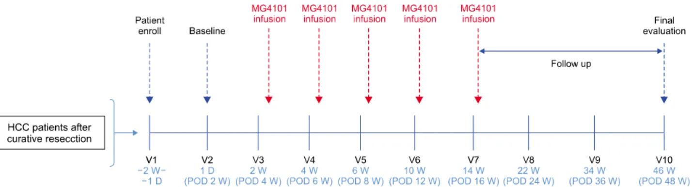
Patients who provided informed consent were screened within 2 weeks following curative liver resection and be- fore the start of immunotherapy. Patients received five treatments with ex vivo-expanded allogenic NK cells over 12 weeks (three treatments every 2 weeks followed by two treatments every 4 weeks). Amounts of 2-3×10⁷ cells/kg were administered to patients for 30 minutes. Patients re- ceived antihistamine for prophylaxis before NK cell in- fusion (Fig. 1).
Preparation and expansion of NK cells
PBMCs were isolated from random healthy donors, and NK cells were expanded as described previously under the conditions of GMP at Green Cross LabCell Corp.17 Briefly, CD3+ T-cell-depleted PBMCs were expanded at a seeding concentration of 2×10⁵ cells/ml in CellGro SCGM serum- free medium (CellGenix) with 1% auto-plasma, 1×10⁶ cells/ml
Fig. 1. Study design for MG4101 infusion and evaluation during follow up period.
irradiated (2,000 rad) autologous PBMCs, 10 ng/ml of monoclonal antibody to CD3 (OKT3; Orthoclon), and 500 IU/ml of IL2 (Proleukin) in an A-350N culture bag (NIPRO). NK cells were fed fresh medium with 500 IU/ml of IL2 every 2 days until they were harvested on day 14. After expansion, the cytotoxicity of MG4101 was evaluated by flow cytometric cytotoxicity assay against K562, SNU387 and Huh7 as described.17 K562 and SNU387 were obtained from the ATCC and cultured in RPMI-1640 medium (GIBCO) supplemented with 10% fetal bovine serum (FBS) (GIBCO). Huh7 was obtained from the Korean Cell Line Bank and also cultured in RPMI-1640 medium (GIBCO) supplemented with 10% FBS (GIBCO).
Flow cytometric analysis of NK cells
The phenotype of expanded NK cells was analyzed by flow cytometry. NK cells were stained with the appro- priate monoclonal antibodies as follows: anti-CD56-PE-Cy5 (B159), anti-CD3-FITC (UCHT1), anti-CD16 (3G8), anti- CD14 (M5E2), anti-CD19 (HIB19), anti-NKp30-PE (P30-15), anti-NKp44-PE (P44-8.1), anti-NKp46-PE (9E2/NKp46), anti-CD226-PE (DX11), anti-CD25-PE (M-A251), anti-CD132- PE (AG184), anti-CD57-PE (NK-1), anti-CXCR3-PE (1C6/
CXCR3) (BD Biosciences), anti-NKG2A-PE (131411), anti-NKG2C-PE (134591), anti-NKG2D-PE (149810), anti- CD69-PE (298614), anti-NKp80-PE (239127), anti-CD122- PE (27302) (R&D), anti-CD96-PE (NK92.39), anti-CD161- PE (HP-3G10), anti-CD62L-PE (DREG-56) (eBioscience), and anti-CD244-PE (C1.7) (Beckman Coulter) Samples were acquired on a BD FACS Canto II or LSR Fortessa and data were analyzed using FlowJo software (TreeStar Inc., Oregon).
Immune monitoring of recipients
Flow cytometric analysis of the change in immune cell populations after MG4101 administration was performed on PBMCs that were serially acquired from recipients.
Various immune cells including T cells (CD3+ CD4+, CD3+
CD8+), B cells (CD19+, CD20+), NK cells (CD3-CD56+, CD56+CD16+), regulatory T cells (Treg, CD4+CD25bright Foxp3+CD127dim) and myeloid-derived suppressor cells (MDSC, Lin−CD14−HLA-DR−CD11b+CD15+) were an- alyzed by Flow cytometry as described.16 Multiple cyto- kines and chemokines in patient plasma were quantified with commercially available cytometric bead-based assays according to the manufacturers’ instructions (FlowCytomix;
eBioscience).
Surveillance and outcomes
The procedures used for surveillance after liver re- section have been described previously.2 All patients were checked every two or three months in the second post- operative year and every 6 months in subsequent years.
Tumors were evaluated by tumor markers such as al- pha-fetoprotein (AFP) and protein induced by vitamin K absence/antagonism-II (PIVKA-II) and radiologic evalua- tions such as contrast-enhanced computed tomography or magnetic resonance imaging every 3 months for 24 months and then every 3-6 months thereafter. Relapse-free survival (RFS) was defined as the interval between the hepatectomy data and the recurrence date or last fol- low-up date, while overall survival (OS) was defined as the interval between the hepatectomy data and the death date or last follow-up date. Follow-up time was the length of time from surgery to final follow-up or death.
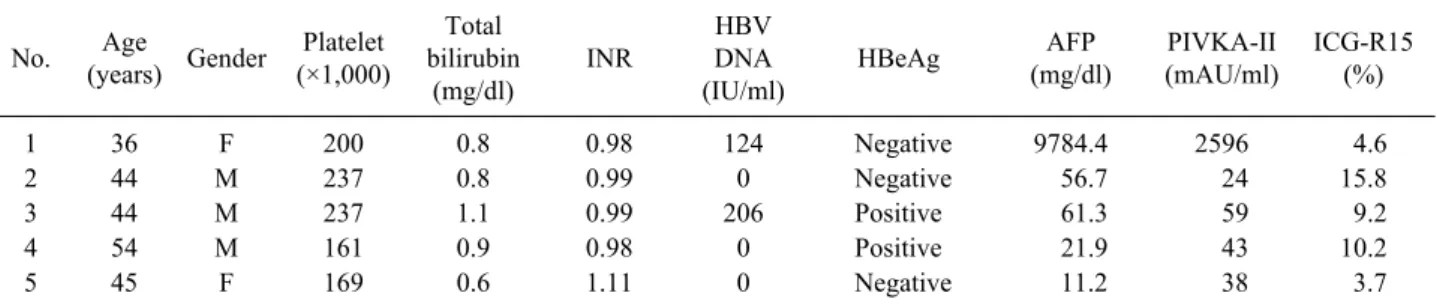
Table 1. Baseline patient characteristics No. Age
(years) Gender Platelet (×1,000)
Total bilirubin
(mg/dl)
INR
HBV DNA (IU/ml)
HBeAg AFP
(mg/dl)
PIVKA-II (mAU/ml)
ICG-R15 (%)
1 36 F 200 0.8 0.98 124 Negative 9784.4 2596 4.6
2 44 M 237 0.8 0.99 0 Negative 56.7 24 15.8
3 44 M 237 1.1 0.99 206 Positive 61.3 59 9.2
4 54 M 161 0.9 0.98 0 Positive 21.9 43 10.2
5 45 F 169 0.6 1.11 0 Negative 11.2 38 3.7
INR, international normalized ratio; AFP, alpha-fetoprotein; PIVKA-II, protein induced by vitamin K absence/antagonism-II;
ICG-R15, indocyanine green retention rate at 15 min centage of subjects. RFS and OS were evaluated with Kaplan-Meier curves using the log-rank test. All tests were two-tailed, and statistical significance was defined as p<0.05.
RESULTS
Patient characteristics
Between 11 August 2014 and 14 December 2015, five patients were screened and enrolled into this study after having met the eligibility criteria. None of the patients had a history of locoregional therapy prior to surgery.
Three patients were male, and The median age was 45 years (range, 36-54). All patients had hepatitis B virus-re- lated HCC, a performance status of 0, and Child-Pugh class A. Median AFP and PIVKA-II were 56.7 mg/dl (range, 11.2-9784.4 mg/dl) and 43 mAU/ml (range, 24-2596 mAU/ml), respectively. Three patients received Entecavir and two received Tenofovir as antiviral agents.
Clinical characteristics are summarized in Table 1.
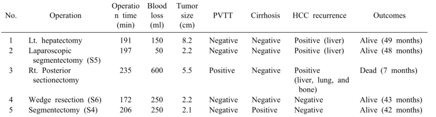
Three patients underwent major liver resection. Median operation time was 197 minutes (range, 172-235 minutes), and median blood loss was 200 ml (range, 50-600 ml).
None developed complications or required transfusion with red blood cells during the surgical procedure and postoperative period. All patients had solitary HCC with a median tumor size of 2.2 cm (range, 2.1-8.2 cm).
Microvascular invasion was seen in all patients, but portal vein tumor thrombosis was seen in only one patient.
the purity of allogeneic, expanded NK cells (CD3 CD56+) was 98.48±1.46% with minimal contamination (less than 1%) of CD3+ T cells (0.57±0.48%), CD14+ monocytes (0.76±0.4%), or CD19+ B cells (0.01±0.02%).
Expanded NK cells exerted strong cytotoxic activity against K562, an NK-sensitive target, and also showed a highly cytotoxic effect against the SNU387 and Hur7 hep- atocellular carcinoma cell lines (Fig. 2B). The surface ex- pression of activating or inhibitory NK receptors was analyzed. Functional markers such as CD16 and NKG2D were highly expressed in expanded NK cells. Expression of activating receptors NKG2C, NCRs, 2B4 (CD244), and DNAM-1 (CD226) was high, while expression of in- hibitory receptor NKG2A was low. CD25, CD62L, and CD69 showed activation status of the expanded NK cells as described (Fig. 2C).
Safety
Mild adverse events developed in only one patient who showed wound seroma discharge in the incisional area.
Across all patients, every infusion was well-tolerated and unassociated with any acute or delayed toxicities. None of the subjects showed drug-related adverse reactions and thus toxicity-related suspension of the MG4101 injection did not occur during our study. There were no increases in aspartate transaminase (AST), alanine transaminase (ALT), alkaline phosphatase (ALP), total bilirubin, inter- national normalized ratio (INR) or HBV DNA after any of the infusions.
Fig. 2. Characterization of ex vivo-expanded NK cells. (A) T cell-depleted PBMCs from heal- thy donors were expanded for 14 days in the GMP-compliant facility. The percentages of CD16+CD56+, CD3+, CD14+, and CD19+ cells were analyzed by flow cytometric analyses.
(B) Cytotoxicity of expanded NK cells against K562, SNU387 and Huh7 cell lines. Each plot represents mean ± standard de- viation (SD). (C) Immunopheno- typic characterization of NK cells was analyzed by flow cytome- tric analyses. The whisker box plots show the percentage of each marker positive NK cells.
All results are calculated as mean ± SD from five different donors.
Table 2. Perioperative characteristics and outcomes
No. Operation
Operatio n time (min)
Blood loss (ml)
Tumor size (cm)
PVTT Cirrhosis HCC recurrence Outcomes
1 Lt. hepatectomy 191 150 8.2 Negative Negative Positive (liver) Alive (49 months) 2 Laparoscopic
segmentectomy (S5)
197 50 2.2 Negative Negative Positive (liver) Alive (48 months)
3 Rt. Posterior sectionectomy
235 600 5.5 Positive Negative Positive
(liver, lung, and bone)
Dead (7 months)
4 Wedge resection (S6) 172 250 2.2 Negative Negative Negative Alive (43 months) 5 Segmentectomy (S4) 206 250 2.1 Negative Positive Negative Alive (42 months) PVTT, portal vein tumor thrombosis; HCC, hepatocellular carcinoma
Efficacy
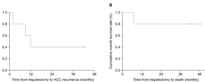
Perioperative characteristics and outcomes are outlined in Table 2. The median follow-up period was 43 months (range, 7-49 months). The RFS and OS rates at 3 years after NK cell infusion were 60% and 80%, respectively (Fig. 3). Three patients were diagnosed with recurrent HCC. One patient (case 3) was diagnosed at 2 months af- ter hepatic resection. The initial recurrence was multiple sites including liver, lung, and bone. He was dead at 7
months after liver resection because of disseminated HCC recurrence. In this patient, HCC recurred at 2 months after curative hepatic resection accompanied with a one-time injection of MG4101. Thus, his recurrence of HCC came before the allogenic NK cells were effective.
The second patient (case 1) was diagnosed with the first intrahepatic HCC recurrence at 7 months after cura- tive liver resection. She was treated with RFA and lived without any further HCC recurrence for 42 months
Fig. 3. RFS and OS of patients treated with MG4101 during the trial period and follow-up study.
Fig. 4. Analysis of T cells in patient peripheral blood. The concentrations of CD4+ T cells (A) and CD8+ T cells (B) were monitored for 10 weeks after MG4101 injection. A higher ratio of CD8+/CD4+ was seen in patient cases 2, 4, and 5 (C).
thereafter. She had a recurrence of HCC after 5 injections of allogenic NK cells. Considering the recurrence of hep- atocellular carcinoma at 7 months after surgery, it may be the case that intrahepatic metastasis was already present at the time of surgery. In the third patient (case 2), HCC recurrence was detected shortly after the study period ended.
Immune monitoring
After repeated injections of MG4101, we did not ob- serve any critical changes in the frequency and activation status of immune cell populations, such as T cells, B cells, NK cells, Treg cells, monocytes, and myeloid-derived suppressor cells (MDSC) in the analysis of the blood sam- ples derived from this clinical trial (data not shown).
However, it is worth noting that the two recurrence-free patients (cases 4 and 5) showed a higher ratio of CD8+
T lymphocyte populations before and after MG4101 ad- ministrations compared with the three patients with re- currence (cases 1, 2, and 3) (Fig. 4).
DISCUSSION
HCC development and progression are related to chron- ic inflammation.18 In addition, patients with HCC demon- strate some dysfunctions in their immune system, includ- ing abnormal innate and adaptive immune responses.19 Once tumors are established, mutual interactions between tumors and immune cells present during inflammation may provide conditions favorable for tumor cell survival.20 Immune suppressor cells including regulatory T cells, myeloid-derived suppressive cells, or tumor-associated macrophages, may facilitate tumor immune evasion.21 If natural tumor-infiltrating lymphocytes are incompletely activated or proliferate as a slower rate, those cells fail to eradicate tumors.22 Therefore, one strategy to reduce tu- mor recurrence is to enhance antitumor immune responses that may induce sufficient inhibitory effects to prevent tu- mor cell growth and survival.
HCC is an ideal tumor for targeting by immune-based therapies.11-13,19 However, the observation of tumor prog- ress in HCC despite the presence of tumor-specific im- mune responses suggests that development of HCC leads to a number of immune suppressor mechanisms.23,24
The number of NK cells in the peripheral blood of HCC patients has been published as significantly pos-
itively correlated with survival rates and the prognosis of liver cancer.25 Given the fact that previous successful treatment of leukemia was achieved with alloreactive hap- loidentical KIR ligand-mismatched NK cells, it was ex- pected that administration of MG4101 would be safe and exhibit enhanced clinical benefit in HCC patients over other therapies. The function of allogeneic NK cells, dis- tinct from major MHC-restricted cytolytic activity of T cells, may play a role in antitumor surveillance through an MHC-unrestricted manner. Together, NK cells contrib- ute to the induction of adaptive immune responses by se- creting cytokines and chemokines. These concepts of allo- geneic NK cell therapy are currently being evaluated in various phases of clinical trials for the treatment of human cancers.
In this clinical trial, we investigated the safety and effi- cacy of ex vivo-expanded allogenic NK cells in five pa- tients who underwent curative liver resection as adjuvant therapy. There were no adverse drug reactions, and all pa- tients, with the exception of one patient (case 3) with ear- ly death due to rapidly-progressing HCC, tolerated the five scheduled injections well. This trial was not con- ducted using the classical designs for phase I dose-escala- tion toxicity trials. Experience has shown that the classic dose escalation trials for vaccine products yield little safe- ty information. The early death patient (case 3) had poor prognostic factors such as increased tumor size, elevated AFP and PIVKA-II, and the presence of portal vein tumor thrombosis (PVTT). The high early recurrence rates of HCC patients with unfavorable characteristics such as PVTT suggest that simple tumor removal would be in- sufficient for microscopic intrahepatic dissemination be- yond the resection field.26
Even though this was a feasibility and safety trial, not a therapeutic efficacy trial, our study showed significant observations. Ex vivo-expanded allogenic NK cell in- fusions demonstrated no significant toxicity, including no worsening of hepatic function and no evidence of ex- acerbation of HBV.27 If early recurrence occurred within 1 year following HCC surgery, patient survival was lower than if recurrence was late.5 Early recurrence has been shown to be associated with tumor-related factors includ- ing large tumor size, increased number of tumors, and high AFP levels.5 Late recurrence seems to be related to background liver disease conditions such as hepatic in-
apeutic effect of allogenic NK cells is likely to have a preventive effect on late recurrence that is related to back- ground liver disease, rather than preventing early re- currence associated with tumor-related factors.
Our study reveals that infusion with allogenic NK cells may be associated with either enhanced innate immunity or the enhancement of the T-cell-mediated adaptive im- mune response. Many studies have shown that CD8+ T lymphocytes in patients are incompletely activated, pro- liferate less vigorously and fail to eradicate tumors.22,29 Additionally, the function of NK cells in patients has been shown to be suppressed or dysfunctional. Based on the positive effect of NK cells on the induction of adaptive immune responses by several mechanisms such as secret- ing cytokines and chemokines, the synergistic effect of CD8+ T cells with NK cells could be proposed to have produced an appropriate anti-tumor immune response in our clinical trial. To this end, additional analysis of if ex vivo-activated NK cells mediated overcoming the less po- tent function of CD8+ T cells will not only provide val- uable information but may also improve efficacy in anti- cancer immunotherapy.
In conclusion, our study shows that patients can tolerate the administration of ex vivo-expanded allogenic NK cells as adjuvant immunotherapy after curative liver resection, without drug-related adverse events. This study not only establishes the safety and feasibility of producing ex vivo-expanded allogenic NK cells as adjuvant im- munotherapy in patients with HBV-related HCC but may also provide preliminary evidence of efficacy in HCC pa- tients after curative liver resection. Thus, expanded allo- genic NK cells may be expected to produce an appropriate anti-tumor activity. In addition, the long-term safety and cost-effectiveness of adjuvant immunotherapy with ex vivo-expanded allogenic NK cells, which were not ad- dressed in this extended follow-up study, need to be fur- ther evaluated. Further large-scale prospective randomized clinical studies are required to evaluate whether the ex vivo-expanded allogenic NK cells immunotherapy ap- proach provides clinical benefit.
ETHICS STATEMENT
The studies involving human participants were re- viewed and approved by the Institutional Review Board of Samsung Medical Center (Seoul, Korea) (SMC-2013- 04-019). The patients/participants provided written informed consent to participate in this study.
ACKNOWLEDGEMENTS
This work was supported by GC LabCell.
We would like to thank the patients and their families for participating in this study.
CONFLICT OF INTEREST
Sung Yoo Cho, Miyoung Jung, Jung Hyun Her, Okjae Lim, and Yu-Kyeong Hwang are GC LabCell employees.
All remaining authors have declared no conflict of interest.
ORCID
Jong Man Kim: https://orcid.org/0000-0002-1903-8354 Sung Yoo Cho: https://orcid.org/0000-0001-7785-1450 Jinsoo Rhu: https://orcid.org/0000-0001-9809-8525 Miyoung Jung: https://orcid.org/0000-0003-0296-3524 Jung Hyun Her: https://orcid.org/0000-0002-1044-2430 Okjae Lim: https://orcid.org/0000-0002-8838-4496 Gyu-Seong Choi: https://orcid.org/0000-0003-2545-3105 Eui-Cheol Shin: https://orcid.org/0000-0002-6308-9503 Yu-Kyeong Hwang: https://orcid.org/0000-0001-5345-6691 Jae-Won Joh: https://orcid.org/0000-0003-4823-6218
AUTHOR CONTRIBUTIONS
Conceptualization: JMK, YKH, JWJ. Data curation:
JMK, SYC, MJ, JHH, OL. Formal analysis: JMK, SYC, JR, MJ, JHH, OL. Funding acquisition: JWJ. Methodolo- gy: JMK, GSC, YKH. Project administration: JMK, YKH, JWJ. Visualization: SYC, JR, GSC, ECS. Writing - origi-
nal draft: JMK, SYC, YKH. Writing - review & editing:
YKH, JWJ.
REFERENCES
1. Yu SJ. A concise review of updated guidelines regarding the management of hepatocellular carcinoma around the world:
2010-2016. Clin Mol Hepatol 2016;22:7-17.
2. Kim JM, Kwon CH, Joh JW, Park JB, Lee JH, Kim SJ, et al.
Outcomes after curative hepatectomy in patients with non-B non-C hepatocellular carcinoma and hepatitis B virus hepato- cellular carcinoma from non-cirrhotic liver. J Surg Oncol 2014;
110:976-981.
3. Kim JM, Kwon CH, Joh JW, Park JB, Lee JH, Kim SJ, et al.
Differences between hepatocellular carcinoma and hepatitis B vi- rus infection in patients with and without cirrhosis. Ann Surg Oncol 2014;21:458-465.
4. Kim JM, Yi NJ, Kwon CHD, Lee KW, Suh KS, Joh JW. Early disseminated recurrence after liver resection in solitary hep- atocellular carcinoma. Ann Surg Treat Res 2018;94:129-134.
5. Jung SM, Kim JM, Choi GS, Kwon CHD, Yi NJ, Lee KW, et al. Characteristics of early recurrence after curative liver re- section for solitary hepatocellular carcinoma. J Gastrointest Surg 2019;23:304-311.
6. Samuel M, Chow PK, Chan Shih-Yen E, Machin D, Soo KC.
Neoadjuvant and adjuvant therapy for surgical resection of hep- atocellular carcinoma. Cochrane Database Syst Rev 2009;2009:
CD001199.
7. Bruix J, Takayama T, Mazzaferro V, Chau GY, Yang J, Kudo M, et al. Adjuvant sorafenib for hepatocellular carcinoma after resection or ablation (STORM): a phase 3, randomised, double- blind, placebo-controlled trial. Lancet Oncol 2015;16:1344-1354.
8. Heimbach JK, Kulik LM, Finn RS, Sirlin CB, Abecassis MM, Roberts LR, et al. AASLD guidelines for the treatment of hep- atocellular carcinoma. Hepatology 2018;67:358-380.
9. European Association for the Study of the Liver. Electronic ad- dress: easloffice@easloffice.eu; European Association for the Study of the Liver. EASL Clinical Practice Guidelines: Manage- ment of hepatocellular carcinoma. J Hepatol 2018;69:182-236.
10. Korean Liver Cancer Study Group (KLCSG); National Cancer Center, Korea (NCC). 2014 KLCSG-NCC Korea Practice Guideline for the management of hepatocellular carcinoma. Gut Liver 2015;
9:267-317.
11. Lee JH, Lee JH, Lim YS, Yeon JE, Song TJ, Yu SJ, et al.
Adjuvant immunotherapy with autologous cytokine-induced kill- er cells for hepatocellular carcinoma. Gastroenterology 2015;148:
1383-1391.e6.
12. Yu R, Yang B, Chi X, Cai L, Liu C, Yang L, et al. Efficacy of cytokine-induced killer cell infusion as an adjuvant immuno- therapy for hepatocellular carcinoma: a systematic review and meta-analysis. Drug Des Devel Ther 2017;11:851-864.
13. Lee JH, Lee JH, Lim YS, Yeon JE, Song TJ, Yu SJ, et al.
Sustained efficacy of adjuvant immunotherapy with cytokine-in-
duced killer cells for hepatocellular carcinoma: an extended 5-year follow-up. Cancer Immunol Immunother 2019;68:23-32.
14. Moretta A, Pende D, Locatelli F, Moretta L. Activating and in- hibitory killer immunoglobulin-like receptors (KIR) in haploiden- tical haemopoietic stem cell transplantation to cure high-risk leukaemias. Clin Exp Immunol 2009;157:325-331.
15. Lim O, Jung MY, Hwang YK, Shin EC. Present and future of allogeneic natural killer cell therapy. Front Immunol 2015;6:286.
16. Yang Y, Lim O, Kim TM, Ahn YO, Choi H, Chung H, et al.
Phase I study of random healthy donor-derived allogeneic natural killer cell therapy in patients with malignant lymphoma or ad- vanced solid tumors. Cancer Immunol Res 2016;4:215-224.
17. Lim O, Lee Y, Chung H, Her JH, Kang SM, Jung MY, et al.
GMP-compliant, large-scale expanded allogeneic natural killer cells have potent cytolytic activity against cancer cells in vitro and in vivo. PLoS One 2013;8:e53611.
18. Grivennikov SI, Greten FR, Karin M. Immunity, inflammation, and cancer. Cell 2010;140:883-899.
19. Hong YP, Li ZD, Prasoon P, Zhang Q. Immunotherapy for hep- atocellular carcinoma: from basic research to clinical use. World J Hepatol 2015;7:980-992.
20. Ungefroren H, Sebens S, Seidl D, Lehnert H, Hass R. Interaction of tumor cells with the microenvironment. Cell Commun Signal 2011;9:18.
21. Zamarron BF, Chen W. Dual roles of immune cells and their factors in cancer development and progression. Int J Biol Sci 2011;7:651-658.
22. Korangy F, Höchst B, Manns MP, Greten TF. Immune responses in hepatocellular carcinoma. Dig Dis 2010;28:150-154.
23. Harding JJ, El Dika I, Abou-Alfa GK. Immunotherapy in hep- atocellular carcinoma: primed to make a difference? Cancer 2016;122:367-377.
24. Aerts M, Benteyn D, Van Vlierberghe H, Thielemans K, Reynaert H. Current status and perspectives of immune-based therapies for hepatocellular carcinoma. World J Gastroenterol 2016;22:253-261.
25. Chew V, Chen J, Lee D, Loh E, Lee J, Lim KH, et al. Chemo- kine-driven lymphocyte infiltration: an early intratumoural event determining long-term survival in resectable hepatocellular car- cinoma. Gut 2012;61:427-438.
26. Lim KC, Chow PK, Allen JC, Chia GS, Lim M, Cheow PC, et al. Microvascular invasion is a better predictor of tumor re- currence and overall survival following surgical resection for hepatocellular carcinoma compared to the Milan criteria. Ann Surg 2011;254:108-113.
27. Kim JM, Kwon CH, Joh JW, Park JB, Lee JH, Kim SJ, et al.
PIVKA-II is a useful marker in patients with modified UICC T3 stage hepatocellular carcinoma. Hepatogastroenterology 2013;60:
1456-1462.
28. Imamura H, Matsuyama Y, Tanaka E, Ohkubo T, Hasegawa K, Miyagawa S, et al. Risk factors contributing to early and late phase intrahepatic recurrence of hepatocellular carcinoma after hepatectomy. J Hepatol 2003;38:200-207.
29. Zarour HM. Reversing T-cell dysfunction and exhaustion in cancer.
Clin Cancer Res 2016;22:1856-1864.