대흉외지 2004;37:809-812 □ 증례보고 □
- 809 - 증 례
환자는 51세 남자로서 내원 8개월 전부터 전흉벽 좌상 부에 촉지되는 종괴가 있었으나 특별한 치료 없이 지내다 1주 전 크기 증가가 심하고 통증이 있어 본원 외래를 통 해 입원하였다. 10년 전 바이러스성 B형간염으로 치료받 은 과거력 이외 특별한 병력은 없었다. 내원 후 흉부 전산 화단층촬영과 전신 골스캔 시행한 결과 좌측 제3번째 늑
골 전궁에 종양 소견과 제4번째 늑골로의 침범 소견을 보 였고(Fig. 1), 당시 알파태아단백 수치는 252.9로 증가된 상태였다. 이에 진단 및 치료를 위해 수술적 절제를 결정 하였다. 수술은 종괴 위 피부를 절개한 후 근육층을 박리 하여 접근하였다. 종괴는 14×10×7 cm에 구형이었으며 혈관이 많이 분포한 피막에 싸여 있었다(Fig. 2A). 종괴의 피막과 흉막의 유착이 심하였고 폐실질에 직접 침범 소견 은 있었으나 근육층이나 종격동에 침범 소견은 없었다.
원발성 종양의 증거 없이 발생한 간세포암종의 흉벽 전이
-1예 보고-
김 혁*․양주민*․강정호*․김영학*․정원상*․전순호**
Chest Wall Metastasis from Unknown Primary Hepatocellular Carcinoma
-A case report
-
Hyuck Kim, M.D.*, Joo Min Yang, M.D.*, Jung Ho Kang, M.D.*, Young Hak Kim, M.D.*, Won Sang Chung, M.D.*, Soon-Ho Chon, M.D.**
Chest wall metastases from malignant tumors are rare and the majority of them are from adjacent structures such as the breast, lung, pleura, and mediastinum. Paticularly, chest wall metastases from distant organs are an even rarer event. There are few reports of chest wall metastasis with obscure or absent primary tumor. A 51-year-old man was diagnosed with metastatic hepatocellular carcinoma after an operation for a palpable mass on his left upper chest wall. At that time, there was no evidence of primary hepatocellular carcinoma in the liver after various examinations. We report a case of chest wall metastasis from unknown primary hepatocellular carcinoma.
(Korean J Thorac Cardiovasc Surg 2004;37:809-812) ꠏꠏꠏꠏꠏꠏꠏꠏꠏꠏꠏꠏꠏꠏꠏꠏꠏꠏꠏꠏꠏꠏꠏꠏꠏꠏꠏꠏꠏꠏꠏꠏꠏꠏꠏꠏꠏꠏꠏꠏꠏꠏꠏꠏꠏꠏꠏꠏꠏꠏꠏꠏꠏꠏꠏꠏꠏꠏꠏꠏꠏꠏꠏꠏꠏꠏꠏꠏꠏꠏꠏꠏꠏꠏꠏꠏꠏꠏꠏꠏꠏꠏꠏꠏꠏꠏꠏꠏꠏꠏꠏꠏ Key words: 1. Chest wall
2. Thorax neoplasm 3. Neoplasm metastasis 4. Liver neoplasm
*한양대학교 의과대학 흉부외과학교실
Department of Thoracic and Cardiovascular Surgery, College of Medicine, Hanyang University
**한양대학교 의과대학 구리병원 흉부외과학교실
Department of Thoracic and Cardiovascular Surgery, College of Medicine, Hanyang University, Guri Hospital
†공지사항: 2003년 35차 추계학술대회에 Ectopic hepatocellular carcinoma로 포스터 발표하였으나 논문 재검토 과정에서 metastatic ca로 정정.
논문접수일:2004년 7월 5일, 심사통과일:2004년 8월 2일
책임저자 : 강정호 (133-792) 서울시 성동구 행당동 산 17번지, 한양대학교병원 흉부외과학교실 (Tel) 02-2290-8464, (Fax) 02-2299-8467, E-mail: [email protected]
본 논문의 저작권 및 전자매체의 지적소유권은 대한흉부외과학회에 있다.
대흉외지 2004;37:809-812
- 810 - 이에 유착된 흉막을 포함하여 종괴를 절제하였고 폐 부분 절제를 동시에 시행하였다. 절제된 종괴의 동결절편검사 상 전이성 간세포암종 소견을 보여 제3번째 늑골과 제2, 4 번째 늑골을 포함하여 광범위 절제술을 시행하였다. 흉벽 의 결손부위는 테플론 펠트를 사용하여 재건하였다(Fig.
2B). 술 후 병리조직검사결과 늑골에 발생한 전이성 간세 포암종으로 최종 판명되었다(Fig. 3). 상기 조직검사 결과 에 따라 복부 전산화단층촬영과 자기공명영상촬영, 간 혈 관촬영 그리고 간의 세침흡인조직검사를 시행하였으나 원발성 간세포암종의 증거는 없었고 단일 단순 낭종과 간 표면에 간경화 초기로 보이는 작은 결절의 소견만 관찰되 었다. 술 후 2주간의 방사선 치료 후 특별한 문제없이 퇴 원하였으나 술 후 2개월째 제3, 4번째 흉추에 전이 소견이
있어 수술을 시행받았고 조직검사상 전이성 간세포암종 으로 진단되었다. 흉추 수술 1개월 후에도 전신 골스캔상 흉골에 전이 소견이 관찰되어 현재 항암 치료 중이다.
고 찰
흉벽에 발생하는 전이성 악성 종양은 드문 질환이며 주 위 장기로부터 전이되는 경우가 대부분으로 유방, 폐, 흉 막, 그리고 종격동 등으로부터 주로 전이되는 것으로 알 려져 있다. 특히 흉부가 아닌 먼 장기로부터 전이되는 경 우는 더욱 드문 것으로 알려져 있다[1]. 흉벽 종양의 진단 및 치료에 있어서 외과의들은 적절한 수술적 절제와 기능 적 손상을 초래하는 해부학적인 결함의 예방 사이에서 고 Fig. 1. Bone scan shows increased uptake in the anterior arc of the left 3rd rib and faint increased uptake in the left 4th rib (A). Chest CT shows heterogenous mass with mottled calcification in the antero- lateral aspect of the left chest wall
A B (B).
Fig. 2. Tumor mass with adhesion between its capsule and visceral pleura is resected from the left 3rd rib (A). Chest wall defect after tumor resection is reconstructed with Teflon felt (B).
A B
김 혁 외 간세포암종의 흉벽 전이
- 811 - 민하게 된다. 현재 흉벽에 발생하는 악성 종양의 경우 주 위 조직을 포함한 광범위 전절제술과 흉벽재건술이 표준 화되어 있고 술 후 방사선 치료를 암종의 종류에 따라 시 행하고 있는 상황이다[2]. 간세포암종은 전 세계적으로, 특히 아시아와 일부 아프리카 국가에서 가장 유행하는 암 종 중 하나이며 다른 장기로의 전이가 빠르고 전이율이 높아 평균 생존기간이 불과 몇 개월에 지나지 않을 정도 로 예후도 좋지 않다[3]. 간세포암종의 골성 전이도 사망 환자의 부검을 포함하여 20%까지 보고되고 있으며 이 증 례에서와 같이 증상 발현이 동반되는 경우도 3∼7% 정도 로 보고되고 있다. 간세포암종의 혈행성 전이는 2가지로 폐순환을 통한 전이와 척추정맥총을 통한 전이로 알려져 있다. 따라서 폐 전이 없이 골성 전이만 되는 경우도 자주 보고되고 있다. 골성 전이의 경우 가장 호발하는 부위는 척추와 늑골로 통증, 피하 종창, 촉지되는 종괴, 신경 증 상, 병적 골절 등으로 증상 발현이 된다고 보고하고 있다 [3,4]. 이 증례와 같이 간 내에 원발성 암종이 없는 경우 전이성 암종으로 볼 것인지 흉벽에 생긴 전위성 간에서 발암과정을 거쳐 발생한 전위성 암종으로 볼 것인지에 대 해서도 논문을[5,6] 토대로 검토해 보았지만 본 환자의 경 우 B형 간염의 과거력과 간경화 초기의 현병력으로 보아 증명되지 않았을 뿐 원발성 간세포암종이 있고 이것이 흉 벽으로 전이되었다고 결론지었다. 이 증례에서와 같이 암 종의 최초 발현이 흉벽 전이로 나타나는 경우는 극히
드물지만 원발성 암종이 밝혀지지 않은 경우 수술적 절제 후 전신적인 진단적 접근이 반드시 뒤따라야 하고 원발성 장기에 대한 집중적인 검사 및 추후 관찰이 필요할 것으 로 생각된다[7].
참 고 문 헌
1. Pairolero PC. Disorders of the sternum and the thoracic wall. In: Sabiston DC Jr, Spencer FJ: Gibbon's Surgery of the chest. Sixth edition. 1996;516-21.
2. Cavanaugh DG, Cabellon S Jr, Peake JB. A logical approach to chest wall neoplasms. Ann thorac Surg 1986;41:436-7.
3. Liaw CC, Ng KT, Chen TJ, Liaw YF. Hepatocellular carcinoma presenting as bone metastasis. Cancer 1989;64:
1753-7.
4. Okazaki N, Yoshino M, Yoshida T, Hirohashi S, Kishi K, Shimosato Y. Bone metastasis in hepatocellular carcinoma.
Cancer 1985;55:1991-4.
5. Asselah T, Condat B, Hatem DC, Hassani Z. Ectopic hepatocellular carcinoma arising in the left chest wall. Eur J Gastroenterol Hepatol 2001;13:873-5.
6. Arakawa M, Kimura Y, Sakata K, Kubo Y, Fukushima T, Okuda K. Propensity of ectopic liver to hepatocarcino- genesis. Hepatology 1999;29:57-61.
7. Hofmann HS, Spillner J, Hammer A, Diez C. A solitary chest wall metastasis from unknown primary hepatocellular carcinoma. Eur J Gastroenterol Hepatol 2002;15:557-9.
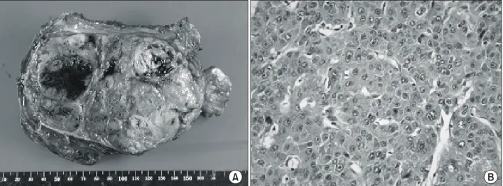
Fig. 3. Gross finding shows a globular tumor mass embeded with segment of rib weighing 650 gm and measuring 14×10×7.5 cm in dimensions (A). Microscopic finding shows metastatic trabecular type of hepatocellular carcinoma (H&E ×200) (B).
A B
대흉외지 2004;37:809-812
- 812 -
=국문 초록=
흉벽에 발생하는 전이성 악성 종양은 드문 질환이며 주위 장기로부터 전이되는 경우가 대부분으로 유방, 폐, 흉막, 그리고 종격동 등으로부터 주로 전이되는 것으로 알려져 있다. 특히 흉부가 아닌 먼 장기로부터 전이되는 경우는 더욱 드문 것으로 알려져 있고 원발성 악성 종양이 없이 흉벽 전이에 의해 발현되는 예는 몇몇의 보고만이 있을 뿐이다. 환자는 51세 남자로 전흉벽 좌상부에 촉지되는 종 괴로 절제 수술을 시행 후 전이성 간세포암종으로 진단 받았고 당시 검사상 간에 원발성 간세포암종 의 증거는 없는 상태였다. 이에 원발성 종양의 증거 없이 간세포암이 흉벽으로 전이된 예를 치험하였 기에 보고하는 바이다.
중심 단어:1. 흉벽 2. 흉벽 종양 3. 전이성 종양 4. 간종양