서론
완전 무치악 환자에서 구강 회복을 위한 총의치나 임 플란트 보철 치료 시, 구치부 배열, 특히 치열궁 폭경을 결정하기 위해 일반적으로 이전 자연치 배열을 찾아 재건 해 주려고 하지만, 실제 임상에서 이를 되찾아 주기란 쉽 지 않다.
Watt1는 완전 무치악 환자의 치열 재건 시, 자연치의 구 개측 치은 변연의 흔적인 상악 무치악 치조능을 기준으 로 제시하면서 이를 상악 구치(구개면) 위치 설정을 위한
가이드로 사용할 수 있음을 강조하였다. 그러나 선천적 결손치 부위, 치조골 인위적 결손이 심한 부위, 치조골 흡 수가 상당히 진행된 부위에서 이를 활용하는 데는 한계 가 있다.
또한 통상적으로 상악과 하악 완전 무치악 환자에서 총의치로 치열을 재건할 경우, 술자의 임상경험, 해부학 적 구조물, 이전 의치의 치아배열 등을 근거로 진행해 왔 고 특히 구치 배열을 위해서는 하악의 후구치 삼각융기 와 잔존 치조능을 참고하여 하악을 먼저 배열하고 이를 기준으로 상악을 결정해 왔다.2 그러나 이는 엄밀히 말하
*Correspondence to: Sang-Chun Oh
Professor, Department of Prosthodontics, College of Dentistry, Wonkwang University, 77 Doonsan-ro, Seo-gu, Daejeon, 35233, Republic of Korea Tel: +82-42-366-1100, Fax: +82-42-366-1115, E-mail: [email protected] Received: October 7, 2019/Last Revision: October 29, 2019/Accepted: October 29, 2019
relationship between inter-condylar width and inter-maxillary first molar width
Sang-Chun Oh*, Hyun-Jun Kong
Department of Prosthodontics, College of Dentistry, Wonkwang University, Iksan, Republic of Korea
Purpose: The aim of this study was to evaluate the correlation between inter-condylar width and inter-maxillary first molar width to present the criteria for prosthetic reconstruction of dental arch width in maxillary and mandibular fully edentulous patients.
Materials and Methods: 120 Koreans (60 males and 60 females) who underwent the cone beam computerized tomography (Cone- beam CT) were selected. The Cone-beam CT images were analysed using Invivo 5.1. After reorientation of axis, inter-maxillary first molar width was measured by clicking both mesio-buccal cusp tip of maxillary first molar. And inter-condylar width was measured by clicking both middle points of condyles. The collected data were analysed with SPSS Version 20.0 and statistical significance of the correlation between inter-condylar width and inter-maxillary first molar width was verified by Pearson’s correlation analysis. Results:
The mean inter-condylar width of Korean was 105.9 mm, and that of male (108.3 mm) was statistically significantly wider than the female (103.4 mm). The inter-maxillary first molar width of Korean was 57.1 mm, and that of male (57.9 mm) was statistically significantly wider than the female (56.2 mm). Pearson’s correlation analysis between inter-condylar width and inter-maxillary first molar width showed a Pearson correlation coefficient of 0.614 and statistically significantly positive correlation. Conclusion: Inter- condylar width and inter-maxillary first molar width showed positive correlation and the average ratio of inter-condylar with and inter-maxillary first molar width was 1:0.54. Based on the results of this limited study, inter-condylar width can be used as a guide for setting up dental arch width in fully edentulous patient. (J Dent Rehabil Appl Sci 2019;35(4):214-9)
Key words: fully edentulous patient; inter-condylar width; dental arch width; cone-beam CT
Copyright© 2019 The Korean Academy of Stomatognathic Function and Occlusion.
It is identical to Creative Commons Non-Commercial License.
cc
면 의사의 임상 경험에 따른 주관적인 판단으로 의사에 의해서 약간씩 달라질 수 있고, 그나마 지표로 쓰고 있는 연조직 해부학적 구조물 또한 치조골 흡수 정도와 연령 증가에 따라 변할 수 있으며, 이는 단지 총의치의 유지와 안정을 우선 고려한 방법이라는 것이다.3
그러나 최근에는 완전 무치악 상태에서도 치조골 증대 술 등을 통해 악골 구조를 적극 개선한 후 임플란트 보철 수복, 즉 임플란트 지지 피개의치와 임플란트 지지 고정 성 보철 치료가 가능하게 되었으며, 따라서 환자 본래 유 치악 상태의 악궁 크기 및 치아 위치를 가늠한 후 그것을 재건하려는 치료 목표가 의미 있게 되었다.
따라서 좀더 과학적이고 객관적인 구치부 악궁 폭경 재건을 위한 가이드의 하나로서 일생을 통해 비교적 변 화가 거의 없는 과두간 폭경을 기준으로 각 환자의 본래 치열궁 폭경을 가늠하기 위하여 이 둘의 비율적 상관관 계를 파악하는 것은 임상적 의미가 크다 할 것이다.
그러나 이에 대한 연구는 거의 전무한 상태이며, 단지 최근에 Debnath 등4 이 과두간 폭경과 상악 견치간 폭경, 상악 제1대구치간 폭경을 조사하였지만, 과두간 폭경을 실측하지 못하고 facebow를 이용한 간접 계측이 이루어 졌으며, 인종간의 치아 및 악궁 크기와 형태 등에 차이가 있기 때문에 과두간 폭경과 상악 제1대구치간 폭경도 인 종 간에 차이가 있을 수 있으므로 한국인을 대상으로 분 석할 필요성이 대두된다.5-8
따라서 본 연구에서는 상악과 하악 완전 무치악 환자 에서 본래 치열궁 폭경을 파악하기 위한 가이드로서 과 두간 폭경의 가치를 파악하기 위해 신속 정확하게 실 측의 효과가 있는 콘빔형 전산화단층영상(Cone-beam computerized tomography)을 사용하여 한국인의 과두 간 폭경과 상악 제1대구치간 폭경을 계측하여 이들의 상 관관계를 평가하였다.
연구 재료 및 방법
Cone-beam CT (PaX-Zenith 3D; Vatech Korea Co., Ltd., Seoul, Korea)를 촬영한 환자 중에서, 한국인으로 제3대구치를 제외한 결손이 없는 영구치열을 갖고 있고, angle 분류 class 1인 환자 120명을 대상으로 하였다. 교 정 치료 또는 악교정 수술 병력이 존재하는 경우, 악관절 장애로 인한 치료 및 과두를 포함하는 외과적 수술 기왕 력이 있는 경우, 상악 및 하악의 중절치에 수복물이 존재 하는 경우, 그 밖의 이유로 계측이 힘든 경우는 연구 대상 에서 배제하였다. 연구 대상에 포함된 120명의 연령별 분 포는 다음과 같다(Table 1). 본 연구는 IRB심사(IRB No.
W1908/001-001)를 거쳐 진행되었다. 연구 대상의 기준 은 Ferrario 등9,10의 연구를 참고하였다.
악궁의 디지털화
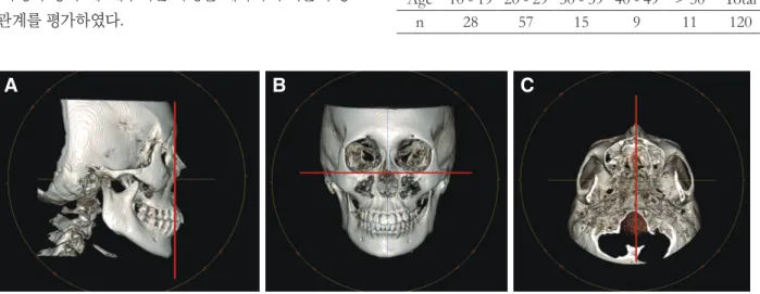
콘빔형 전산화단층영상을 통해 얻어진 이미지를 digi- tal imaging and communications in medicine (DICOM) format 으로 변환하였으며, 영상은 Invivo 5.1 (Anatom- age, San Jose, USA)을 사용해서 분석하였다. Bayome 등11 의 연구를 참고하여 영상의 기준 축 설정을 시행했다. 먼 저 시상영상에서 수직선을 비근점과 전비극을 지나는 선 으로 설정하였다(Fig. 1A). 다음으로 관상영상으로 선택 한 후 수평선을 양측 안와점을 지나는 선으로 설정하였 다(Fig. 1B). 마지막으로 축영상에서 전비극과 후비극을 지나는 수직선을 기준으로 하였다(Fig. 1C).
Table 1. Distribution of patients by age
Age 10 - 19 20 - 29 30 - 39 40 - 49 > 50 Total
n 28 57 15 9 11 120
Fig. 1. Reorientation of axis. (A) Sagittal view, (B) Coronal view, (C) Axial view.
A B C
계측 기준
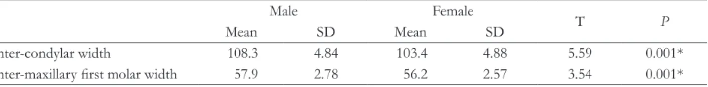
상악 제1대구치간 폭경을 계측하기 위해 축영상에서 영상조절을 치아로 설정하였다. 이후 5 mm 간격으로 조 절하면서 상악 제1대구치의 최대 협설 폭경이 보이는 상 을 선택한 후 Alam등12의 연구를 참고하여 상악 제1대구 치의 근심협측 교두정 부위를 클릭하여 상악 제1대구치 간 폭경을 측정하였다(Fig. 2)
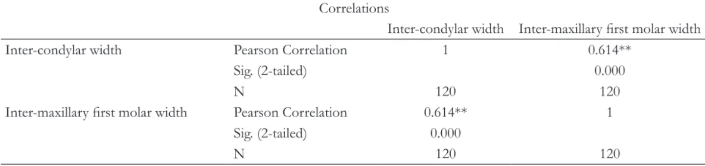
다음으로 과두간 폭경을 계측하기 위해 관상영상에서 영상을 경조직으로 선택한 후, 5 mm 간격으로 조절하면 서, 양측 과두의 최대 외연이 보이는 상을 선택하였다. 이 후 양측 과두의 최대 외연과 내연의 중심점을 선택하였으 며, 양측 중심점을 클릭하여 과두간 폭경을 계측하였다 (Fig. 3). 계측점의 설정은 Nagi 등13의 연구를 참고하였다.
통계 분석
과두간 폭경과 상악 제1대구치간 폭경의 성별에 대한
통계적 유의성을 검증하기 위하여 독립표본 t 검정을 시 행하였고, 이 둘의 상관관계를 분석하기 위해 Pearson 의 상관분석을 시행하였다. 통계처리에는 SPSS Version 20.0 (IBM Inc., Armonk, USA)을 이용하였다.
결과
한국인의 평균 과두간 폭경은 105.9 mm였으며, 남성 (108.3 mm)이 여성(103.4 mm)에 비해 통계학적으로 유 의하게 크게 나타났다(P < 0.001). 평균 상악 제1대구치 간 폭경은 57.1 mm 였으며, 남성(57.9 mm)이 여성(56.2 mm)에 비해 통계학적으로 유의하게 크게 나타났다(P <
0.001, Table 2). 한국인의 평균 과두간 폭경과 상악 제1 대구치간 폭경은 1:0.54의 비율을 보였다.
과두간 폭경과 상악 제1대구치간 폭경의 상관관계를 분석하기 위해 Pearson의 상관분석을 시행한 결과(Table 3), Pearson의 상관계수(r)는 0.614로 나타났으며 통계적 으로 유의한 양의 상관관계를 보였다.
Table 2. Independent t-test for differences of inter-condylar width and Inter-maxillary first molar width between male and female
Male Female T P
Mean SD Mean SD
Inter-condylar width 108.3 4.84 103.4 4.88 5.59 0.001*
Inter-maxillary first molar width 57.9 2.78 56.2 2.57 3.54 0.001*
*: P < 0.001
Fig 2. Inter-maxillary first molar width. Fig. 3. Inter-condylar width.
50.61 mm
101.54 mm
고찰
일반적으로 상악과 하악 완전 무치악에서 총의치를 통 한 악궁 재건을 위해 교합평면 높이는 구치는 하악의 후 구치 삼각융기를14 전치는 구각부를 활용하고,15 교합평 면 주행 방향은 Camper’s line과 나란하도록 기준이 정립 되었으나16 악궁의 폭에 관련된 과학적인 기준은 부족하 며 통상 임상가의 경험에 근거하여 임의적으로 결정하고 있다.
그러나 무치악 악궁을 의치나 임플란트를 통해 재건할 경우, 악궁의 폭을 임의적으로 결정하게 되면 본래의 크 기보다 지나치게 넓거나 좁게 될 수 있고, 이는 각 과두에 서 구치까지 거리를 본래보다 길거나 짧게 만들어, 결국 구치의 교합면 형태, 즉 교두융선과 구의 방향 그리고 상 악 전치부 설면 형태 부여에 영향을 주게 된다.17
따라서 가급적 각 환자의 두부, 즉 과두간 폭경에 맞는 악궁 폭경을 재현해 주어야 하는데, 이러한 이유에서 과 두간 폭경에 따른 악궁 폭경의 비율 및 상관관계를 파악 하는 것은 의미 있는 일이다. 그러나 지금까지는 단지 과 두간 폭경, 악궁의 크기 등을 해부학적 차원의 계측 수준 의 연구만 있었을 뿐, 이 둘의 상관관계에 관련된 연구는 매우 미흡한 상황이었다.
이 둘의 관계를 연구한 Debnath 등4은 과두간 거리는 상악 견치간 거리의 3.17배, 상악 제1대구치간 거리의 2 배, 안면수직고경의 1.7배를 보인다고 하였고, Keshvad18 는 과두간 거리가 상악 견치간 거리의 3.39배, 상악 제1 대구치간 거리의 2.19배였다고 보고하였다. 그러나 이들 은 과두간 폭경을 facebow의 hinge axis locator를 이용 하여 계측하였으며, 본 연구에서는 3차원 영상 속에서 실 측이 가능한 Cone-beam CT를 이용하여 보다 신속하고
정확한 계측 값을 얻을 수 있었다. 그 결과 과두간 폭경 은 상악 제1대구치간 폭경의 1.85배를 나타내었고, 이는 계측방법의 차이와 인종간의 차이가 반영된 값이라 할 수 있다.
또한 Keshvad는 과두간 폭경과 상악 견치간 거리, 상 악 제1대구치간 거리, 하악 견치간거리, 하악 제1대구치 간 거리 등의 상관관계를 분석한 결과, 과두간 폭경과 상 악 제1대구치간 거리 사이에서 가장 큰 상관관계를 보였 다고 보고하였다. 본 연구에서 한국인의 과두간 폭경과 상악 제1대구치간 폭경에 대한 Pearson의 상관분석을 시 행한 결과 Pearson 상관계수(r)가 0.614로 나타났으며, 이는 Taylor의 연구에 의하면 중등도의 양의 상관관계를 보인다고 해석할 수 있다.19 이러한 결과는 과두간 폭경 의 측정을 통해 상악 구치간 폭경을 유추할 수 있고, 기 준점이 없는 상악과 하악 완전 무치악 환자에서 구강재 건 시 치열궁 폭경 설정에 도움을 줄 수 있으며, 이는 기 존에 환자가 가졌던 자연치열과 더욱 유사한 치아배열이 가능해 질 수 있음을 의미한다.
본 연구는 콘빔형 전산화단층영상을 촬영한 환자 중 연구기준에 부합되는 120명을 대상으로 하였으나 이 중 85명이 30세 이하로, 특정 연령층에 연구 대상이 편중되 어 있는 한계점을 가진다. 또한 연구 방법에서 5 mm 단 위로 영상을 조절하여, 정확한 계측 기준 설정에 오차가 발생할 수 있다.
향후, 상악과 하악의 완전 무치악 환자에서 치열궁 재 건을 위한 지표로서 CBCT 등의 디지털영상장비를 이용 한 과두간 폭경에 대한 악궁의 전후방적 거리의 관계나 수직고경과의 관계, 그리고 이 입체영상을 통해 두개골 에 대한 악궁의 3차원적 위치적 관계 등을 포함한 추가적 인 연구도 가능할 것으로 사료된다.
Table 3. Pearson correlation analysis for inter-condylar width and Inter-maxillary first molar width Correlations
Inter-condylar width Inter-maxillary first molar width Inter-condylar width Pearson Correlation 1 0.614**
Sig. (2-tailed) 0.000
N 120 120
Inter-maxillary first molar width Pearson Correlation 0.614** 1 Sig. (2-tailed) 0.000
N 120 120
**: Correlation is significant at the 0.01 level.
결론
한국인의 과두간 폭경과 상악 제1대구치간 폭경의 상 관관계를 알아보기 위해 120명의 Cone-beam CT 영상 으로 과두간 폭경과 상악 제1대구치간 폭경을 분석한 결 과, 한국인의 평균 과두간 폭경은 105.9 ± 5.44 mm, 상 악 제1대구치간 폭경은 57.1 ± 2.80 mm이었다. 과두간 폭경과 상악 제1대구치간 폭경은 1:0.54의 비율을 보였 으며, 중등도의 양의 상관관계를 보였다(r = 0.614). 성 별에 따른 과두간 폭경과 상악 제1대구치간 폭경은 양쪽 모두 남성이 여성에 비해 통계학적으로 유의성 있게 크 게 나타났다(P < 0.001). 본 제한된 연구의 결과를 근거 로 상악과 하악의 완전 무치악 환자에서 과두간 폭경은 본래의 상악 치열궁 폭경 설정을 위한 가이드로서 가치 가 있을 것으로 사료된다.
Acknowledgements
This paper was supported by Wonkwang University in 2018.
ORCID
Sang-Chun Oh https://orcid.org/0000-0001-5496-126X Hyun-Jun Kong https://orcid.org/0000-0001-9331-3572
References
1. Watt DM, McGregor AR. Designing complete den- ture. 2nd ed. Philadelphia; WB Saunders Co.; 1986. p.
22-8.
2. Pound E. Recapturing esthetic tooth position in the edentulous patient. J Am Dent Assoc 1957;55:181-91.
3. Lang BR. Complete denture occlusion. Dent Clin North Am 2004;48:641-65.
4. Debnath N, Gupta R, Meenakshi A, Kumar S, Hota S, Rawat P. Relationship of inter-condylar distance with inter-dental distance of maxillary arch and oc- clusal vertical dimension: a clinical anthropometric study. J Clin Diagn Res 2014;8:ZC39-43.
5. Lee KY, Lee DJ. A comparative study of teeth and dental arch of Korean and Caucasian. Oral Biol Res 1993;71:1-15.
6. Song WC, Yun KH, Koh KS. Facial flatness of Ko- rean: using facial depth. Korean J Anat 2003;36:499- 506.
7. Kook YA, Nojima K, Moon HB, McLaughlin RP, Sinclair PM. Comparison of arch forms between Korean and north American white populations. Am J Orthod Dentofacial Orthop 2004;126:680-6.
8. Hwang HS, Kim WS, McNamara JA Jr. Ethnic dif- ferences in the soft tissue profile of Korean and European-American adults with normal occlusions and well-balanced faces. Angle Orthod 2002;72:72- 80.
9. Ferrario VF, Sforza C, Miani A Jr, Colombo A, Tar- taglia G. Mathematical definition of the curve of Spee in permanent healthy dentitions in man. Arch Oral Biol 1992;37:691-4.
10. Ferrario VF, Sforza C, Miani A Jr, Tartaglia G.
Mathematical definition of the shape of dental arches in human permanent healthy dentitions. Eur J Orthod 1994;16:287-94.
11. Bayome M, Park JH, Kook YA. New three-dimen- sional cephalometric analyses among adults with a skeletal Class I pattern and normal occlusion. Ko- rean J Orthod 2013;43:62-73.
12. Alam MK, Shahid F, Purmal K, Ahmad B, Khamis MF. Tooth size and dental arch dimension measure- ment through cone beam computed tomography: Ef- fect of age and gender. Res J Recent Sci 2014;3:85-94.
13. Naji P, Alsufyani NA, Lagravère MO. Reliability of anatomic structures as landmarks in three-dimen- sional cephalometric analysis using CBCT. Angle Orthod 2014;84:762-72.
14. Boucher CO. Swenson’s complete dentures. 5th ed.
St. Louis; Mosby Company; 1964. p. 246-51.
15. Lunquist DO, Luther WW. Occlusal plane determi- nation. J Prosthet Dent 1970;23:489-98.
16. Boucher CO. Current clinical dental terminology.
2nd ed. St. Louis; Mosby Company; 1974. p. 229.
17. Lee SH. Fixed prosthodontics. 2nd ed. Seoul; Jee- seung Publisuing co.; 2007. p. 102.
18. Keshvad A, Winstanley RB, Hooshmand T. Inter- condylar width as a guide to setting up complete denture teeth. J Oral Rehabil 2000;27:217-26.
19. Taylor R. Interpretation of the correlation coeffi- cient: A basic review. JDMS. 1990;6:35-9.
*교신저자: 오상천
(35233)대전광역시 서구 둔산로 77 원광대학교 대전치과병원 치과보철과 Tel: 042-366-1100|Fax: 042-366-1115|E-mail: [email protected] 접수일: 2019년 10월 7일|수정일: 2019년 10월 29일|채택일: 2019년 10월 29일
과두간 폭경과 상악 제1대구치간 폭경 사이의 관계
오상천*, 공현준
원광대학교 치과대학 치과보철학교실
목적:본 연구의 목적은 상악과 하악의 완전 무치악 환자에서 보철물로 상실된 치열궁 폭경 재건을 위한 기준을 제시하기 위하여 과두간 폭경에 대한 상악 제1대구치간 폭경의 상관관계를 평가하기 위함이다.
연구 재료 및 방법: 콘빔형 전산화단층영상(Cone-beam CT)을 촬영한 환자 중, 본 연구의 기준에 부합하는 한국인 120명 (남성 60명, 여성 60명)을 선택하였으며 Invivo 5.1을 이용해 Cone-beam CT scan을 분석하였다. 축의 방향설정 후, 상악 제1대구치의 근심 협측 교두정간 거리를 측정하였으며, 과두간 폭경은 각 과두의 중심점을 지정하여 측정하였다. 수집된 자료는 SPSS Version 20.0으로 분석하였고, Pearson의 상관분석을 이용하여 과두간 폭경과 상악 제1대구치간 폭경의 상 관관계의 통계적 유의성을 검증하였다.
결과: 한국인의 평균 과두간 폭경은 105.9 mm였으며, 남성이 108.3 mm 여성이 103.4 mm였다. 평균 상악 제1대구치간 폭경은 57.1 mm였으며, 남성이 57.9 mm 여성이 56.2 mm였다. 과두간 폭경과 상악 제1대구치간 폭경에 대한 Pearson 의 상관분석 결과, 0.614의 Pearson 상관계수로 통계적으로 유의한 양의 상관관계를 보였다.
결론: 과두간 폭경과 상악 제1대구치간 폭경은 양의 상관관계를 보였으며, 한국인의 과두간 폭경과 상악 제1대구치간 폭경의 평균 비율은 1:0.54를 보였다. 본 제한된 연구의 결과를 근거로 상악과 하악의 완전 무치악 환자에서 과두간 폭경 을 본래의 상악 치열궁 폭경 설정을 위한 가이드로서 사용이 가능할 것이다.
(구강회복응용과학지 2019;35(4):214-9)
주요어: 완전 무치악; 과두간 폭경; 치열궁 폭경; 콘빔형 전산화단층영상