https://doi.org/10.14734/PN.2018.29.1.13 pISSN 2508-4887•eISSN 2508-4895
Sae Mi Yang, MD1, O Kyu Noh, MD, PhD2,3, Jang Hoon Lee, MD, PhD1, Moon Sung Park, MD, PhD1
1Department of Pediatrics, Ajou University School of Medicine, Suwon, 2Department of Radiation Oncology, Ajou University School of Medicine, Suwon, 3Department of Biomedical Informatics, Ajou University School of Medicine, Suwon, Korea
Objective: To investigate the incidence of early severe hypophosphatemia, and examine the associated clinical factors and outcomes, in extremely low birth weight infants (ELBWI) who received early amino acid administration.
Methods: Medical records of 82 ELBWI were retrospectively reviewed. Severe hypophosphatemia was defined as a serum phosphate level <2 mg/dL during the first week after birth.
Results: Nineteen ELBWI (23.2%) experienced severe hypophosphatemia. The supplementation of phosphorus was started significantly later in the hypophosphatemia group compared to that in the control group (P=0.036). Small for gestational age infants (SGAI) (P=0.006) and bronchopulmonary dysplasia (P=0.016) were more prevalent in the hypophosphatemia group compared to that in the control group.
Conclusion: Early severe hypophosphatemia is common in ELBWI. Late and insufficient supplemen
tation of phosphorus and SGAI were associated with severe hypophosphatemia.
Key Words: Infant, Extremely low birth weight, Hypophosphatemia, Bronchopulmonary dysplasia
Introduction
Premature infants, including extremely low birth weight infants (ELBWI) miss the last trimester, which involves rapid intrauterine growth and nutrition accretion.1 Optimal nu
tritional support through parenteral nutrition (PN) is important for survival and postnatal growth, as most ELBWI cannot tolerate enteral nutrition. In the last few decades, nutrition guidelines have emphasized early and aggressive PN with high amino acid supplementation immediately after birth, to minimize growth restriction and reduce adverse outcomes due to nutrition deficiency.2,3
However, a recent study reported that early aggressive amino acid administration causes hypophosphatemia.4 A highprotein diet accelerates protein synthesis and tissue anabo lism.
In addition, the intracellular influx of serum phosphate and synthesis of adenosine tripho
sphate and creatine phosphate are increased, resulting in serum phosphorus deple tion in ELBWI, especially in small for gestational age infants (SGAI).58 Unfortunately, the literature is limited, as wide variations exist in the definition of hypophosphatemia, and assessments regarding the impact of hypophosphatemia on the major outcomes in the neonatal intensive care unit (NICU) are lacking. In addition, few studies have examined whether delayed and Received: 18 September 2017
Revised: 12 October 2017 Accepted: 23 October 2017 Correspondence to Jang Hoon Lee, MD, PhD Department of Pediatrics, Ajou University School of Medicine, 164 Worldcupro, Yeongtonggu, Suwon 16499, Korea
Tel: +82312195167 Fax: +82312195169 E-mail: [email protected] Copyright© 2018 by The Korean Society of Perinatology
This is an Open Access article distributed under the terms of the Creative Com
mons Attribution NonCommercial License (http://creativecommons.org/
license/bync/4.0/), which permits unrestricted noncommercial use, distribution, and reproduction in any
Late and Insufficient Phosphorus Sup
ple mentation is Associated with Early
Severe Hypophosphatemia in Extremely
Low Birth Weight Infants with Early
Amino Acid Administration
early amino acid administration.9 There fore, the present study investigated the incidence of severe hypophosphatemia during the first week after birth, and examined the associated clinical factors (including phosphorus supplementation) and outcomes, in ELBWI who received same policy of early amino acid admi
nistration.
Methods
1. Study population
ELBWI (birth weight less than 1,000 g) born at Ajou Univer
sity Hospital from January 2013 to December 2016 were in
cluded. Exclusion criteria included death within seven days after birth and severe congenital malformations, resulting in the exclusion of five of the 87 reviewed ELBWI. Nineteen of the included 82 ELBWI experienced severe hypophosphatemia (serum phosphate level <2 mg/dL) during the first week after birth (hypophosphatemia group); the remaining 63 ELBWI were classified as the control group (Fig. 1). This study was approved by the Institutional Review Board of Ajou University Hospital (M2017C046000003) and we received permission to waive parental consent.
2. Data collection
Clinical data was collected retrospectively from a medical record review. The collected data included perinatal and neo
natal characteristics such as sex, gestational age, birth weight, SGAI status, use of antenatal steroids, whether delivered via caesarean section, Apgar scores at 1 and 5 minutes of life, exi
stence of histologic chorioamnionitis, preterm premature rup
ture of membrane (PPROM), patent duct arteriosus (PDA) liga
tion, intraventricular hemorrhage (IVH), sepsis, extrauterine growth retardation (EUGR), necrotizing enterocolitis (NEC), duration of PN (days), retinopathy of prematurity (ROP), bron
chopulmonary dysplasia (BPD), duration of mechanical ventila
tion, and mortality. Serum phosphate and calcium levels, and the amounts of amino acid, phosphorus, calcium, and fluid admi
nistration during the first week after birth were also collected.
3. Definitions
Early severe hypophosphatemia was defined as a serum phosphate level less than 2 mg/dL during the first week after birth. Hypercalcemia was defined as a serum calcium level higher than 11 mg/dL. Treatment was performed when hyper
calcemia persisted for more than two days or when there was an electrocardiography change.
BPD was defined as the need for supplemental oxygen for at least 28 days after birth, and the severity was graded according to the respiratory support required at 36 postmenstrual weeks or discharge, whichever came first.10 If the infants died before 36 weeks, they were excluded from the BPD classification.
NEC was defined as Bell’s stage II or greater.11 IVH was de
fined as grade III or IV on a cranial ultrasonography, based on the Papile grading system.12 ROP was defined using Interna
tional Classification of Retinopathy of Prematurity criteria.13 PPROM was defined as a rupture of membranes before the onset of uterine contractions before 37 weeks, SGAI was de
fined as <10th percentile for gestational age at birth and EUGR was defined as <10th percentile for the corrected gestational age at 36 weeks.14
According to our NICU policy, PN was started on postnatal day 1. Carbohydrates were given at a minimum of 4 mg/kg/min on day 1. Amino acids (Primene® 10%; Baxter, Grosotto, Italy) were given at 1 g/kg/day starting on day 1, and were increased 0.51.0 g/kg/day daily to a maximum of 3.54 g/kg/day on day 7. The amount of administered amino acids was guided by the serum blood urea nitrogen level and the development of severe me
tabolic acidosis. Lipids (SMOF lipid® 20%; Fresenius Kab, Up
psala, Sweden) were given at a minimum of 0.5 g/kg/day start ing on day 1, and were increased to a maximum of 3 g/kg/day. Cal
cium (Calcium Gluconate Injection®; Daihan Pharm, Seoul, Fig. 1. The patient enrollment flow chart. BW, birth weight.
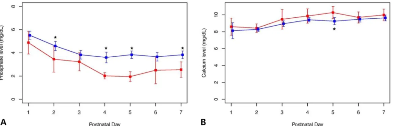
2, 4, 5, and 7 were significantly lower in the hypophospha temia group compared to those in the control group (P<0.05) (Fig. 2A).
The serum calcium level on day 5 was significantly higher in the hypophosphatemia group compared to that in the control group (10.0 [9.7; 10.8] vs. 9.2 [8.6; 9.9] mg/dL, P=0.012) (Fig. 2B).
The incidence of hypercalcemia was significantly higher in the hypophosphatemia group (52.6%, 10/19) compared to that in the control group (14.3%) (P=0.002). A total of 36.8% (7/19) and 3.2% (2/63) of the infants in hypophosphatemia and control groups, respectively, required treatment for hypercalcemia (such as hydration and systemic steroids), with no significant group difference (P=0.55).
2. Demographic characteristics
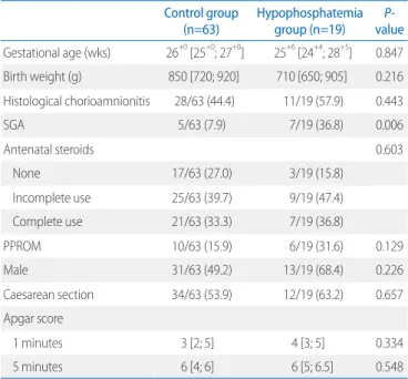
Demographic characteristics are presented in Table 1.
There were no significant group differences in sex, gestational age, birth weight, histologic chorioamnionitis, use of antenatal steroid therapy, caesarean sections, and Apgar scores at 1 and 5 minutes. However, the incidence of SGAI was significantly higher in the hypophosphatemia group (36.8%, 7/19), compared to that in the control group (7.9%, 5/63) (P=0.006).
3. Supplementation of amino acids, phosphorus, and fluid Amino acids were administered at a rate of 1 g/kg/day start
ing on day 1, with no significant group differences during the first week after birth (P=0.95) (Fig. 3A). The supplementation of phosphorus started significantly later in the hypophospha
Korea) infusion was started on day 1 at 6080 mg/kg/day (1.5
2 mmol/kg/day). Phosphorus supplementation (Pho sten Injec
tion®, JW Pharma, Seoul, Korea) began on days 24, with con
sideration of the infants’ condition, and were prescrib ed by the physician at an adequate dose of 15.546.5 mg/kg/day (0.5
1.5 mmol/kg/day), as guided by the stability of the PN. Fluid administration was started on day 1 at a rate of 80 mL/kg/day and thereafter was determined by the monitoring of vital signs, urination, and daily weight.
4. Statistical analysis
Data are presented as the median [25; 75 percentile] for continuous variables, and as the number (%) for categorical va
riables. Group differences were evaluated using the Student’s ttest or KruskalWallis rank sum test for continuous variables and the chisquare test or Fisher’s exact test for categorical variables. Pvalues <0.05 were considered statistically signifi
cant. All statistical analyses were performed using R software (R Foundation for Statistical Computing, Vienna, Austria).15
Results
1. Phosphate and calcium levels
The serum phosphate level in the hypophosphatemia group on day 1 did not differ from that in the control group (5.5 [2.9; 6.6]
vs. 5.8 [4.5; 6.5] mg/dL, P=0.389); however, the levels on days
A B
Fig. 2. The serum phosphate and calcium levels during the first week after birth are shown for the hypophosphate mia and control groups. (A) Mean phosphate levels during the first week after birth: there was no group difference in phosphate level on day 1, but there were significant group differences on day 2 (3.5±1.9 vs. 4.6±1.34 mg/dL, P=0.015), day 4 (1.9 [1.8; 2.2] vs.
3.5 [2.9; 4.3] mg/dL, P<0.001), day 5 (2.0±10.6 vs. 3.8±1.0 mg/dL, P<0.001), and day 7 (2.5±1.2 vs. 3.8±1.0 mg/dL, P<0.001).
(B) Mean calcium levels during the first week after birth: on day 5 (10.0 [9.7; 10.8] vs. 9.2 [8.6; 9.9] mg/dL, P=0.012), there was a significant group difference in calcium levels.
temia group compared to that in the control group (73 [61;
88.5] vs. 85 [71.5; 142.0] hours after birth, P=0.036). Although there was no significant group difference in the amount of sup
plied phosphorus on days 1, 2, and 3, the amount on days 4 and 5 was significantly less in the hypophosphatemia group com
pared to that in the control group (all P<0.05) (Fig. 3B). There was no significant group difference in the amount of admini
stered fluid during the first week after birth (Fig. 3C).
4. Clinical outcomes during the neonatal intensive care unit stay
Clinical outcomes during the NICU stay are provided in Table 2. There were no significant group differences in the rates of PDA ligation, IVH, sepsis, EUGR, NEC, duration of PN, ROP, duration of mechanical ventilation, and mortality. How
ever, the incidence of BPD was greater in the hypophosphatemia group compared to that in the control group (100% [11/11] vs.
59.6% [31/52], P=0.016).
Discussion
The purpose of the present study was to investigate the inci
dence of early severe hypophosphatemia, as well as the asso
ciated risk factors and outcomes among ELBWI who received Table 1. Patient Characteristics
Control group
(n=63) Hypophosphatemia group (n=19) P-
value Gestational age (wks) 26+0 [25+0; 27+0] 25+6 [24+4; 28+5] 0.847 Birth weight (g) 850 [720; 920] 710 [650; 905] 0.216 Histological chorioamnionitis 28/63 (44.4) 11/19 (57.9) 0.443
SGA 5/63 (7.9) 7/19 (36.8) 0.006
Antenatal steroids 0.603
None 17/63 (27.0) 3/19 (15.8)
Incomplete use 25/63 (39.7) 9/19 (47.4)
Complete use 21/63 (33.3) 7/19 (36.8)
PPROM 10/63 (15.9) 6/19 (31.6) 0.129
Male 31/63 (49.2) 13/19 (68.4) 0.226
Caesarean section 34/63 (53.9) 12/19 (63.2) 0.657
Apgar score
1 minutes 3 [2; 5] 4 [3; 5] 0.334
5 minutes 6 [4; 6] 6 [5; 6.5] 0.548
Values are presented as median [25; 75 percentile] or number (%).
Abbreviations: SGA, small for gestational age; PPROM, preterm premature rupture of membrane.
A B
C
Fig. 3. The supplementation of amino acid, phosphorus, and fluid during the first week after birth is shown for the hypo- phosphatemia and control groups. (A) Amino acids supplemen- tation during the first week after birth: there was no group difference in the amount of amino acid administration during the first week after birth. (B) Phosphorus supplementa tion during the first week after birth: there were significant group differences in the amount of phosphorus administered on day 4 (0.0 [0.0; 0.5] vs. 0.6 [0.0; 1.0] mmol/kg/day, P=0.37) and day 5 (0.5 [0.0; 1.0] vs. 1.0 [0.8; 1.0] mmol/kg/day, P=0.017). (C) Fluid supplementation during the first week after birth: there was no group difference in the amount of fluid administration.
same policy of early amino acids administration. The hypopho
sphatemia group had a later starting time and received a smal
ler amount of phosphorus supplementation compared to that in the control group. In addition, the hypophosphatemia group showed a higher morbidity of SGAI and incidence of BPD com
pared to that in the control group.
An absolute level for hypophosphatemia has not been de fined for preterm infants. In this study, severe hypophospha temia was defined as a serum phosphate level <2 mg/dL. Brener Dik et al.4 defined severe hypophosphatemia in a similar manner and reported that the incidence of hypophosphatemia in very low birth weight infants was 34%, which is higher than that in the present study. This difference may result from dif ferences in amino acid administration policies (3 mg/kg/day starting on day 1 in the previous study). In the present study, supplied amino acids were not increased when the serum blood urea nitrogen level was 30 mg/dL or more. In a study by Boubred et al.,8 80% (48/60) of ELBWI experienced hypopho sphatemia, defined as a serum phosphate level <1.6 mmol/L (4.95 mg/dL), which corresponds to relatively milder hypopho sphatemia com
pared to that in the present study. Bonsante et al.16 showed a clear association between high amino acid admi nistration and hypophosphatemia; hypophosphatemia was more frequent in the high amino acid group (12.5%) than in the medium (4.6%) and low amino acid groups (0%). Similarly, in the study of Ichi
kawa et al.,7 higher parenteral amino acid admi nistration was
associated with hypophosphatemia on day 8. Thus, knowledge regarding the factors that enhance or re duce the risk of hypo
phosphatemia in ELBWI who receive early amino acid admini
stration is needed. The present results sug gest that the timing and amount of phosphorous supplementation is important.
SGAI who faced with chronic malnutrition in the fetal period have metabolic and electrolyte imbalances, which can be expl
ain ed by the concept of refeeding syndrome.17 Small for gesta
tional age ELBWI were shown to have lower phosphate levels on day 8 compared to that in ELBWI who were appro priate for gestational age in study by Ichikawa et al.7 Similarly, Boubred et al.8 reported that hypophosphatemia is more likely to occur in SGAI among ELBWI who receive high amounts of amino acids.
The present results are consistent with results from these studies.
A few previous studies have attempted to define the associa
tion between phosphorus administration and hypophosphatemia.
For example, Moe et al.9 compared the incidence of hypopho
sphatemia in three groups of ELBWI (less than 28 weeks of age) who received three types of PN containing different amounts of phosphorus and amino acids; phosphorus levels were lowest in the group receiving PN containing a low amount of phosphorus and a high amount of amino acids. How ever, the authors were unable to investigate whether phos phorus administration af
fected the development of hypopho sphatemia regardless of the amount of administered amino acids. In the present study, the time to the start of phosphorus administration and the amount of supplemented phosphorus were associated with the develop
ment of hypophosphatemia, in the clinical setting of a shared PN policy among those who did and did not develop hypophospha
temia. There is no consensus on the optimal requirements for phosphorus in preterm infants. In this study, most physicians usually decide to supply phos phorus when preterm infants enter a diuretic phase and re covery of hyperkalemia, and it seems to be later and insufficient than actual demand. An earlier and adequate supply of phos phorus should be considered in the care of preterm infants.
The manifestations of hypophosphatemia may occur in the cardiovascular system, skeletal muscles, nervous system, and hematoimmunologic system in the critical care setting.18 Hy
pophosphatemia may increase bone resorption in associa tion with bone metabolism in preterm infants.9 In the present study, Table 2. Clinical Outcomes in the Neonatal Intensive Care Unit
Control group
(n=63) Hypophosphatemia group (n=19) P-
value
PDA ligation 30/63 (47.6) 11/19 (57.9) 0.601
IVH (≥grade III) 11/63 (18.0) 7/19 (36.9) 0.475
EUGR 18/63 (28.6) 7/19 (36.8) 0.688
Sepsis 22/63 (34.9) 11/19 (57.9) 0.128
NEC (≥stage IIa) 8/63 (12.7) 4/19 (21.1) 0.594
Duration of TPN (days) 37 [26; 53] 37 [17.5; 60.5] 0.899
ROP (≥stage I) 24/63 (38.1) 5/19 (36.8) 0.346
BPD (≥moderate) 31/52 (59.6) 11/11 (100) 0.016
Duration of mechanical ventilation (days)
15 [6; 42.5] 20 [9; 62] 0.361
Mortality 11/63 (17.5) 8/19 (42.1) 0.055
Values are presented as median [25; 75 percentile] or number (%).
Abbreviations: SGA, small for gestational age; PPROM, preterm premature rupture of membrane.
hypercalcemia, but not treatment for hypercalcemia, was more common in the hypophosphatemia group. Although hypopho
sphatemia can cause growth retardation in prematurity,19 there was no significant group difference in EUGR. Hypophosphate
mia has been reported as a risk factor for the prolongation of mechanical ventilation and the hospital stay in children and adults.20,21 In our study, the duration of invasive mechanical ventilation was not different between the two groups; however, the prevalence rate of BPD, duration of oxygen therapy, was higher in the hypophosphatemia group compared to that in the control group. Although the effect of hypophosphatemia on the respiratory system remains unclear, hypophosphatemia may cause or exacerbate respiratory failure by interfering with weaning from the mechanical ventilator and impairing the con
tractility of the diaphragm.2225
Previous studies have reported that early aggressive amino acid administration causes hypophosphatemia and is associated with small for gestational age. On the other hand, the present study provides meaningful information regarding the relation
ship between hypophosphatemia and phosphorus supplemen
tation in ELBWI with same policy of early amino acid admini
stration; however, several limitations should be acknowledged.
Mainly, the present study involved a retrospective review in a single institution and small study population. Also, we cannot investigate to a direct relationship between hypophosphatemia and BPD. Further studies are needed to explain the relationship between hypophosphatemia and BPD.
In conclusion, early severe hypophosphatemia is common in ELBWI who receive early amino acid administration. Late sup
plementation of phosphorus and SGAI were associated with severe hypophosphatemia in ELBWI.
Acknowledgments
This work was supported by the clinical research fund of Ajou University School of Medicine, Suwon, Korea.
Conflict of interest
No potential conflict of interest relevant to this article was
reported.
References
1) Valentine CJ, Fernandez S, Rogers LK, Gulati P, Hayes J, Lore P, et al. Early amino-acid administration improves preterm infant weight. J Perinatol 2009;29:428-32.
2) Koletzko B, Goulet O, Hunt J, Krohn K, Shamir R. Guidelines on paediatric parenteral nutrition of the european society of paediatric gastroen- terology, hepatology and nutrition (ESPGHAN) and the European So- ciety for Clinical Nutrition and Metabolism (ESPEN), supported by the European Society of Paediatric Research (ESPR). J Pediatr Gastroen terol Nutr 2005;41 Suppl 2:S1-87.
3) Thureen PJ, Melara D, Fennessey PV, Hay WW Jr. Effect of low versus high intravenous amino acid intake on very low birth weight infants in the early neonatal period. Pediatr Res 2003;53:24-32.
4) Brener Dik PH, Galletti MF, Fernández Jonusas SA, Alonso G, Mariani GL, Fustiñana CA. Early hypophosphatemia in preterm infants receiving aggressive parenteral nutrition. J Perinatol 2015;35:712-5.
5) Skipper A. Refeeding syndrome or refeeding hypophosphatemia: a systematic review of cases. Nutr Clin Pract 2012;27:34-40.
6) Ross JR, Finch C, Ebeling M, Taylor SN. Refeeding syndrome in very-low- birth-weight intrauterine growth-restricted neonates. J Perinatol 2013;
33:717-20.
7) Ichikawa G, Watabe Y, Suzumura H, Sairenchi T, Muto T, Arisaka O. Hypo- pho sphatemia in small for gestational age extremely low birth weight infants receiving parenteral nutrition in the first week after birth. J Pediatr Endocrinol Metab 2012;25:317-21.
8) Boubred F, Herlenius E, Bartocci M, Jonsson B, Vanpee M. Extremely preterm infants who are small for gestational age have a high risk of early hypophosphatemia and hypokalemia. Acta Paediatr 2015;104:
1077-83.
9) Moe K, Beck-Nielsen SS, Lando A, Greisen G, Zachariassen G. Administer- ing different levels of parenteral phosphate and amino acids did not influence growth in extremely preterm infants. Acta Paediatr 2015;104:
894-9.
10) Jobe AH, Bancalari E. Bronchopulmonary dysplasia. Am J Respir Crit Care Med 2001;163:1723-9.
11) Neu J. Necrotizing enterocolitis: the search for a unifying pathogenic theory leading to prevention. Pediatr Clin North Am 1996;43:409-32.
12) Papile LA, Burstein J, Burstein R, Koffler H. Incidence and evolution of subependymal and intraventricular hemorrhage: a study of infants with birth weights less than 1,500 gm. J Pediatr 1978;92:529-34.
13) International Committee for the Classification of Retinopathy of Prema- turity. The international classification of retinopathy of prematurity revisited. Arch Ophthalmol 2005;123:991-9.
14) Fenton TR. A new growth chart for preterm babies: Babson and Benda's chart updated with recent data and a new format. BMC Pediatr 2003;
3:13.
15) Team RC. R: a language and environment for the statistical computing.
[accessed on 16 Oct 2017]. Available at https://cran.r-project.org/
mirrors.html.
16) Bonsante F, Iacobelli S, Latorre G, Rigo J, De Felice C, Robillard PY, et al.
Initial amino acid intake influences phosphorus and calcium homeo- stasis in preterm infants--it is time to change the composition of the early parenteral nutrition. PLoS One 2013;8:e72880.
17) Mizumoto H, Mikami M, Oda H, Hata D. Refeeding syndrome in a small- for-dates micro-preemie receiving early parenteral nutrition. Pediatr Int 2012;54:715-7.
18) Marik PE, Bedigian MK. Refeeding hypophosphatemia in critically ill patients in an intensive care unit. A prospective study. Arch Surg 1996;
131:1043-7.
19) Dreyfus L, Fischer Fumeaux CJ, Remontet L, Essomo Megnier Mbo Owono MC, Laborie S, Maucort-Boulch D, et al. Low phosphatemia in extremely low birth weight neonates: a risk factor for hyperglycemia?
Clin Nutr 2016;35:1059-65.
20) Zhao Y, Li Z, Shi Y, Cao G, Meng F, Zhu W, et al. Effect of hypopho- sphatemia on the withdrawal of mechanical ventilation in patients with acute exacerbations of chronic obstructive pulmonary disease.
Biomed Rep 2016;4:413-6.
21) Kilic O, Demirkol D, Ucsel R, Citak A, Karabocuoglu M. Hypophospha- temia and its clinical implications in critically ill children: a retrospective study. J Crit Care 2012;27:474-9.
22) Gustavsson CG, Eriksson L. Acute respiratory failure in anorexia nervosa with hypophosphataemia. J Intern Med 1989;225:63-4.
23) Hasselstrøm L, Wimberley PD, Nielsen VG. Hypophosphatemia and acute respiratory failure in a diabetic patient. Intensive Care Med 1986;
12:429-31.
24) Liu PY, Jeng CY. Severe hypophosphatemia in a patient with diabetic ketoacidosis and acute respiratory failure. J Chin Med Assoc 2004;67:
355-9.
25) Aubier M, Murciano D, Lecocguic Y, Viires N, Jacquens Y, Squara P, et al.
Effect of hypophosphatemia on diaphragmatic contractility in patients with acute respiratory failure. N Engl J Med 1985;313:420-4.