INTRODUCTION
The complement system is a biochemical cascade which helps clear pathogens from a host organism. However, the complement system also has the potential to be extremely detrimental to host tissues, meaning its activation must be tightly regulated to prevent developing inflammatory reac- tions (1). Complement 1 (C1) inhibitor is a naturally occur- ring serine proteinase inhibitor that inhibits activated serine proteinases, C1s and C1r, from the classical pathway of com- plement, and is a main inhibitor of activated factor FXII from the contact system, and also is an inhibitor of kallikrein and activated factor XI (2). Furthermore, C1 inhibitor is an acute phase protein, the plasma level of which may increase 2- to 2.5-fold during an inflammatory condition, such as an infec- tion or rheumatoid arthritis (3, 4). Arteriolosclerosis or arte- rial stiffness is any hardening of arteries due to loss of elastic- ity. On the other hand, atherosclerosis is an inflammatory dis- ease of the artery and shows hardening of artery specifically due to an atheromatous plaque (5-8). Atherosclerosis is the most common form of arteriosclerosis, thus the risk factors of both are similar (6, 7). Recently it has been reported that arterial stiffness is strongly associated with atherosclerosis and accelerated by risk factors such as aging, hypertension, dia- betes, and renal insufficiency (6, 7, 9). The non-invasive test for arterial stiffness, brachial ankle pulse wave velocity (ba-
PWV), is used in large scale populations and has become avai- lable in clinical settings (10). As arterial stiffness is closely rel- ated to atherosclerosis, an inflammatory disease of arterial wall (9, 11), we postulated that if the arterial stiffness is progress- ing, the inflammation of the arterial wall will corresponding- ly deteriorate. Thus, we hypothesize that if baPWV increas- es, the serum level of the acute phase protein, C1 inhibitor, may also rise to counteract the inflammation. To investigate this hypothesis, we measured baPWV and C1 inhibitor. At the same time, to evaluate the clinical relevance of baPWV, we also measured the conventional risk factors of arterioscle- rosis and/or atherosclerosis (6-8, 12), such as age, gender, hy- pertension, smoking, body mass index (BMI), total choleste- rol, low-density lipoprotein (LDL), high-density lipoprotein (HDL), triglycerides (TG), hemoglobin A1c (HbA1c), and the parameters of inflammation (erythrocyte sedimentation rate [ESR], C-reactive protein [CRP], and C3).
MATERIALS AND METHODS Study population
One hundred consecutive subjects, 18-77 yr of age, from the Department of Surgery of our hospital were enrolled in this study between June and December 2008, and analyzed
831
Yong Min Chae and Jong Kwon Park
Department of Surgery, Masan Samsung Hospital, Sungkyunkwan University School of Medicine, Masan, Korea
Address for correspondence Jong Kwon Park, M.D.
Department of Surgery, Masan Samsung Hospital, 50 Hapseong 2-dong, Masan 630-723, Korea Tel : +82.55-290-6586, Fax : +82.55-290-6586 E-mail : [email protected]
This study was supported by a Samsung Biomedical Research Institute grant (# SBRI C-A9-309).
DOI: 10.3346/jkms.2009.24.5.831
The Relationship between Brachial Ankle Pulse Wave Velocity and Complement 1 Inhibitor
Complement 1 (C1) inhibitor is an acute phase protein with anti-inflammatory prop- erties. The aim of the present study was to elucidate the relationship between bra- chial ankle pulse wave velocity (baPWV), the parameter of arterial stiffness, and C1 inhibitor. One hundred subjects were randomly enrolled in this study. Data about baPWV, age, gender, hypertension, smoking, and body mass index (BMI) were mea- sured. Blood tests for total cholesterol, low density lipoprotein, high density lipopro- tein, triglycerides, hemoglobin A1c, erythrocyte sedimentation rate, C-reactive pro- tein, complement 3, and C1 inhibitor were performed. Based on the Pearson cor- relation, the C1 inhibitor showed a positive relation to the baPWV (P<0.001). Multi- ple regression analysis revealed the significant predictors of baPWV were not only the conventional risk factors of arteriosclerosis and/or atherosclerosis, such as age (P<0.001), gender (P<0.001), hypertension (P<0.001), and BMI (P=0.006), but also the acute phase protein, C1 inhibitor (P=0.025). In conclusion, C1 inhibitor is associ- ated with arterial stiffness through its association with increased inflammation.
Key Words : Complement 1 Inhibitor; Pulse Wave Velocity; Stiffness
Received : 6 April 2009 Accepted : 25 June 2009
cross-sectionally at the end of the study. All the subjects were enrolled voluntarily and randomly without any inclusion or exclusion criteria, however all the patients had surgical dis- eases such as varicose vein, hemorrhoid, and inguinal hernia (Table 1). A total of 40.5% of male (17/42) and 20.7% of fe- male (12/58) patients had hypertension. They had calcium channel blocker (14 patients), angiotensin-2 receptor inhibitor (10 patients), beta blocker (4 patients), and diuretics (1 pati- ent). A total of 19% of male (8/42) and 13.8% of female (8/
58) patients had diabetes, and all of them administered oral hypoglycemic agents. Two patients with hypercholesterolemia had HMG CoA reductase inhibitor. In this study, hyperten- sion was defined as a systolic blood pressure ≥140 mmHg and a diastolic pressure ≥90 mmHg, or for the subject who was taking antihypertensive medications, regardless of the current pressure. Smokers included the subjects who were smoking at the time of the measurement and ex-smokers.
BMI was calculated as the body weight in kilograms divid- ed by the height in meters squared. Informed consent was obtained from all the subjects. This study was approved by the Institutional Review Board of Masan Samsung Hospital of Sungkyunkwan University School of Medicine in Masan, Korea.
Blood samples
Blood samples were obtained from an antecubital vein in the morning after 12 hr of overnight fasting. An automatic analyzer (Modular System; Roche, Mannheim, Germany) was used to measure total cholesterol, LDL, HDL, and TG. HbA1c was measured by an automatic machine (HLC723-G7; TO- SOH, Tokyo, Japan). CRP and C3 were analyzed by an auto- matic analyzer (COBAS Integra 800; Roche, Rotkreuz, Swiss).
C1 inhibitor was measured using a kit (C1 inactivator; RID, San Diego, CA, U.S.A.). ESR was measured manually with the Wintrobe tube method (Espette, Kormed, Seoul, Korea).
Instrument and assessment of inter-observer and intra-observer reliability
The baPWV was measured by a volume plethysmograph- ic apparatus (Vasoguard, model P84; Nicolet Vascular, Gold- en, CO, U.S.A.). For the assessment of inter-observer reliabili- ty (reproducibility) and intra-observer reliability (repeatabili- ty), 10 volunteers (5 males and 5 females; 26-67 yr of age ran- ge; mean age, 45 yr; 95% CI, 34-56 yr) were measured con- secutively by 3 observers (technicians of the vascular labora- tory) with 10 min intervals. Cuffs were rewrapped at every measurement. The same measurements were repeated the next day; hence there were a total of 60 measurements. All the measurements were performed in the morning after over- night fasting. To obtain the reproducibility and repeatabili- ty of measuring baPWV by Vasoguard, the intraclass corre- lation coefficient (ICC) was calculated using the data of the right baPWV of the volunteers.
Brachial ankle pulse wave velocity
In this study, 100 subjects were examined for baPWV in the morning after 10 min of rest in a temperature-controlled warm room (24±1℃). They did not take any medications on the day of the examination. Measurements were performed in the supine position. Waveforms were obtained from vol- ume plethysmographic sensors in cuffs on the right brachi- um and both ankles. The time intervals ( Tba) between the wave at the right brachium and at both ankles were record- ed automatically by the machine. The distance between the sampling points of baPWV was calculated using the follow- ing equations: the length from the suprasternal notch to the right brachium (Lb)=0.2195×height (cm)-2.0734, and the length from the suprasternal notch to the ankle (La)=0.8129
×height (cm)+12.328. The baPWV=(La-Lb)/ Tba (10).
The average values of the right and left baPWVs were used in the statistical analysis.
Statistics
Intraclass correlation coefficient (ICC) was calculated by SAS (version 9.1; SAS Institute Inc., Cary, NC, U.S.A.). ICC values <0.4 represent poor reliability, and ICC values between 0.4 and 0.75 indicate fair-to-good reliability, whereas an ICC value >0.75 represents excellent reliability (13). The relation between C1 inhibitor and other inflammatory markers was tested by Spearman Correlation. The age distribution was test- ed by the Kolmogorov-Smirnov test of normality. The differ- ences between males and females were compared by an inde- pendent sample t-test and the chi-square test. The univariate analysis between baPWV and age, BMI, total cholesterol, LDL, HDL, TG, HbA1c, C1 inhibitor, C3, ESR, and CRP was performed by Pearson and Spearman Correlation depend- ing on the distribution patterns of the variables. Using the
Diseases Number
Varicose vein 24
Hemorrhoid 18
Inguinal hernia 16
Gallbladder stone 11
Atherosclerosis of extremity 4
Buerger’s disease 3
Sebaceous cyst 3
Breast fibroadenoma 6
Gastric ulcer 1
Thyroid cancer 8
Breast cancer 2
Colon cancer 2
Stomach cancer 1
Gallbladder cancer 1
Table 1. The surgical diseases of the patients
independent sample t-test, the differences of baPWV were measured as a function of gender, smoking, and hypertension.
Multiple regression analysis using the stepwise method was performed to identify the predictors of baPWV. The indepen- dent variables included in the multiple regression analysis were age, gender, hypertension, BMI, total cholesterol, TG, HbA1c, C3, ESR, CRP, and C1 inhibitor. LDL, HDL choles- terol, and smoking were not included in the multiple regres- sion analysis, because LDL and HDL cholesterol showed mul- ticollinearity with total cholesterol (VIF 13.84, 3.40, and 15.19 respectively), and there were marked difference in smoking rate between males (80.9%, 34/42) and females (1.7%, 1/58).
Statistical analysis was done using SPSS (version 11.5; SPSS Inc., Chicago, IL, U.S.A.). P values <0.05 were considered statistically significant.
RESULTS
Inter-observer and intra-observer reliability, calculated by the ICC were 0.8783 and 0.8927, respectively, thus indicat- ing that measuring baPWV with the Vasoguard machine has excellent reproducibility and repeatability (Fig. 1). The nor- mality assumption for the distribution of age for 100 subjects and for males and females were not rejected by Kolmogorov- Smirnov test (P value=0.200, 0.110, and 0.200, respectively).
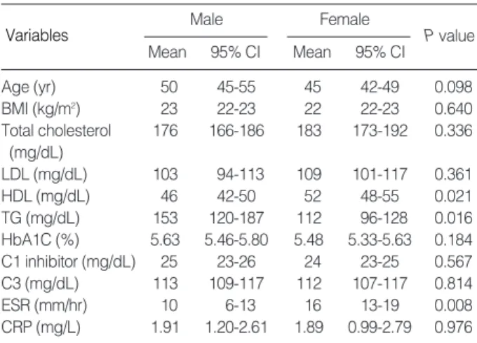
The mean and 95% confidence interval of age for 100 subjects were 47.81 and 44.92-50.70 yr, and its values ranged from 18 to 77. There was no age difference between the males and females (Table 2); however, the smoking habit (male 80.9%
vs. female 1.7%) and hypertension rate (male 40.5% vs. female 20.7%) were higher in males than females (P value <0.001
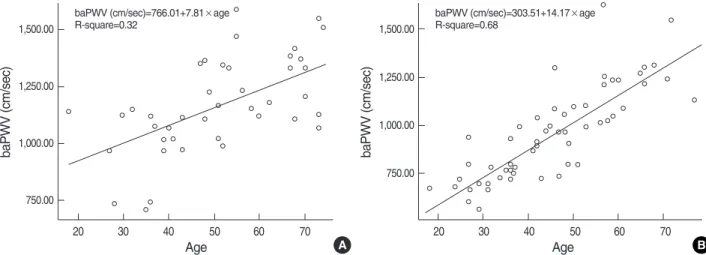
and 0.044, respectively by chi-square test). Fig. 2 depicts that baPWVs increase in proportion to the advancing age in both males and females. In Table 3, male gender, smokers, and hypertensive subjects had higher baPWVs than females, non- smokers, and non-hypertensive subjects. The univariate anal- ysis indicated that baPWV had a positive correlation with age, BMI, TG, HbA1c, C1 inhibitor, C3, and CRP; howev- er, HDL had a negative correlation (Table 4). The multiple regression analysis (stepwise method) showed not only the conventional risk factors of arteriosclerosis and/or atheroscle-
Independent sample t-test; P<0.05 is significant.
CI, confidence interval; BMI, body mass index; LDL, low density lipopro- tein; HDL, high density lipoprotein; TG, triglycerides; HbA1c, hemoglobin A1c; C1, complement 1; C3, complement 3; ESR, erythrocyte sedimen- tation rate; CRP, C-reactive protein.
Male Female
P value Mean 95% CI Mean 95% CI
Age (yr) 50 45-55 45 42-49 0.098
BMI (kg/m2) 23 22-23 22 22-23 0.640
Total cholesterol 176 166-186 183 173-192 0.336 (mg/dL)
LDL (mg/dL) 103 94-113 109 101-117 0.361
HDL (mg/dL) 46 42-50 52 48-55 0.021
TG (mg/dL) 153 120-187 112 96-128 0.016
HbA1C (%) 5.63 5.46-5.80 5.48 5.33-5.63 0.184 C1 inhibitor (mg/dL) 25 23-26 24 23-25 0.567
C3 (mg/dL) 113 109-117 112 107-117 0.814
ESR (mm/hr) 10 6-13 16 13-19 0.008
CRP (mg/L) 1.91 1.20-2.61 1.89 0.99-2.79 0.976 Table 2. The difference of clinical characteristics between the male and female participants
1,300
1,200
1,100
1,000
900
800
700
700 800 900 1,000 1,100 1,200 1,300
ICC=0.8783 ICC=0.8927
r1 vs. r2 r1 vs. r3 r2 vs. r3 baPWV (cm/sec)
1,300
1,200
1,100
1,000
900
800
700
700 800 900 1,000 1,100 1,200 1,300 baPWV (cm/sec)
Trial 2
Trial 1
A B
Fig. 1. Inter-observer and intra-observer reliability of measurements of the brachial ankle pulse wave velocity (cm/sec). (A) Inter-observer reliability (reproducibility) depicts the relationship between three independent measurements by three observers, r1 (observer 1), r2 (observ- er 2), and r3 (observer 3). The ICC value is 0.8783. (B) Intra-observer reliability (repeatability) shows the relationship between two differ- ent measurements by one observer. The ICC value is 0.8927.
Variables
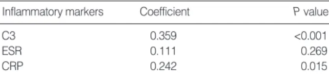
rosis, such as age, gender, hypertension, and BMI but also the acute phase protein, C1 inhibitor, was a significant pre- dictor of baPWV (Table 5, Fig. 3). C1 inhibitor revealed pos- itive correlation with other inflammatory markers, C3 and
CRP (Table 6).
Variables Coefficient P value
Age 0.713 <0.001*
BMI 0.362 <0.001*
Total cholesterol 0.070 0.484*
LDL 0.037 0.710*
HDL -0.243 0.014�
TG 0.349 <0.001�
HbA1C 0.414 <0.001�
C1 inhibitor 0.329 <0.001*
C3 0.329 <0.001*
ESR 0.129 0.199�
CRP 0.324 <0.001�
Table 4. Univariate analysis between brachial ankle pulse wave velocity and the variables
*Pearson correlation (Variables have normal distribution.); �Spearman cor- relation (Variables do not have normal distribution.); P<0.05 is significant.
BMI, body mass index; LDL, low density lipoprotein; HDL, high density lipoprotein; TG, triglycerides; HbA1c, hemoglobin A1c; C1, complement 1; C3, complement 3; ESR, erythrocyte sedimentation rate; CRP, C-reac- tive protein.
Variables Regression coefficient P value R2
Constant 657.06 <0.001 0.695
Age 7.365 <0.001
Gender -137.521 <0.001
Hypertension -143.066 <0.001
BMI 14.605 0.006
C1 inhibitor 6.367 0.025
Table 5. Predictors for brachial ankle pulse wave velocity in mul- tiple regression analysis
Multiple regression analysis (stepwise method); gender (male=1, female=
2); hypertension (yes=1, no=2); P<0.05 is significant.
BMI, body mass index; C1, complement 1.
baPWV (cm/sec)=766.01+7.81×age R-square=0.32
baPWV (cm/sec)
1,500.00
1,250.00
1,000.00
750.00
20 30 40 50 60 70
Age
baPWV (cm/sec)=303.51+14.17×age R-square=0.68
baPWV (cm/sec)
1,500.00
1,250.00
1,000.00
750.00
20 30 40 50 60 70
A Age B
Fig. 2. Linear regression graphs between baPWV and age in both males and females. (A) male, (B) female, baPWVs increase in propor- tion to the age in both males and females.
baPWV (cm/sec)=652.98+15.59×C1 inhibitor R-square=0.11
baPWV (cm/sec)
1,500.00
1,250.00
1,000.00
750.00
15.00 20.00 25.00 30.00 35.00 C1 inhibitor (mg/dL)
Fig. 3. Linear regression graph between baPWV and C1 inhibitor.
Brachial ankle pulse wave velocity showed positive relationship to the serum level of C1 inhibitor. baPWV increases according to the increasing serum level of C1 inhibitor.
Independent-sample t-test; P<0.05 is significant.
Variables Number Mean (cm/sec)
95% CI
(cm/sec) P value
Gender Male 42 1,161 1,097-1,226 <0.001 Female 58 952 888-1,015
Smoking Yes 35 1,126 1,054-1,198 0.010
No 65 993 929-1,057
Hypertension Yes 29 1,288 1,222-1,354 <0.001
No 71 938 891-985
Table 3. Comparison of brachial ankle pulse wave velocity as a function of gender, smoking, and hypertension
DISCUSSION
In the current study, baPWV had a significant association with the conventional risk factors of arteriosclerosis and/or atherosclerosis, such as age, gender, hypertension, and BMI in both univariate and multivariate studies. This means that baPWV is a clinically relevant method to evaluate the arter- ies, and this result is compatible with the other studies on the relation between baPWV and risk factors (6, 7, 10, 14, 15). According to our hypothesis, we measured the relative- ly novel inflammatory marker, C1 inhibitor. In the result, the serum level of C1 inhibitor was positively related to baPWV, and C1 inhibitor was one of the significant predictors of ba- PWV. The serine proteinase inhibitor, C1 inhibitor, is syn- thesized and secreted from the several cell types, including hepatocytes, mononuclear phagocytes, fibroblasts, and umbil- ical vein endothelial cells (16-18). The major function of the C1 inhibitor is inhibition of the complement system to pre- vent spontaneous activation, in which the amount of C1 in- hibitor in the serum is crucial. A minimal C1 inhibitor con- centration of approximately 5.5 mg/dL (22% of the normal human serum level) is required to control C1-autoactivation (19). Originally, C1 inhibitor was shown to be associated with angioedema, and its deficiency was identified as the cause of hereditary angioedema (20, 21). However, C1 inhibitor be- longs to a group of acute phase proteins, and its production and release increase during inflammation (3, 4). In our study, C1 inhibitor revealed positive correlation with C3 and CRP, thus indicating that C1 inhibitor was a member of acute phase proteins. Recently, it has been suggested that all the risk fac- tors of atherosclerosis contribute to the development of athe- rosclerosis by aggravating the underlying inflammatory pro- cess (5, 8, 9, 11, 22-25). Although the exact mechanism is unknown, it has been reported that the inflammatory cyto- kine, interferon-gamma (IFN-gamma) is highly expressed in atherosclerotic lesions (26), moreover, IFN-gamma also enhances the expression of C1 inhibitor mRNA, primarily due to an enhanced transcription rate (27). Other cytokines, such as tumor necrosis factor alpha, interferon-alpha, mono- cyte colony stimulating factor, and interleukin-6 were also identified to stimulate the synthesis of C1 inhibitor (2). De- termination of baPWV involves a simple and non-invasive method, and its validity, reproducibility, and clinical signif- icance have been evaluated and confirmed in a previous study
(10). In this study, we also demonstrated that the reproduci- bility and repeatability of measuring baPWV with Vasoguard equipment is reliable. In recent years, baPWV has been used more widely in assessing arterial stiffness, and has become available in a clinical setting as a simple predictor of the prog- nosis of patients (9, 14, 15, 28-30). In current study, we mea- sured baPWV as a parameter of arterial stiffness and C1 in- hibitor as a counteracting anti-inflammatory substance. Our result showed that baPWV had positive relation with C1 in- hibitor; the higher the baPWV, the greater the serum level of C1 inhibitor. In summary, C1 inhibitor was positively relat- ed to baPWV, and was one of the significant predictors of ba- PWV. The present study suggests that C1 inhibitor is asso- ciated with arterial stiffness through its association with incre- ased inflammation.
REFERENCES
1. Zanker KS. General introduction to innate immunity: Dr. Jekyl/Mr.
Hyde quality of the innate immune system. Contrib Microbiol 2008;
15: 12-20.
2. Caliezi C, Wuillemin WA, Zeerleder S, Redondo M, Eisele B, Hack CE. C1-Esterase inhibitor: an anti-inflammatory agent and its poten- tial use in the treatment of diseases other than hereditary angioede- ma. Pharmacol Rev 2000; 52: 91-112.
3. Kalter ES, Daha MR, ten Cate JW, Verhoef J, Bouma BN. Activation and inhibition of Hageman factor-dependent pathways and the com- plement system in uncomplicated bacteremia or bacterial shock. J Infect Dis 1985; 151: 1019-27.
4. Woo P, Lachmann PJ, Harrison RA, Amos N, Cooper C, Rosen FS.
Simultaneous turnover of normal and dysfunctional C1 inhibitor as a probe of in vivo activation of C1 and contact activatable proteases.
Clin Exp Immunol 1985; 61: 1-8.
5. Kostner KM, Fahti RB, Case C, Hobson P, Tate J, Marwick TH. In- flammation, complement activation and endothelial function in sta- ble and unstable coronary artery disease. Clin Chim Acta 2006; 365:
129-34.
6. Jani B, Rajkumar C. Ageing and vascular ageing. Postgrad Med J 2006; 82: 357-62.
7. Wykretowicz A, Gerstenberger P, Guzik P, Milewska A, Krauze T, Adamska K, Rutkowska A, Wysocki H. Arterial stiffness in relation to subclinical atherosclerosis. Eur J Clin Invest 2009; 39: 11-6.
8. Ridker PM, Silvertown JD. Inflammation, C-reactive protein, and atherothrombosis. J Periodontol 2008; 79 (8 Suppl): 1544-51.
9. van Popele NM, Grobbee DE, Bots ML, Asmar R, Topouchian J, Re- neman RS, Hoeks AP, van der Kuip DA, Hofman A, Witteman JC.
Association between arterial stiffness and atherosclerosis: the Rot- terdam Study. Stroke 2001; 32: 454-60.
10. Yamashina A, Tomiyama H, Takeda K, Tsuda H, Arai T, Hirose K, Koji Y, Hori S, Yamamoto Y. Validity, reproducibility, and clinical significance of noninvasive brachial-ankle pulse wave velocity mea- surement. Hypertens Res 2002; 25: 359-64.
11. Nicoletti A, Caligiuri G, Hansson GK. Immunomodulation of athero-
Inflammatory markers Coefficient P value
C3 0.359 <0.001
ESR 0.111 0.269
CRP 0.242 0.015
Table 6. Relation of inflammatory markers with C1 inhibitor
Spearman correlation; P<0.05 is significant.
C1, complement 1; C3, complement 3; ESR, erythrocyte sedimentation rate; CRP, C-reactive protein.
sclerosis: myth and reality. J Intern Med 2000; 247: 397-405.
12. Kullo IJ, Gau GT, Tajik AJ. Novel risk factors for atherosclerosis.
Mayo Clin Proc 2000; 75: 369-80.
13. Rosner B. Fundamentals of Biostatistics. 4th ed. Belmont, California, USA: Duxbury Press 1995.
14. Kim DH, Kim J, Kim JM, Lee AY. Increased brachial-ankle pulse wave velocity is independently associated with risk of cerebral isch- emic small vessel disease in elderly hypertensive patients. Clin Neu- rol Neurosurg 2008; 110: 599-604.
15. Xu Y, Wu Y, Li J, Ma W, Guo X, Luo Y, Hu D. The predictive value of brachial-ankle pulse wave velocity in coronary atherosclerosis and peripheral artery diseases in urban Chinese patients. Hypertens Res 2008; 31: 1079-85.
16. Lappin D, Whaley K. Regulation of C1-inhibitor synthesis by inter- ferons and other agents. Behring Inst Mitt 1989: 180-92.
17. Zuraw BL, Lotz M. Regulation of the hepatic synthesis of C1 inhibitor by the hepatocyte stimulating factors interleukin 6 and interferon ga- mma. J Biol Chem 1990; 265: 12664-70.
18. Prada AE, Zahedi K, Davis AE 3rd. Regulation of C1 inhibitor syn- thesis. Immunobiology 1998; 199: 377-88.
19. Windfuhr JP, Alsenz J, Loos M. The critical concentration of C1-es- terase inhibitor (C1-INH) in human serum preventing auto-activation of the first component of complement (C1). Mol Immunol 2005; 42:
657-63.
20. Johnson AM, Alper CA, Rosen FS, Craig JM. C1 inhibitor: evidence for decreased hepatic synthesis in hereditary angioneurotic edema.
Science 1971; 173: 553-4.
21. Donaldson VH, Rosen FS. Action of complement in hereditary an- gioneurotic edema: the role of C’1-esterase. J Clin Invest 1964; 43:
2204-13.
22. Natali A, L’Abbate A, Ferrannini E. Erythrocyte sedimentation rate, coronary atherosclerosis, and cardiac mortality. Eur Heart J 2003;
24: 639-48.
23. Buono C, Come CE, Witztum JL, Maguire GF, Connelly PW, Car- roll M, Lichtman AH. Influence of C3 deficiency on atherosclerosis.
Circulation 2002; 105: 3025-31.
24. Mallika V, Goswami B, Rajappa M. Atherosclerosis pathophysiolo- gy and the role of novel risk factors: a clinicobiochemical perspective.
Angiology 2007; 58: 513-22.
25. Bisoendial RJ, Kastelein JJ, Stroes ES. C-reactive protein and athero- genesis: from fatty streak to clinical event. Atherosclerosis 2007; 195:
e10-8.
26. McLaren JE, Ramji DP. Interferon gamma: a master regulator of atherosclerosis. Cytokine Growth Factor Rev 2009; 20: 125-35.
27. Zahedi K, Prada AE, Davis AE 3rd. Transcriptional regulation of the C1 inhibitor gene by gamma-interferon. J Biol Chem 1994; 269:
9669-74.
28. Tomiyama H, Koji Y, Yambe M, Shiina K, Motobe K, Yamada J, Shido N, Tanaka N, Chikamori T, Yamashina A. Brachial--ankle pul- se wave velocity is a simple and independent predictor of prognosis in patients with acute coronary syndrome. Circ J 2005; 69: 815-22.
29. Hung CS, Lin JW, Hsu CN, Chen HM, Tsai RY, Chien YF, Hwang JJ. Using brachial-ankle pulse wave velocity to associate arterial stiff- ness with cardiovascular risks. Nutr Metab Cardiovasc Dis 2009; 19:
241-6.
30. Farrar DJ, Green HD, Bond MG, Wagner WD, Gobbee RA. Aortic pulse wave velocity, elasticity, and composition in a nonhuman pri- mate model of atherosclerosis. Circ Res 1978; 43: 52-62.